Medical treatment method and apparatus

a technology of insertion support and insertion portion, which is applied in the direction of surgical instrument support, catheter, application, etc., can solve the problems of large frictional force between, difficult to smoothly insert or retract the insertion portion, and no established medical treatment for treating the lesioned part of the organ using such a support tool

Inactive Publication Date: 2006-01-31
OLYMPUS CORP
View PDF4 Cites 330 Cited by
  • Summary
  • Abstract
  • Description
  • Claims
  • Application Information

AI Technical Summary

Problems solved by technology

Therefore, although the balloon portion is formed of such a material that has low friction with the insertion portion of the endoscope, a large frictional force is generated between the insertion portion of the endoscope and the

Method used


Image

  • Medical treatment method and apparatus
  • Medical treatment method and apparatus
  • Medical treatment method and apparatus

Examples


first embodiment

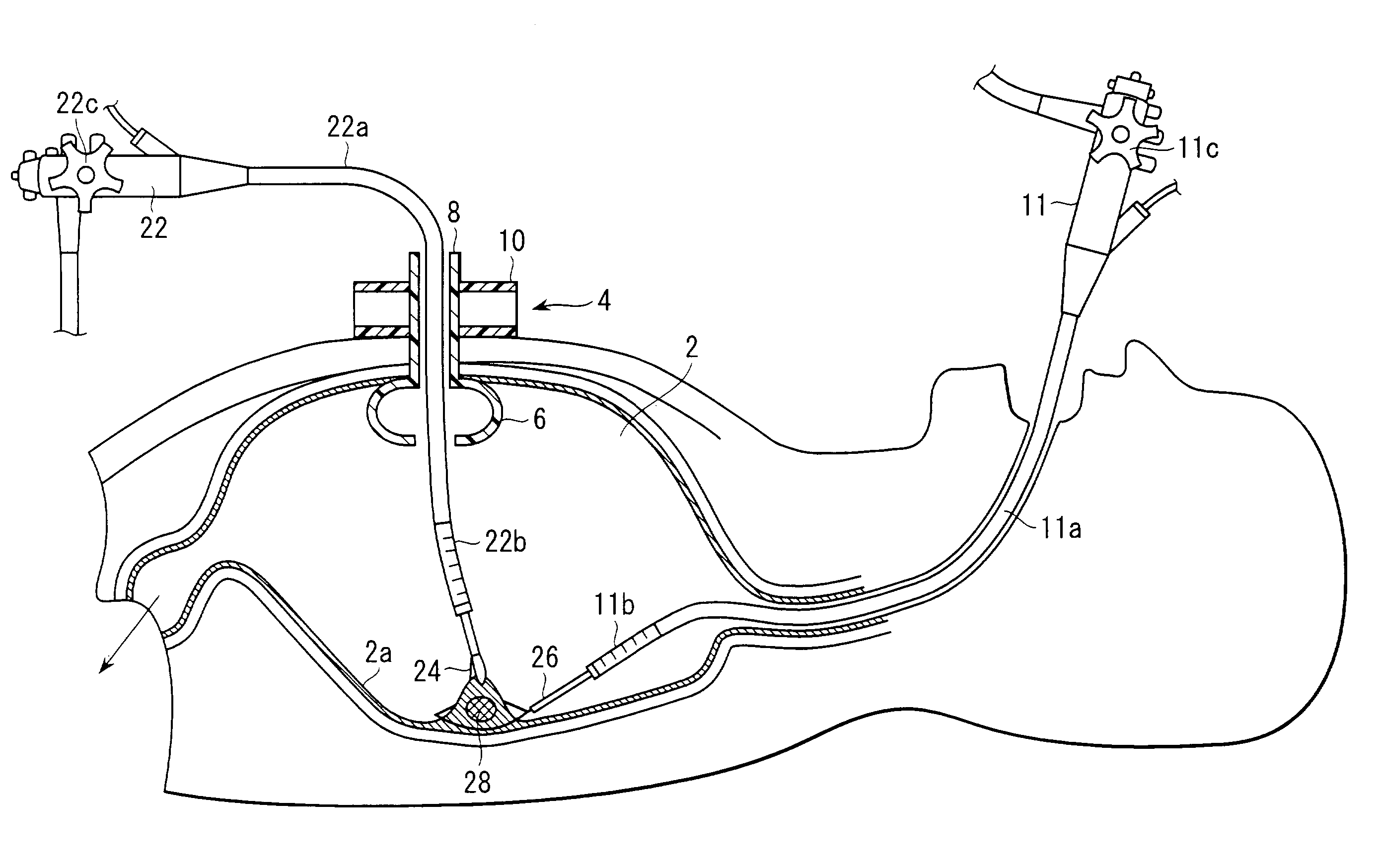
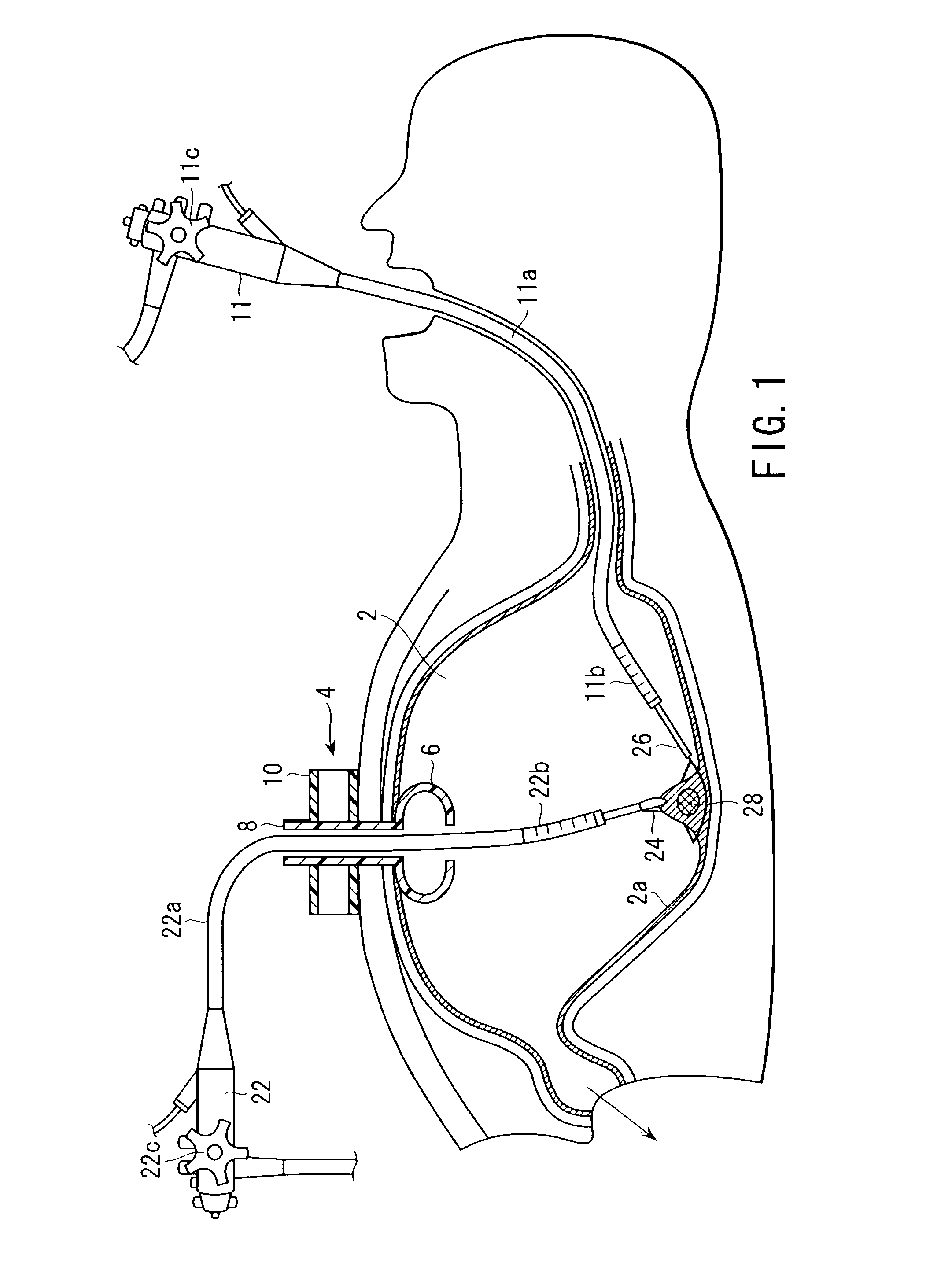
[0071]A first embodiment will be first explained in connection with FIGS. 1 to 3.

[0072]As shown in FIG. 1, a treatment apparatus according to this embodiment includes two flexible endoscopes 11 and 22 each having flexible insertion portions 11a and 22a which can be inserted into a body cavity as insertion members. Curved portions 11b and 22b which can be bent are provided at respective ends of the insertion portions 11a and 22a of the flexible endoscopes 11 and 22. The curved portions 11b and 22b are operated by operation portions 11c and 22c respectively provided in main bodies of the endoscopes 11 and 22. Although not shown, at least one of a forceps channel, an air supply / water supply / suction channel and an observation optical system is provided to each of the insertion portions 11a and 22a of the endoscopes 11 and 22. In this embodiment, at least one forceps channel and an observation optical system are provided to each of the insertion portions 11a and 22a of the respective end...

seventh embodiment

[0141]Moreover, like the seventh embodiment, the seal member 98 is fitted to the upper end portion of the gastric fistula formation tube 4. Into respective holes or slits 98a and 98b of the seal member 98 are inserted the insertion portion 22a of the second endoscope 22 and the grasping forceps 102 which is a rigid treatment tool (insertion member), respectively. An opening / closing operation portion 102a and a swiveling operation portion 102b are provided to the rigid grasping forceps 102 and, as shown in FIG. 11B, the end of this grasping forceps is formed so as to be capable of being opened / closed and swiveling.

[0142]When the lesioned part 28 exists on the gastric mucosa in the stomach 2 on the back side, the lesioned part 28 is treated by the two flexible endoscopes 11 and 22 as follows.

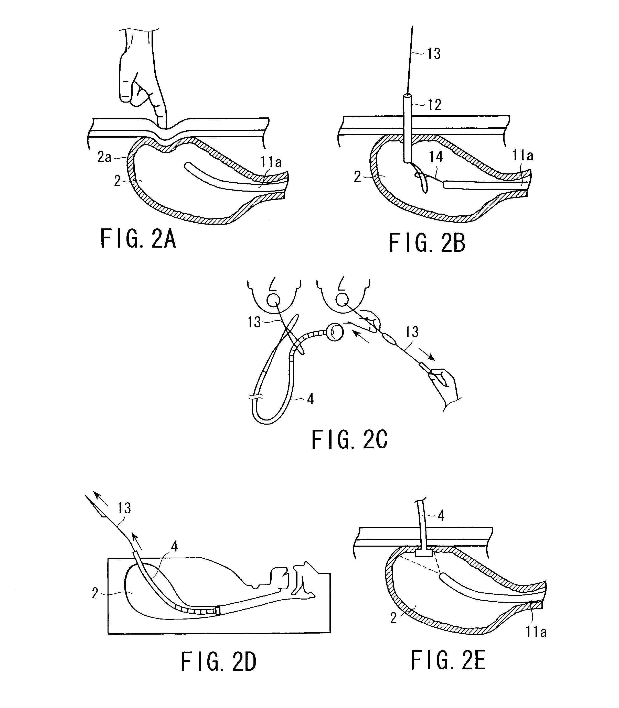
[0143]Like the first embodiment, after confirming a position and a size of the lesioned part 28 in the stomach 2 by using the first endoscope 11, the gastric fistula formation tube 4 is positioned...

eighth embodiment

[0234]When the lesioned part 28 exists on the gastric mucosa in the stomach 2 on the back side, the same medical treatment as that described in connection with the eighth embodiment is given to the lesioned part 28 by these two flexible endoscopes 11 and 22.

[0235]Therefore, the following can be said with respect to this embodiment. By leading the first endoscope 11 toward the lesioned part 28 by using the rigid grasping forceps 102, an operator of the second endoscope 22 can readily issue an instruction to an operator of the first endoscope 11.

[0236]A 21st embodiment will now be described with reference to FIGS. 28A to 28C. This embodiment is a modification of the 16th embodiment, and like reference numerals denote like or corresponding parts, thereby omitting the detailed explanation.

[0237]As shown in FIG. 28A, in a medical treatment apparatus according to this embodiment, the first and second flexible endoscopes 11 and 22 are inserted into the stomach 2. A clip with a thread 152 i...


PUM


Abstract

A medical treatment method comprises orally inserting a first endoscope having a first treatment tool provided thereto into a body cavity, inserting a first insertion member insertion support tool from at least one of abdominal and transdermal cavities into the body cavity, inserting a second endoscope having a second treatment tool provided thereto into the body cavity through the first insertion member insertion support tool, grasping a lesioned part by the second treatment tool inserted into the body cavity by the second endoscope, cutting the lesioned part by the first treatment tool inserted into the body cavity by the first endoscope, and collecting the lesioned part.

Description

CROSS-REFERENCE TO RELATED APPLICATIONS[0001]This application is based upon and claims the benefit of priority from the prior Japanese Patent Application No. 2002-005106, filed Jan. 11, 2002, the entire contents of which are incorporated herein by reference.BACKGROUND OF THE INVENTION[0002]1. Field of the Invention[0003]The present invention relates to a medical treatment method for treating a lesioned part in an organ, and an apparatus using this method.[0004]2. Description of the Related Art[0005]For example, as disclosed in U.S. Pat. No. 5,545,179, in regard to an insertion support tool used to insert an endoscope into a body cavity, an insertion portion of the endoscope is inserted from an insertion opening of this insertion support tool. In this case, the inside of the insertion opening, namely, a balloon portion in the body cavity is inflated and the insertion portion of the endoscope is inserted into the body while being appressed against the balloon portion. The horizontal c...

Claims


Application Information

Patent Timeline
IPC IPC(8): A61B1/00A61B1/01A61B1/018A61B1/31A61B1/313A61B17/00A61B17/34A61B18/14A61B19/00
CPCA61B1/00073A61B1/015A61B1/00154A61B1/01A61B1/313A61B17/3415A61B17/3421A61B1/018A61B1/31A61B19/26A61B2017/00269A61B2017/00296A61B2017/3445A61B2018/1407A61B1/00149A61B1/0014A61B90/50
Inventor NAKAZAWA, MASAAKINAKAMURA, TOSHIOKIMURA, HIDENOBUFURUKAWA, TATSUYAYAMAYA, KOJIMATSUURA, NOBUYUKIYABE, HISAO
Owner OLYMPUS CORP
Why Eureka
Try Eureka