Atherectomy catheter with aligned imager

a catheter and imager technology, applied in the field of atherectomy catheters, can solve the problems of user's first scan of a large area, limited surgeon's view ability, and inability to accurately diagnose the area,

Inactive Publication Date: 2006-02-14
TYCO HEALTHCARE GRP LP
View PDF2 Cites 199 Cited by
  • Summary
  • Abstract
  • Description
  • Claims
  • Application Information

AI Technical Summary

Benefits of technology

[0008]In a first aspect, the present invention provides a catheter for use in excising material from a body lumen. The catheter comprises a catheter body, a cutting blade, and an imaging device. The cutting blade is mounted on the catheter body and configured to move between a material capture (open) position and a closed position. When the cutting blade is in the closed position, the blade substantially closes an aperture or “cutting window” on the catheter body. The imaging device which is usually mounted on or coupled to the cutting blade is configured to be in an imaging position when the cutting blade substantially closes the aperture or cutter window. This allows the imaging device to survey material within a “cutting zone” of the atherectomy catheter, preferably precisely at the location where severing of the material will occur, most preferably being aligned with the location of a cutting edge on the cutting blade when the blade is in the capture configuration. By allowing the imaging device to view materials within this cutting zone, preferably precisely at the spot where severing of the material will occur or be initiated, the material may be imaged and then removed from the body lumen without having to ...

Problems solved by technology

Atheromatous and other vascular deposits restrict blood flow and can cause ischemia which, in acute cases, can result in myocardial infarction.
Although some existing catheters provide imaging capability, these devices generally require repositioning of the catheter and the cutting element between imaging and cutting procedures.
This undesirably limits the ability of the surgeon to view the exact area of occlusive material to be removed prior to the cut.
Such known devices also typically cause the user to first scan a large, less precise area of body lumen.
Additionally, the repositioning of the en...

Method used


Image

  • Atherectomy catheter with aligned imager
  • Atherectomy catheter with aligned imager
  • Atherectomy catheter with aligned imager

Examples


Embodiment Construction

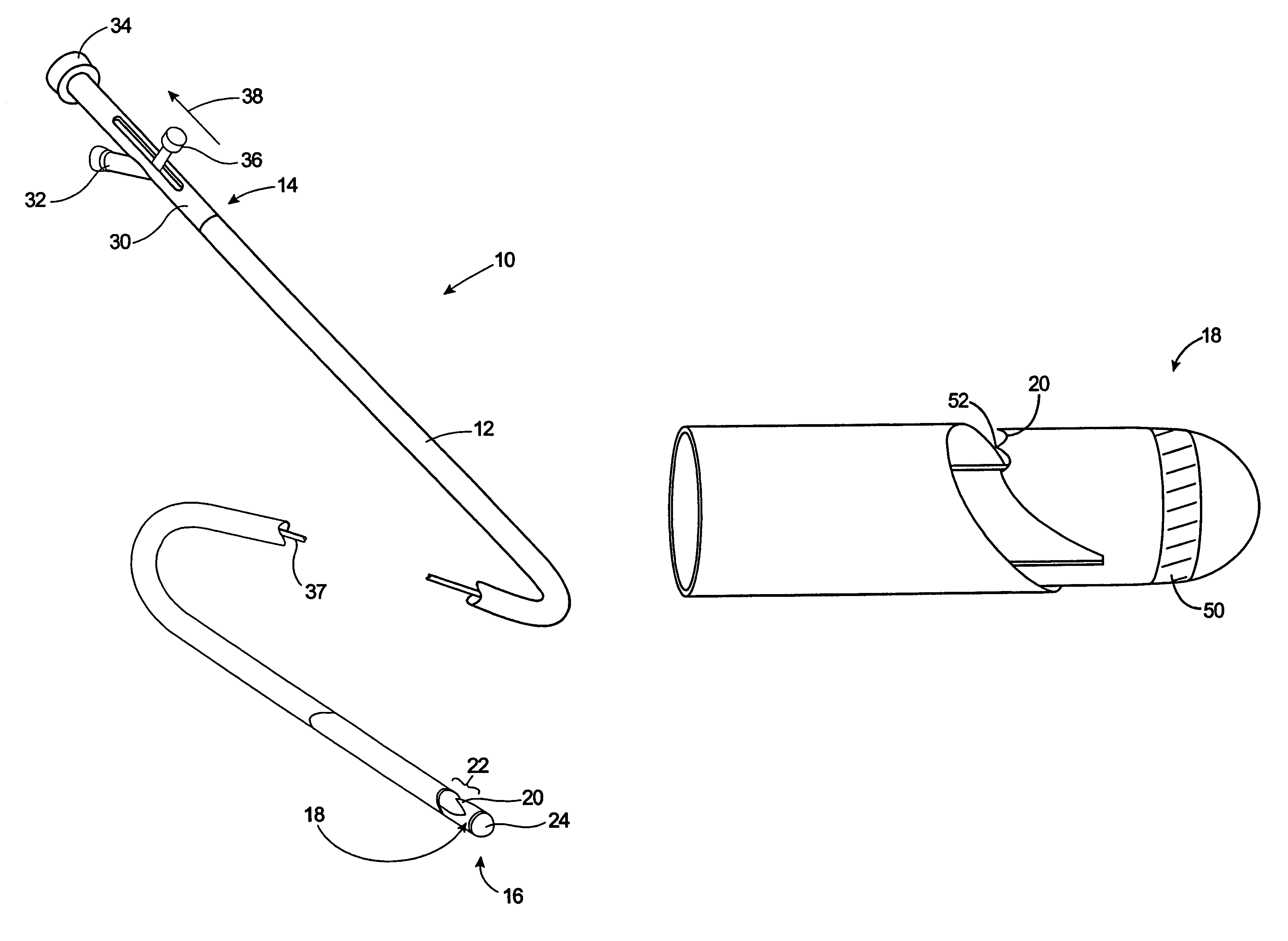
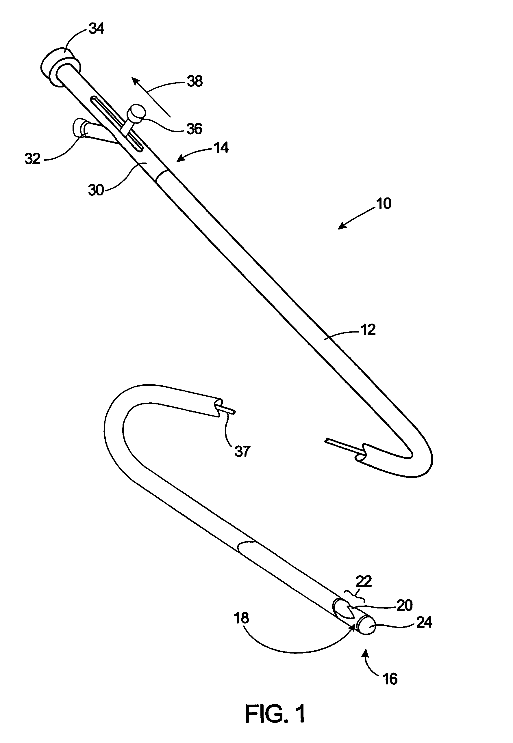
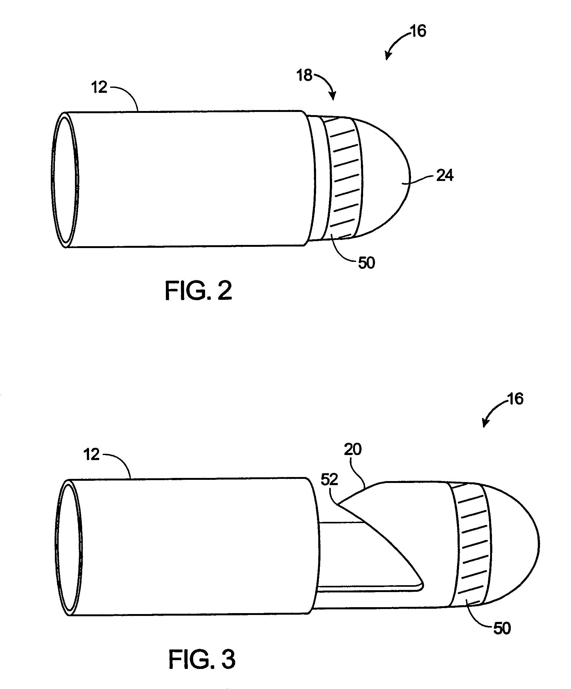
[0021]The present invention is generally directed to excising material from a body lumen. More particularly, the present invention provides catheters, methods, and kits for imaging material to be removed from a body lumen prior to performing the removal or cutting procedure. The present invention advantageously allows for the imaging of material to be cut prior to the cutting or removal procedure. Furthermore, the material may be imaged and then cut without requiring the repositioning of the catheter body as commonly required in conventional intravascular catheters.

[0022]Apparatus according to the present invention will comprise catheters having catheter bodies adapted for intraluminal introduction to the target body lumen. The dimensions and other physical characteristics of the catheter bodies will vary significantly depending on the body lumen which is to be accessed. In the exemplary case of atherectomy catheters intended for intravascular introduction, the catheter bodies will ...


PUM


Abstract

An atherectomy catheter is provided for excising and imaging material in a body lumen. In one embodiment, the catheter comprises a catheter body, a cutting blade, and an imaging device. The cutting blade is mounted on the catheter body and configured to move between a first position and a second position relative to an aperture or cutting window. When the cutting blade is in the second position, the blade substantially closes the aperture on the catheter body. The imaging device which is mounted on or otherwise coupled to the cutting blade is configured to be in an imaging position when the cutting blade substantially closes the aperture or cutter window. This allows the imaging device to survey material within the cutting zone of the atherectomy catheter. By allowing the imaging device to view materials within this cutting zone, material may be imaged and then removed from the body lumen without having to reposition the catheter between each step.

Description

CROSS-REFERENCES TO RELATED APPLICATIONS[0001]This application is a divisional of U.S. Ser. No. 09 / 930,372; filed on Aug. 14, 2001 now U.S. Pat. No. 6,623,496, which is a continuation of U.S. Ser. No. 09 / 378,224; filed on Aug. 19, 1999, now U.S. Pat. No. 6,299,622, each of which is expressly incorporated herein in its entirety by reference.BACKGROUND OF THE INVENTION[0002]The present invention relates generally to apparatus and methods for removing occluding materials from body lumens. More particularly, the present invention relates to the construction and use of atherectomy catheters with an ability to view atheroma and other materials to be excised from blood vessels.[0003]Cardiovascular disease frequently arises from the accumulation of atheromatous material on the inner walls of vascular lumens, particularly arterial lumens of the coronary and other vasculature, resulting in a condition known as atherosclerosis. Atherosclerosis occurs naturally as a result of aging, but may als...

Claims


Application Information

Patent Timeline
IPC IPC(8): A61B17/22A61B19/00
CPCA61B17/32075A61B17/320783A61B2017/320791A61B2019/5217A61B2017/00353A61B2019/5276A61B2019/528A61B2017/00022A61B2017/00292A61B2019/5231A61B2090/3614A61B2090/373A61B2090/378A61B2090/3784
Inventor SNOW, DAVID W.FLOM, JAMES R.
Owner TYCO HEALTHCARE GRP LP
Why Eureka
Try Eureka