Lasso for pulmonary vein mapping and ablation

a technology of pulmonary veins and lassos, applied in the field of electrophysiological mapping of intracardiac sites, can solve the problems of asynchronous rhythm and disruption of normal cardiac cycle, and achieve the effect of increasing accuracy

Inactive Publication Date: 2005-12-06
BIOSENSE WEBSTER INC
View PDF54 Cites 197 Cited by
  • Summary
  • Abstract
  • Description
  • Claims
  • Application Information

AI Technical Summary

Benefits of technology

[0026]It is an object of some aspects of the present invention to provide apparatus and methods to increase the accuracy with which electrophysiological properties of the heart are mapped.
[0028]It is a further object of some aspects of the present invention to provide improved apparatus and methods for real-time, accurate, simultaneous determination of the location of a plurality of electrophysiological sensing elements mounted on an intracardiac catheter.
[0029]It is yet a further object of some aspects of the present invention to provide apparatus and methods for reducing the time required for mapping electrophysiological properties of the heart.
[0030]It is still a further object of some aspects of the present invention to provide apparatus and methods for intracardiac mapping which can readily be integrated into existing mapping support systems and methods, in order to increase the accuracy of these systems and methods.
[0032]Advantageously, the single-coil position sensors, which are generally substantially smaller than the multi-coil position sensor, are fixed to the curved section, which preferably is relatively small in order to be readily placed in the PV. The larger multi-coil position sensor is fixed to the base section, which can be relatively large because it remains within the atrium. Six-dimensional information from the multi-coil position sensor, combined with the generally known angle of the joint and predetermined flexing behavior of the curved section, compensates for the more limited information from the single-coil position sensors. The resulting accurate position information for the sensing electrodes enables high-resolution electrophysiological mapping of the PV and accurate location of regions of abnormal electrical behavior.
[0034]Alternatively, in some preferred embodiments of the present invention, the sensing electrodes on the mapping catheter are adapted to additionally perform ablation of selected sites, or the mapping catheter further comprises ablation elements, thereby eliminating the need for separate ablation apparatus. Preferably, the termination of the abnormal electrical activity is confirmed using the sensing electrodes prior to removal of the mapping catheter.

Problems solved by technology

Cardiac arrhythmia, such as atrial fibrillation, occurs when regions of cardiac tissue abnormally conduct electric signals to adjacent tissue, thereby disrupting the normal cardiac cycle and causing asynchronous rhythm.

Method used


Image

  • Lasso for pulmonary vein mapping and ablation
  • Lasso for pulmonary vein mapping and ablation
  • Lasso for pulmonary vein mapping and ablation

Examples


Embodiment Construction

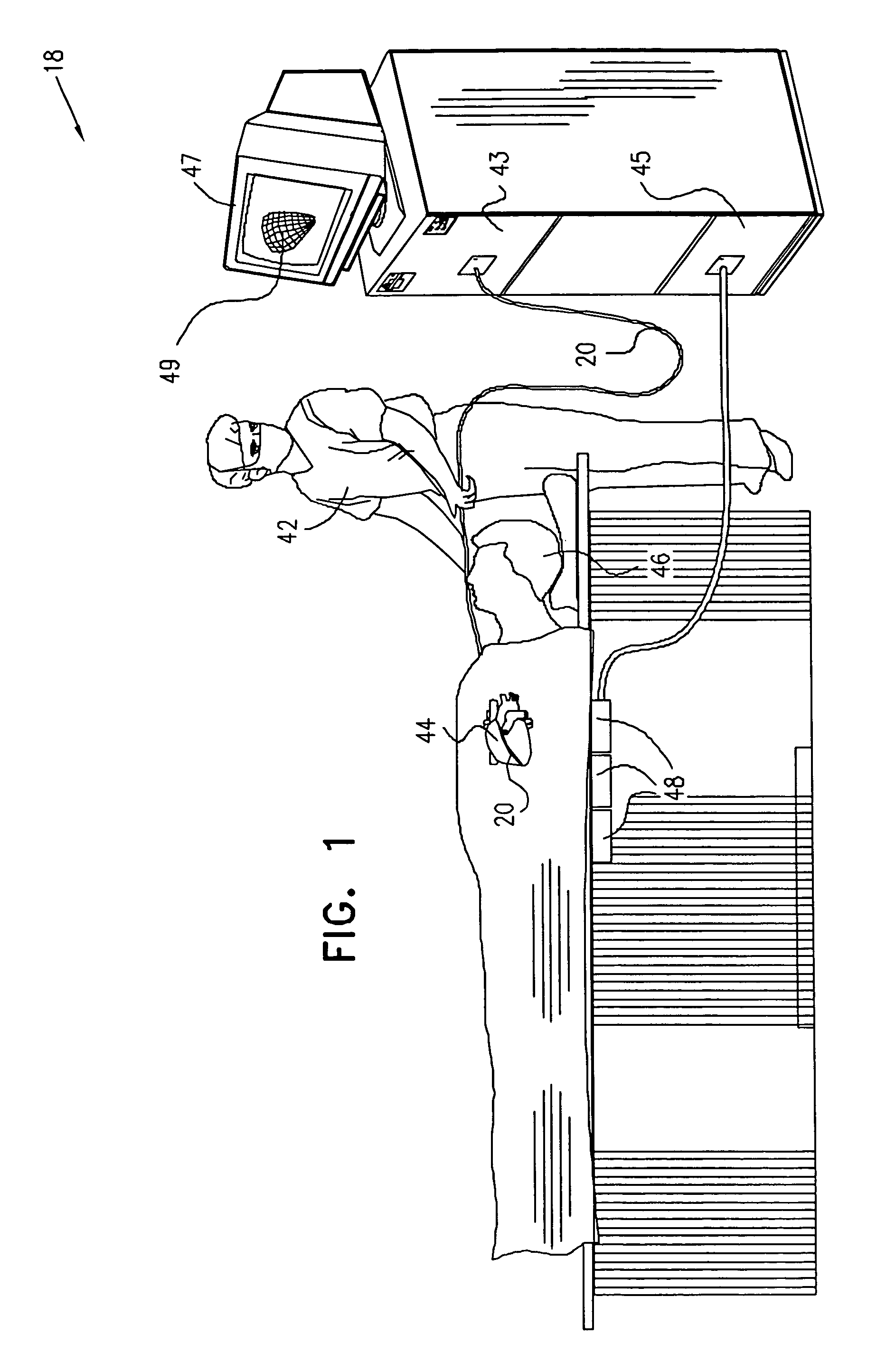
[0072]FIG. 1 is a schematic, pictorial illustration of a mapping system 18, for mapping of electrical activity in a pulmonary vein of a heart 44 of a subject 46, in accordance with a preferred embodiment of the present invention. System 18 comprises a catheter 20, which is inserted by a user 42 through a vein or artery of the subject into a pulmonary vein of the heart. Preferably, system 18 further comprises a console 43.

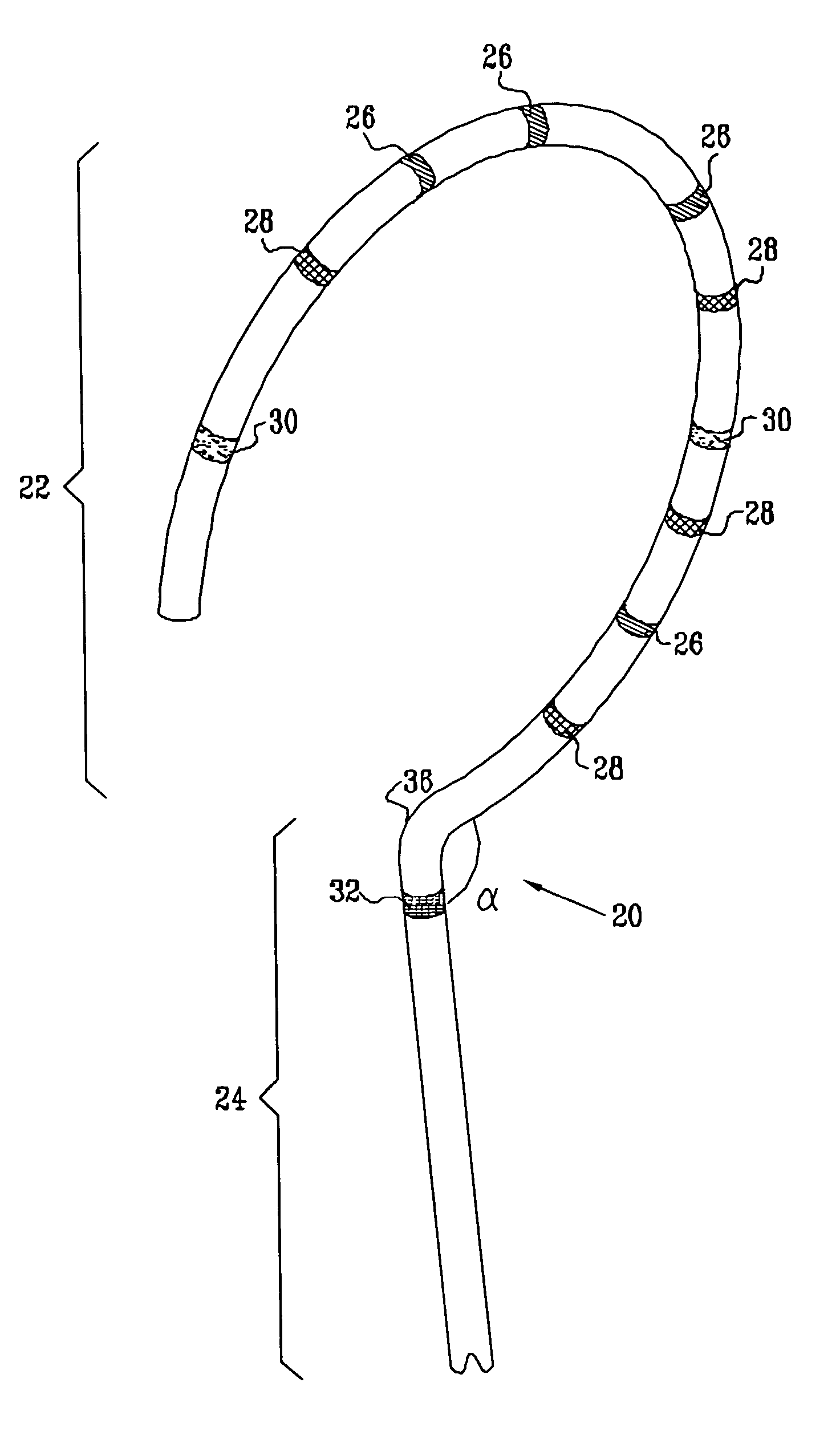
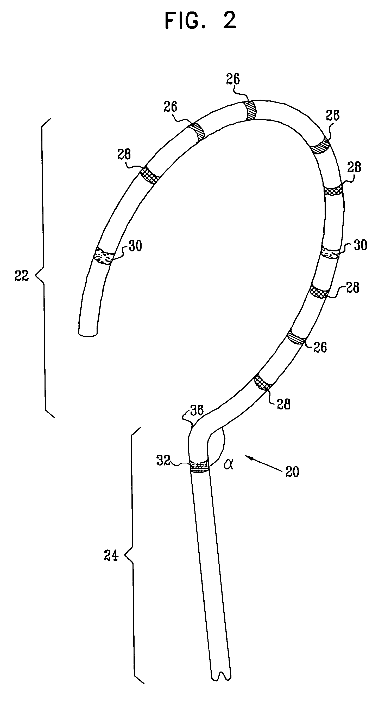
[0073]FIG. 2 is a schematic, pictorial illustration showing a distal portion of catheter 20, for facilitating accurate electrophysiological mapping of a pulmonary vein (PV) in order to enable therapeutic ablation. Catheter 20 comprises a curved section 22 joined at a generally known or range-restricted angle alpha to a base section 24, at a joint 36. Joint 36 may define the point where two initially-separate members (sections 22 and 24) are joined, or, alternatively, the joint may define the point on catheter 20 where a single member is bent, so as to form base sect...


PUM


Abstract

A method is provided for electrical mapping of a pulmonary vein of a heart, including introducing into the heart a catheter having a curved section and a base section, the base section having a distal end attached to a proximal end of the curved section. At a location on the curved section, a first position signal is generated having fewer than six dimensions of position and orientation information. At a vicinity of the distal end of the base section, a second position signal is generated having six dimensions of position and orientation information. The method also includes measuring, at one or more locations on the curved section, an electrical property of the pulmonary vein.

Description

FIELD OF THE INVENTION[0001]The present invention relates generally to intrabody mapping systems, and specifically to methods and devices for electrophysiological mapping of intracardiac sites to facilitate therapeutic procedures.BACKGROUND OF THE INVENTION[0002]Cardiac arrhythmia, such as atrial fibrillation, occurs when regions of cardiac tissue abnormally conduct electric signals to adjacent tissue, thereby disrupting the normal cardiac cycle and causing asynchronous rhythm. The primary sources of undesired signals are located in the tissue region along the pulmonary veins of the left atrium and in the superior pulmonary veins. After unwanted signals are generated in the pulmonary veins or conducted through the pulmonary veins from other sources, they are conducted into the left atrium where they can initiate or continue arrhythmia.[0003]Procedures for treating arrhythmia include surgically disrupting the origin of the signals causing the arrhythmia, as well as disrupting the con...

Claims


Application Information

Patent Timeline
Patent Type & Authority Patents(United States)
IPC IPC(8): A61B5/06A61B5/296A61B18/14A61N1/05
CPCA61B5/053A61B5/06A61B18/1492A61B2018/00375A61B2018/00839A61B2018/1467A61B5/062
Inventor GOVARI, ASSAF
Owner BIOSENSE WEBSTER INC
Why Eureka
Try Eureka