Bifurcated biological pulmonary valved conduit

a biological and valved technology, applied in blood vessels, medical science, prosthesis, etc., can solve the problems of ineffective valve operation, ineffective valve replacement, and inability to operate effectively

Inactive Publication Date: 2006-04-04
MEDTRONIC INC
View PDF15 Cites 82 Cited by
  • Summary
  • Abstract
  • Description
  • Claims
  • Application Information

AI Technical Summary

Problems solved by technology

Second, after months of usage, a layer of fibrinous material formed in the inner lumen over time from the accumulation of blood proteins.
This layer of fibrinous material, referred to as “intimal peel,” often separated from the inner wall and interfered with the effective operation of the valve.
An ineffective valve required re-operation to replace the conduit and valve, if caught in time.
In some instances, if valve replacement came too late, the results were fatal.
Moreover, for children suffering from congenital pulmonary atresia or pulmonary insufficiency, the use of pulmonary valved conduits were not always effective.
As the child grew, the prosthetic was less effective due to its inadequate size, requiring frequent replacement.
Also, for children especially, anticoagulation requirements are difficult to regiment.
While the desired result was achieved, there was still the problem of separation of the “intimal peel” from the inner lumen.
The intimal peel clogged the leaflets of the biological valve.
Moreover, while a porcine aortic valve functioned relatively well as a replacement aortic valve, where the blood pressures from the left ventricle are substantially high (80–120 mmHg), it did not function nearly as effective as a replacement pulmonary valve, where the blood pressures from the right ventricle are quite low (0–15 mmHg).
Opening at low pressures becomes more difficult.
Also, the flow therethrough becomes turbulent, which undesirably promotes the degeneration of the biological material.
Even this arrangement is limited in that the naturally formed donor conduits have normally minimum diameters of about 22 mm.

Method used


Image

  • Bifurcated biological pulmonary valved conduit
  • Bifurcated biological pulmonary valved conduit
  • Bifurcated biological pulmonary valved conduit

Examples


Embodiment Construction

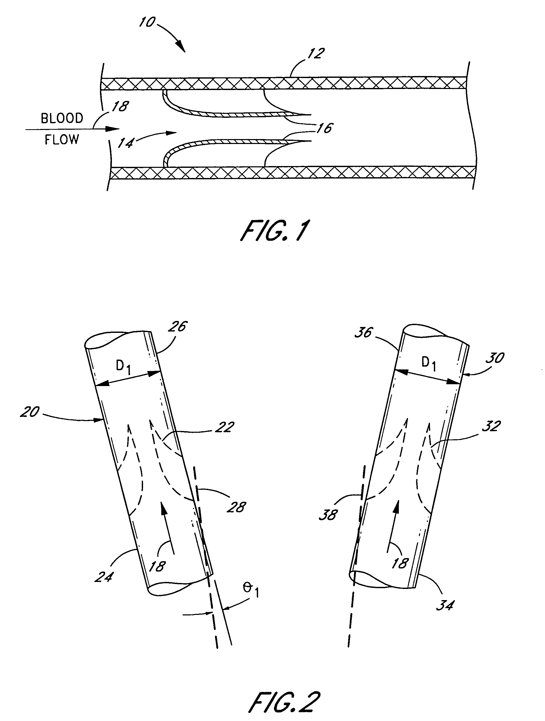
[0015]With reference to FIG. 1, the present invention improves upon the use of a naturally-formed venous valvular conduit 10 consisting of a blood vessel 12 housing a biological valve 14. The valve 14 consists of a plurality of leaflets 16 (two shown) that easily open upon the flow of low pressure blood 18, but closely tightly against a relatively low backpressure of blood; remaining sealed even against backpressures as high as 200–300 mmHg. Such venous valvular conduits may be found, as indicated above, in one of many quadrupeds. Preferably, the donor venous valvular conduit is a section of the jugular vein of a quadruped, the diameters of which approach about 22 mm. While humans have naturally-formed venous valvular conduits, none approach that diameter.

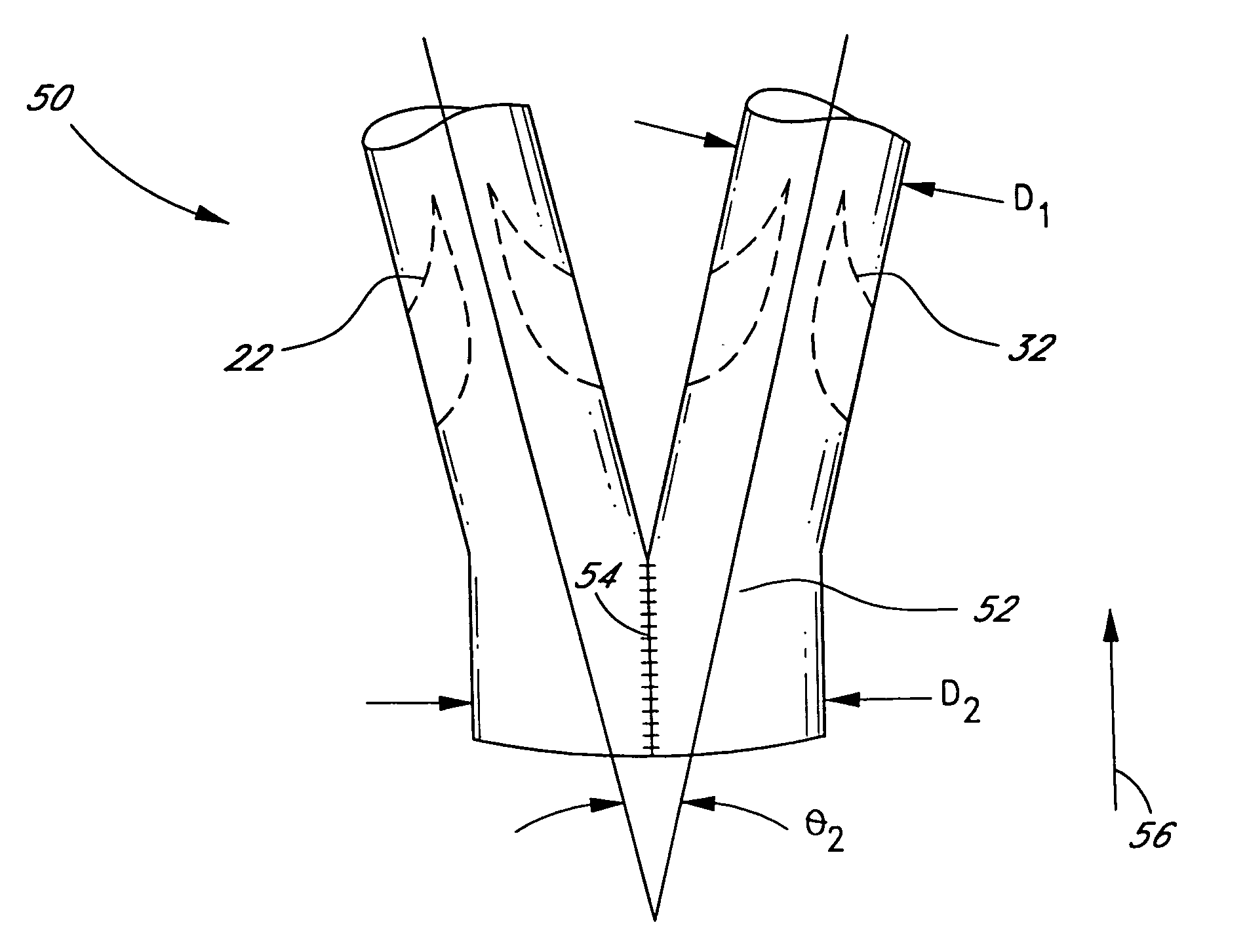
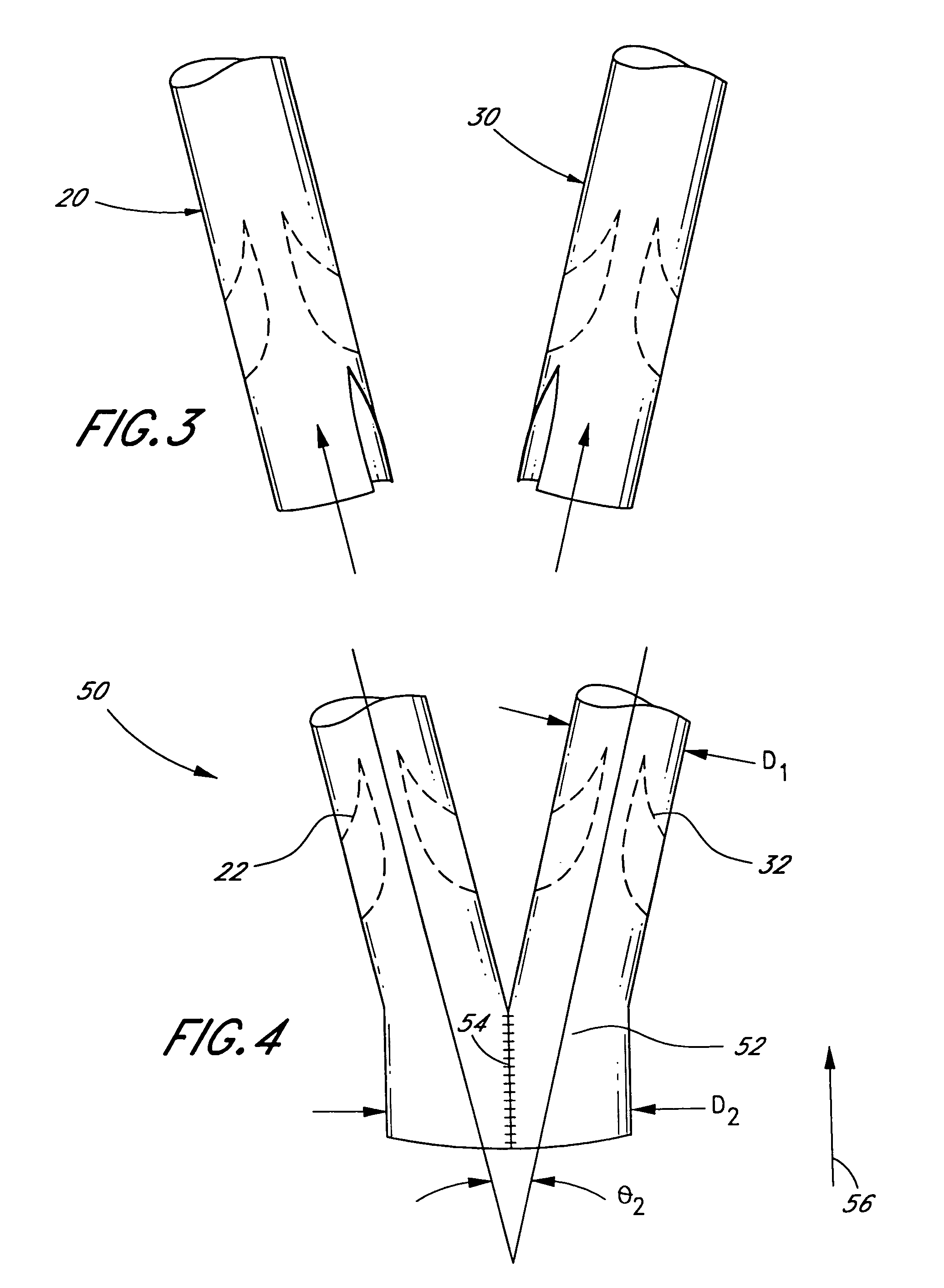
[0016]With reference to FIGS. 2–4, the present invention comprises a method of making a vascular prosthetic comprising a valvular conduit suitable for pulmonary valve replacement, wherein the conduit has a diameter greater than 22 ...


PUM


Abstract

The present invention comprises a pulmonary valved conduit that permits inflow diameters greater than about 22 mm while still maintaining the advantages of a naturally-formed biological valved conduit. Specifically, the present invention comprises a valved vascular prosthetic having an inflow conduit comprising a manifold formed from the sealed attachment of at least two venous valvular conduits.

Description

[0001]This is a continuation of U.S. patent application Ser. No. 09 / 252,333, filed Feb. 18, 1999, now U.S. Pat. No. 6,110,201.BACKGROUND OF THE INVENTION[0002]Pulmonary valved conduits, used to replace diseased arteries such as the pulmonary artery, have been used for the last thirty years. These pulmonary valved conduits have been generally effective at treating pulmonary atresia, pulmonary stenosis, or pulmonary insufficiency. In early embodiments, the conduits consisted of crimped woven or knit polyester tubes housing a valve fabricated from metal components or metal / plastic polymer components. Later valves consisted of metal frames suspending ceramic flat or curved (convex or concave) discs or hemidiscs that were passive to the flow of blood exiting the chambers of the heart.[0003]There were at least two disadvantages of these pulmonary valved conduits. First, there was a need to preclot the polyester tubes in order to preclude blood from seeping through the pores of the knit or...

Claims


Application Information

Patent Timeline
Patent Type & Authority Patents(United States)
IPC IPC(8): A61F2/06A61F2/24
CPCA61F2/2412A61F2002/065
Inventor QUIJANO, RODOLFO C.LOYA, ROBERT
Owner MEDTRONIC INC
Why Eureka
Try Eureka