Apparatus for retaining a radiographic sensor during dental x-ray imaging

a radiographic sensor and apparatus technology, applied in the field of dental x-ray equipment, can solve the problems of difficult size adjustment, clasp mechanism subject to disengagement, and difficult to make close tolerance adjustments to the clasp, so as to eliminate the ratcheting retention of the second jaw member, the effect of easy and accurate alignmen

Active Publication Date: 2006-04-25
OP D OP
View PDF3 Cites 41 Cited by
  • Summary
  • Abstract
  • Description
  • Claims
  • Application Information

AI Technical Summary

Benefits of technology

[0019]Compliant materials, such as compressible material pads, or the like, may be joined to at least a portion of the inner surfaces of one or both jaw members to distribute the compression forces being applied to the radiographic sensor and for preventing the sensor from slipping out from the jaws.
[0020]An alignment guide may optionally extend from the bite block as a positional reference so that one may readily and accurately align a radiographic imaging emission source with the radiographic sensor whose position is substantially obscured within the mouth of the patient. The alignment guide may comprise an elongated member that is either permanently or removably joined to the bite block.
[0021]The sensor retention device of the present invention is an improvement of radiographic sensor holders which utilize ratcheted retention positions for friction-based securement. The improvement may be implemented by eliminating the ratcheted retention of the second jaw member; adapting the second jaw member for continuously variable slidable engagement with the bite block; and engaging a threaded member between the second jaw member and the bite block, inclusive of the first jaw member which extends from the bite block. The threaded member within the improved sensor retention apparatus provides continuously variable adjustment of the distance between the jaw members to control retention pressure applied to the radiographic sensor. The apparatus may further be configured with alignment guides for guiding the orientation of a radiographic emission source.
[0022]An object of the invention is to provide secure retention of a radiographic sensor between the upper and lower teeth of a patient while x-ray images of dental structures are being registered.

Problems solved by technology

Utilizing this ratcheted form of friction retention mechanism results in a number of drawbacks.
It is difficult to make close tolerance adjustments to the clasp due to the discrete positioning provided by the ratchet teeth.
Size adjustment is difficult while manually holding a radiographic sensor within the clasp while it is being secured.
Furthermore, the clasp mechanism is subject to disengagement, wherein the radiographic sensor may fall loose within the mouth of the patient, or may even fall to the floor prior to being positioned in the patient's mouth.

Method used


Image

  • Apparatus for retaining a radiographic sensor during dental x-ray imaging
  • Apparatus for retaining a radiographic sensor during dental x-ray imaging
  • Apparatus for retaining a radiographic sensor during dental x-ray imaging

Examples


Embodiment Construction

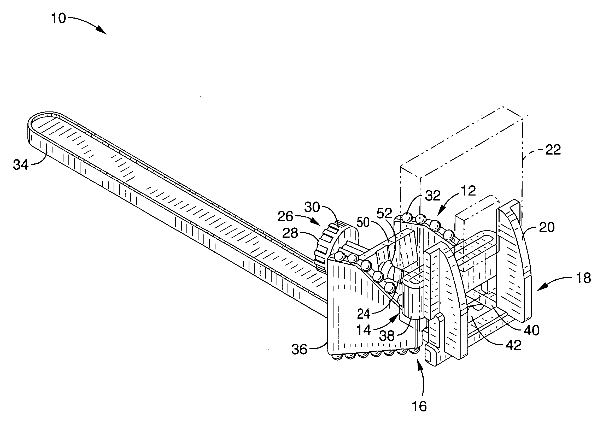
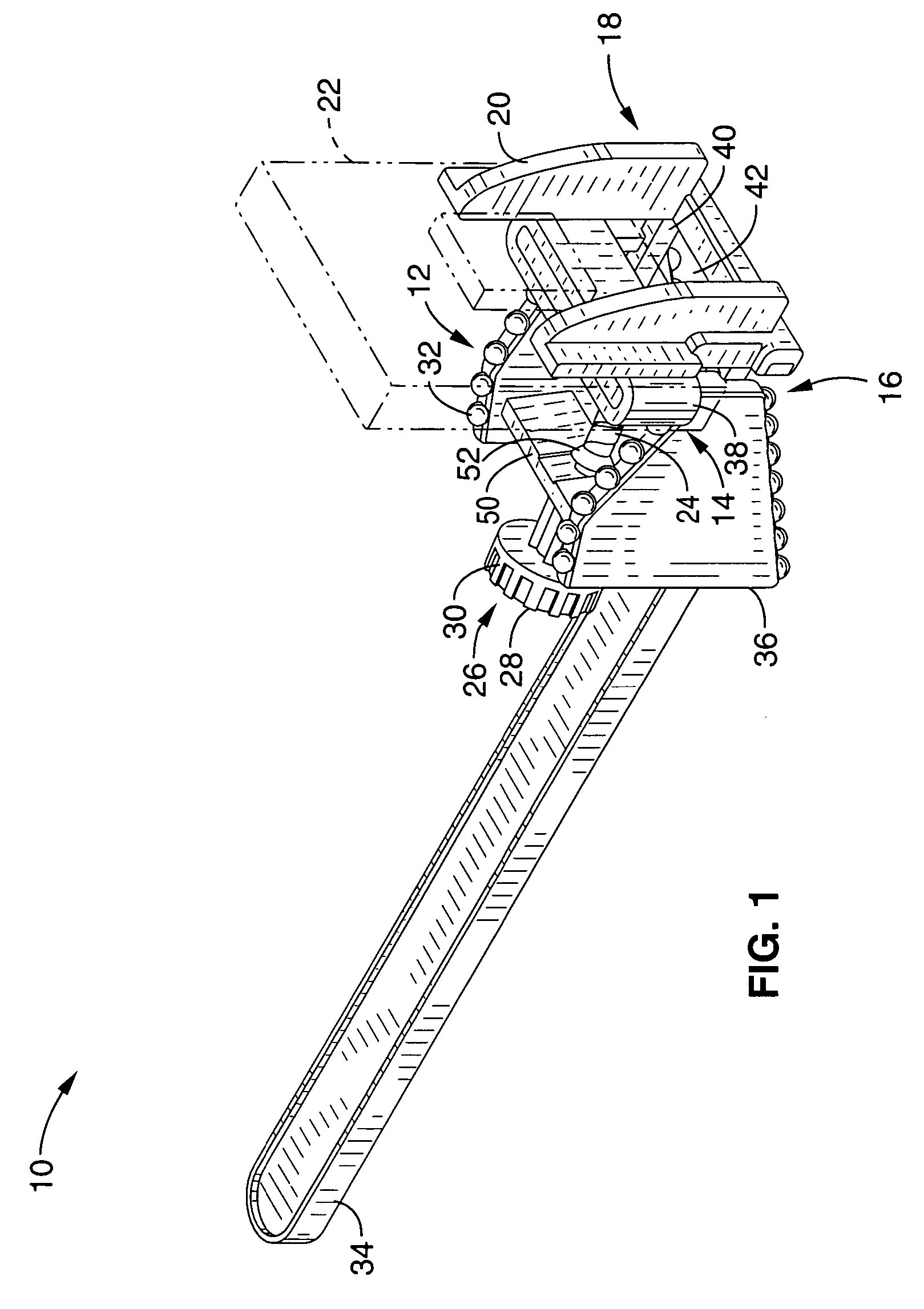
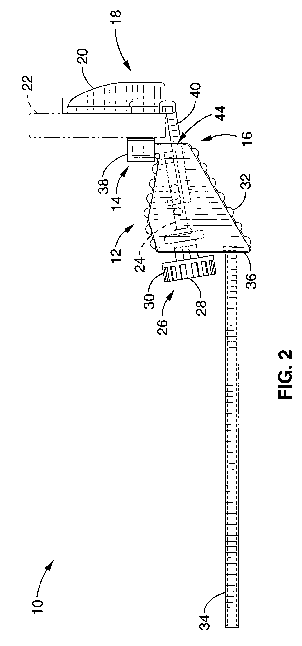
[0042]Referring more specifically to the drawings, for illustrative purposes the present invention is embodied in the apparatus generally shown in FIG. 1 through FIG. 10. It will be appreciated that the apparatus may vary as to configuration and as to details of the parts, and that the method may vary as to the specific steps and sequence, without departing from the basic concepts as disclosed herein.

[0043]The present invention comprises an apparatus for holding a radiographic sensor positioned within the mouth of a dental patient. The sensor retention apparatus is configured for being held within the mouth of the patient in response to the biting force applied between upper and lower dental surfaces.

[0044]FIG. 1 through FIG. 3 depict a radiographic retention apparatus 10 comprising a bite block 12 from which extends first jaw member 14 from a distal end 16 of bite block 12. A movable jaw assembly 18, is shown movably engaged with distal end 16 of bite block 12, preferably slidably ...


PUM


Abstract

A radiographic sensor retention apparatus for retaining a radiographic sensor and allowing it to be properly positioned within the mouth of a dental patient during x-ray image registration. The apparatus is held in position within the mouth of the patient in response to biting pressure from the upper and lower dental surfaces, and comprises a bite block with an extending first jaw member, a second jaw member slidably engaged with the bite block, and a threadable member connecting the second jaw member to either the bite block or the first jaw member, wherein the separation between the jaws is continuously variable in response to the rotation of the threadable member. Compliant members are preferably joined to one or both of the jaw members to aid in distributing the retention forces and to reduce sensor slippage. The bite block is preferably configured for the attachment of at least one alignment guide.

Description

CROSS-REFERENCE TO RELATED APPLICATIONS[0001]This application claims priority from U.S. provisional application Ser. No. 60 / 429,721 filed on Nov. 27, 2002, incorporated herein by reference in its entirety.STATEMENT REGARDING FEDERALLY SPONSORED RESEARCH OR DEVELOPMENT[0002]Not ApplicableREFERENCE TO A COMPUTER PROGRAM APPENDIX[0003]Not ApplicableBACKGROUND OF THE INVENTION[0004]1. Field of the Invention[0005]The present invention generally pertains to dental x-ray equipment, and more particularly to an apparatus for retaining a radiographic sensor adjacent to a bite block held between the teeth of a patient during dental x-ray image registration.[0006]2. Description of the Background Art[0007]The registration of radiographic data during dental x-ray imaging utilizes a radiographic sensor that must be retained in a desired position within the mouth of the patient and in a fixed relationship to the dental structures for which images are sought. A radiographic emitter (x-ray source uni...

Claims


Application Information

Patent Timeline
Patent Type & Authority Patents(United States)
IPC IPC(8): A61B6/14G03B42/04
CPCA61B6/145A61B6/4435G03B42/042A61B6/4423
Inventor LANDIS, TIMOTHY J.ALLEN, CLAY D.
Owner OP D OP
Why Eureka
Try Eureka