Light source for ophthalmic use

a technology for ophthalmic use and light sources, applied in the field of surgical devices, can solve the problems of insufficient illumination of eye segments, additional problems, and difficulty in distinguishing using conventional illumination, and achieve the effect of improving the visual perception of eye tissu

Inactive Publication Date: 2006-06-20
JOHNSON & JOHNSON SURGICAL VISION INC
View PDF4 Cites 21 Cited by
  • Summary
  • Abstract
  • Description
  • Claims
  • Application Information

AI Technical Summary

Benefits of technology

[0014]In addition, a color filter may be provided in order to enhance visual perception of eye tissue.

Problems solved by technology

Sufficient illumination of segments of the eye is difficult with typical microscope lights.
Ocular structures such as the cornea, lens and inner ocular fluids are nearly transparent and accordingly difficult to distinguish using conventional illumination due to the small amount of light scatter which they produce when diffusely lit.
Additional problems may be presented in insuring the physician has proper visualization of a surgical device which may be blurred or partially obscured when blood, scar tissue, or other debris is present.
Alternatively, multiple surgical openings in the eye and multiple illuminators may be used, however this adds to the risk of complications and may increase the difficulty of the surgical procedure.

Method used


Image

  • Light source for ophthalmic use
  • Light source for ophthalmic use
  • Light source for ophthalmic use

Examples


Embodiment Construction

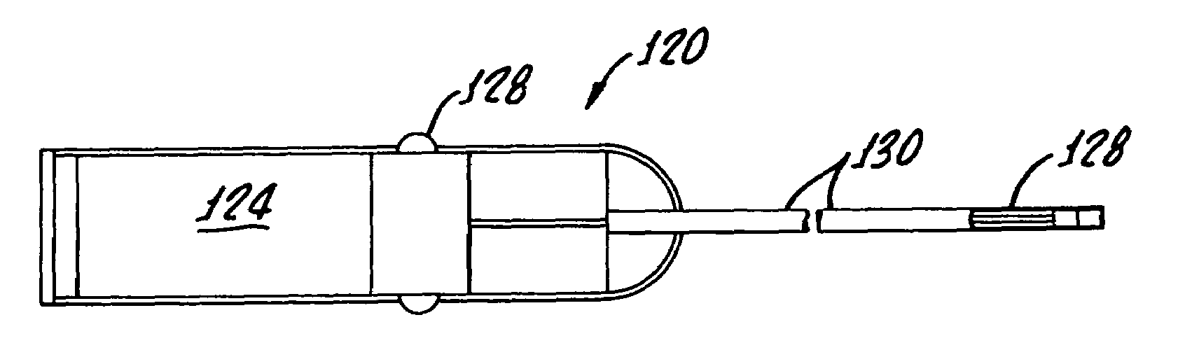
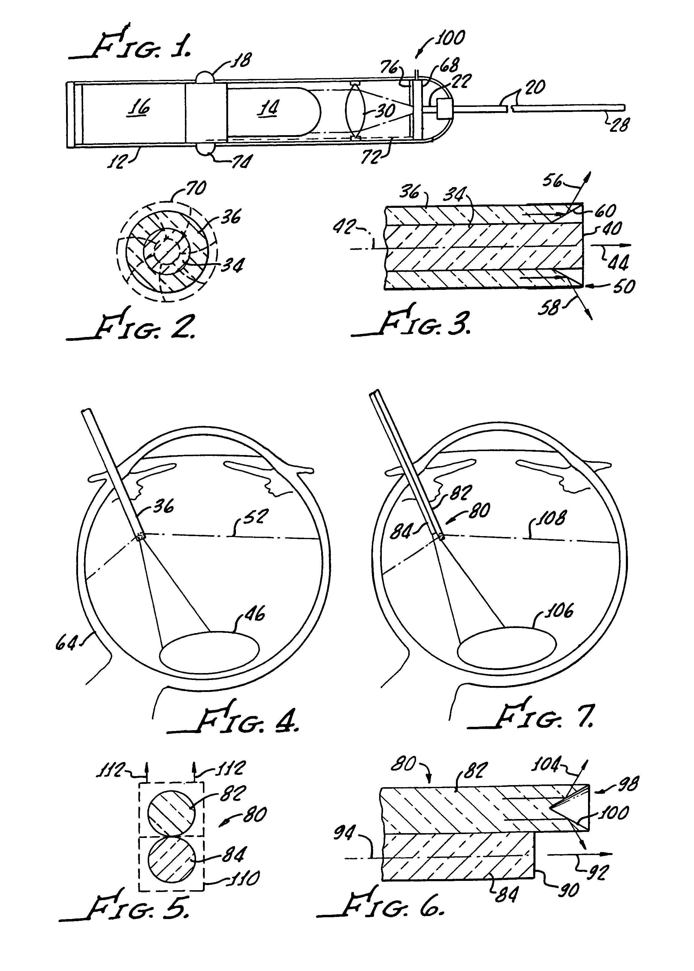
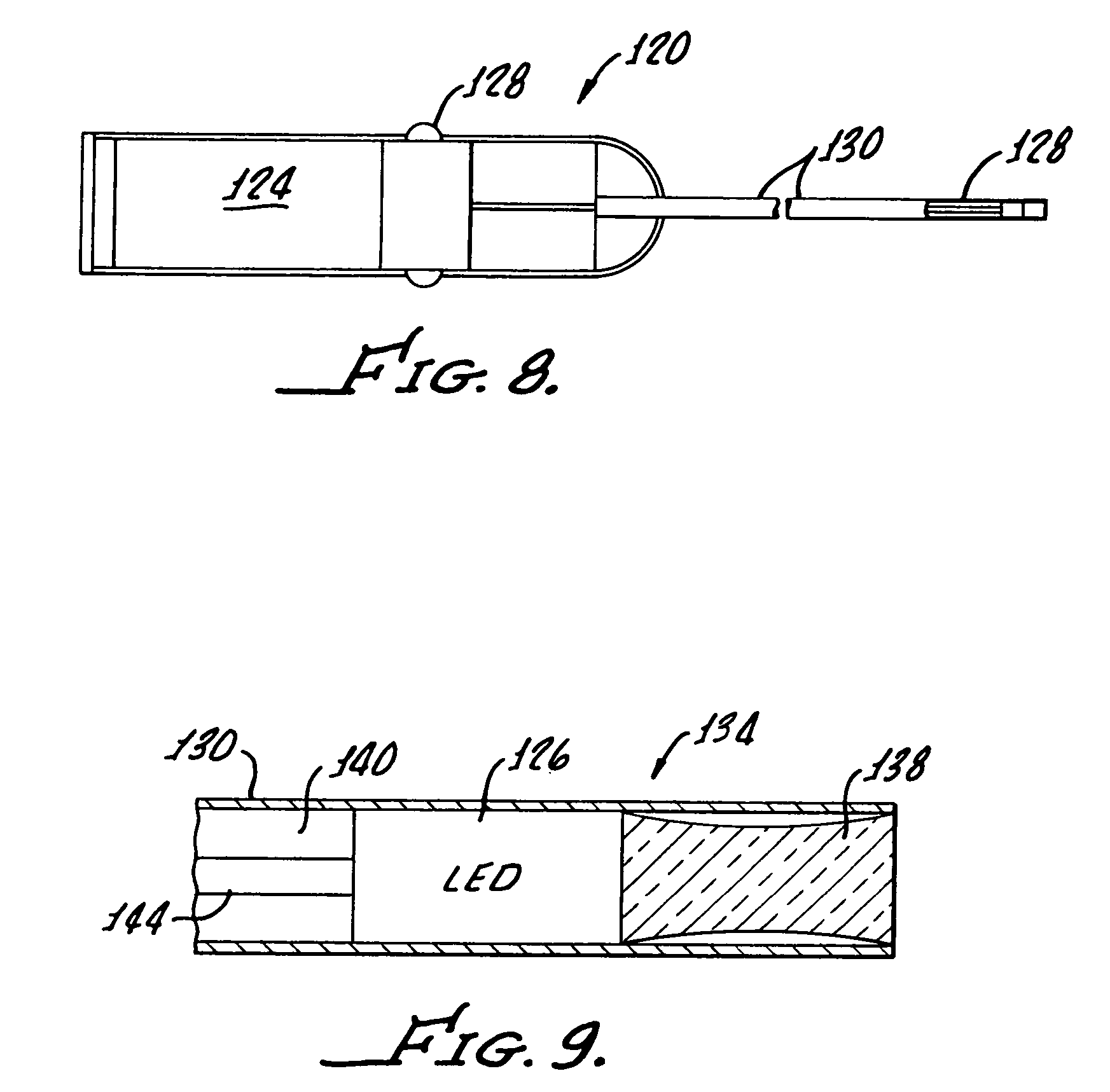
[0026]With reference to FIG. 1 there is shown an instrument 10 in accordance with the present invention for providing illumination of intraocular tissue (not shown) during surgery. A housing 12 incases a light source 14, preferably an LED, a battery power source 16 and a control switch 18 for interconnecting the LED 14 with the power source.

[0027]As hereinafter discussed in greater detail, at least one fiber optic 20 is provided with a proximal end 22 in light communication with the LED and a distal end 28 sized for insertion into an eye (not shown) for illumination of intraocular tissue (not shown). A lens and / or filter 30 may be used to focus light onto the fiber optic proximal end 22 or direct coupling may be utilized. In addition a shutter arrangement 68 may be utilized for controlling light passage into the fiber optic 20. Thus in accordance with the present invention the LED is utilized instead of the standard Xenon or Halogen bulb in a console (not shown) to provide illuminat...


PUM


Abstract

An instrument for providing illumination of intraocular tissue during surgery generally includes a hand held light source which includes a light emitting diode, a power source for driving the diode and a control switch for interconnecting the diode with the power source. At least one fiber optic having a proximal end in light communication with the LED and a distal end size for insertion into an eye for illumination of intraocular tissue is provided.

Description

[0001]The present application claims priority under 35 U.S.C. §120, and is a Continuation of U.S. application Ser. No. 10 / 189,675 filed Jul. 3, 2002, now U.S. Pat, No. 6,786,628, which is incorporated by reference herein in its entirety.[0002]Present invention generally relates to the surgical devices and more particularly to devices for illumination of anterior and posterior segments of the eye during surgery. Multiple instruments are typically utilized in performing intraocular surgery which include instruments designed for irrigation, emulsification or cutting in the aspiration tissue.[0003]Sufficient illumination of segments of the eye is difficult with typical microscope lights. Ocular structures such as the cornea, lens and inner ocular fluids are nearly transparent and accordingly difficult to distinguish using conventional illumination due to the small amount of light scatter which they produce when diffusely lit.[0004]Heretofore light pipes have been used to deliver additio...

Claims


Application Information

Patent Timeline
Patent Type & Authority Patents(United States)
IPC IPC(8): A61B18/18A61B3/00A61B19/00A61F9/007
CPCA61B3/0008A61B5/6848A61F9/007A61B2019/5206A61B2090/306
Inventor STEEN, MARK E.KADZIAUSKAS, KENNETH E.
Owner JOHNSON & JOHNSON SURGICAL VISION INC
Why Eureka
Try Eureka