Electroporation microneedle and methods for its use

a micro-needle and electro-enhanced technology, applied in the field of electro-enhanced delivery and intracellular delivery, can solve the problems that electro-enhanced delivery might have a very difficult time entering the cells, and achieve the effect of enhancing electro-enhanced delivery and enhancing intracellular delivery

Active Publication Date: 2006-10-24
MERCATOR MEDSYST
View PDF23 Cites 35 Cited by
  • Summary
  • Abstract
  • Description
  • Claims
  • Application Information

AI Technical Summary

Benefits of technology

[0009]The present invention provides improved methods and apparatus for the electrically-enhanced delivery of substances to cells in solid target tissues. The application of an alternating electric field provides “electroporation” of the individual cells in the target tissue, which in turn causes a temporary opening of pores in the membranes of those cells. Such pores enhance the intracellular delivery of genes, nucleic acids, proteins, and other large molecules, which in the absence of electroporation might have a very difficult time entering into the cells. While the present invention is particularly useful for the delivery of such large molecules, it will also be useful for the delivery of small molecule drugs, typically having molecular weights below 2 kD, which for a variety of reasons, might have difficulty entering particular target cells.

Problems solved by technology

Such pores enhance the intracellular delivery of genes, nucleic acids, proteins, and other large molecules, which in the absence of electroporation might have a very difficult time entering into the cells.

Method used


Image

  • Electroporation microneedle and methods for its use
  • Electroporation microneedle and methods for its use
  • Electroporation microneedle and methods for its use

Examples


Embodiment Construction

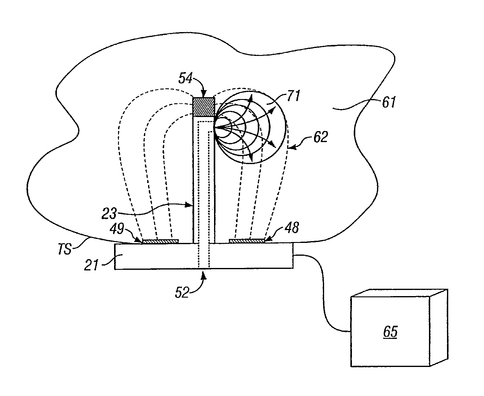
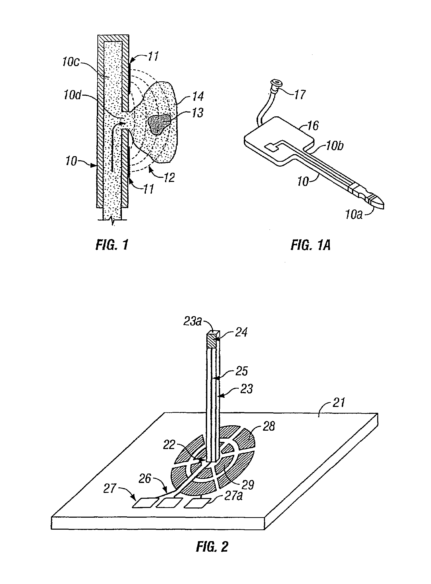
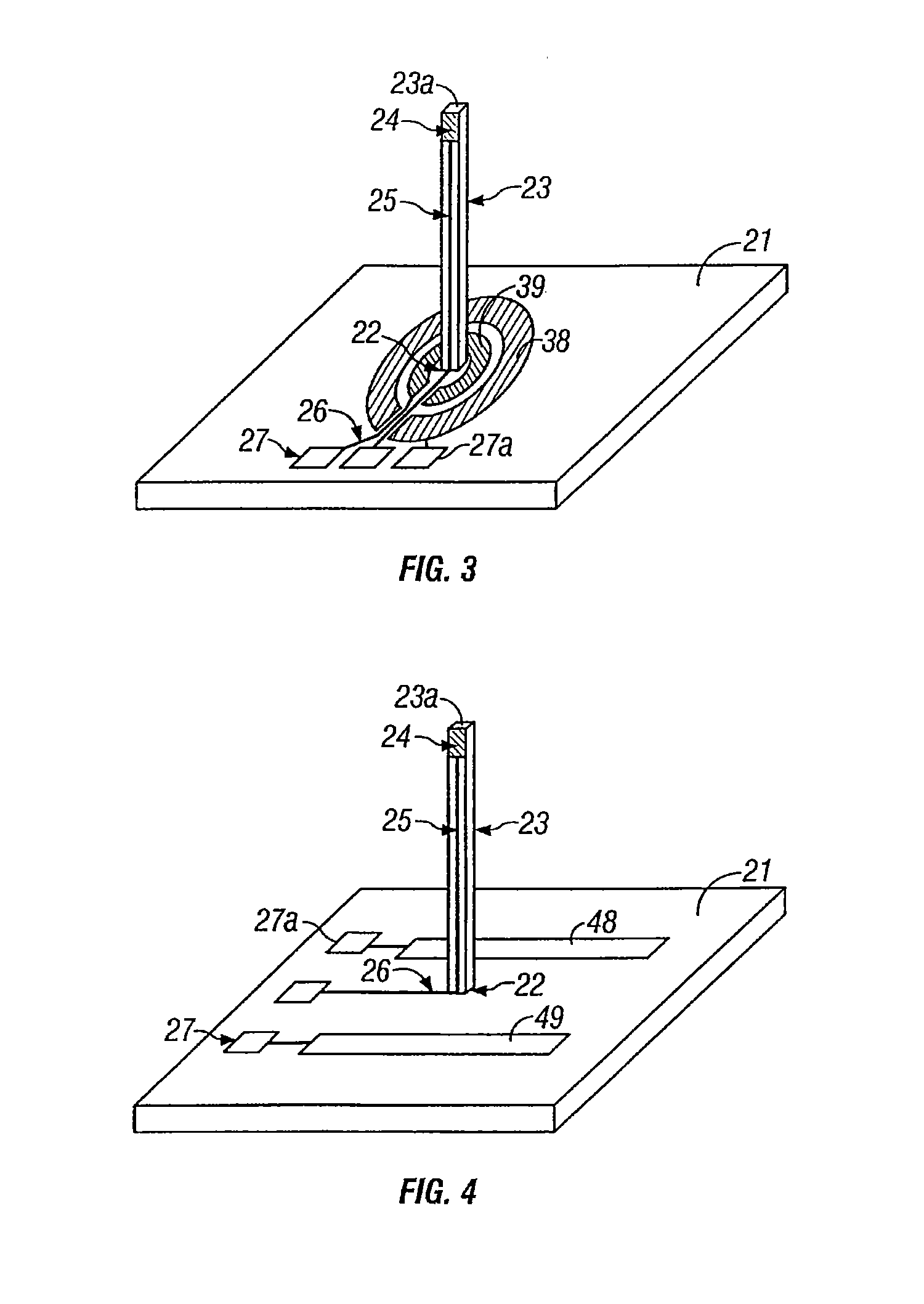
[0032]A first exemplary electroporation apparatus constructed in accordance with the principles of the present invention is illustrated in FIGS. 1 and 1A. The apparatus comprises a shaft 10 having a distal end 10a and proximal end 10b attached to a base 16 at its proximal end. The apparatus of FIGS. 1 and 1A may be formed by vapor deposition of a material into a mold or by chemical etching of a material. Suitable materials include metals, polymers, and semi-conductors Specific methods for fabricating apparatus as illustrated in FIGS. 1 and 1A are described in copending application Ser. No. 09 / 877,653, the full disclosure of which has previously been incorporated by reference.

[0033]The shaft 10 has a substance-delivery lumen 10c which terminates in an opening 10d in the wall of the shaft. The lumen 10c is connected to a luer or other conventional fitting 17 which permits connection of the lumen 10c to a conventional pressurized-delivery device, such as a syringe, pump, or the like. T...


PUM


Abstract

Electroporation of cells in target tissues is performed using microneedles having integral electrode structures. In a first embodiment, a pair of electrode structures are disposed on opposite sides of a substance delivery opening on a needle or other tissue-penetrating shaft. In a second embodiment, a first electrode structure is disposed on the needle and a second electrode structure is disposed on an attached base. The electrode structures are spaced closely together and relatively low voltages are applied to achieve electroporation-enhanced substance delivery.

Description

CROSS-REFERENCES TO RELATED APPLICATIONS[0001]This application claims the benefit of prior provisional application No. 60 / 297,418, filed on Jun. 11, 2001, the full disclosure of which is incorporated herein by reference.BACKGROUND OF THE INVENTION[0002]1. Field of the Invention[0003]Drug and gene delivery to solid tissues is of significant medical and scientific interest. Most simply, drugs and genes can be injected into muscle, organs, and other solid tissues using a needle under pressure, typically provided by a syringe. While many small molecule drugs are adequately taken up by such straight-forward injection methods, the delivery and uptake of large molecules (such as proteins) and genes (and other nucleic acids) is often more problematic. In particular, the successful delivery of functional genes to solid tissues such that the genes are expressed can often be quite difficult.[0004]Various methods for enhancing the delivery of genes and other large molecules have been proposed. ...

Claims


Application Information

Patent Timeline
Patent Type & Authority Patents(United States)
IPC IPC(8): A61N1/30A61M5/32A61M37/00A61N1/32
CPCA61M37/0015A61N1/325A61N1/327A61M2025/0093A61M2025/105A61M2037/0023
Inventor SEWARD, KIRK PATRICK
Owner MERCATOR MEDSYST
Why Eureka
Try Eureka