Method and apparatus for enhancing an image

Inactive Publication Date: 2006-12-12
GENERAL ELECTRIC CO
View PDF14 Cites 31 Cited by
  • Summary
  • Abstract
  • Description
  • Claims
  • Application Information

AI Technical Summary

Benefits of technology

[0004]In one aspect, a method for facilitating an enhancement of a visibility of an object in an x-ray image is provided. The method includes generating an x-ray image including at least one object, generating an estimate of a background surrounding the at least one object, subtracting the background estimate from the x-ray image to generate an estimate of pixel intensities due the object, mapping the estimate of pixel intensities due to the object, and combining the mapped estimate of pixel intensities due to the object and the x-ray image to generate an enhanced image.
[0005]In another aspect, a medical imaging system for facilitating an enhancement of a

Problems solved by technology

However, some microcalcifications are relatively small and can be virtually imperceptible to the human eye

Method used


Image

  • Method and apparatus for enhancing an image
  • Method and apparatus for enhancing an image
  • Method and apparatus for enhancing an image

Examples


Example

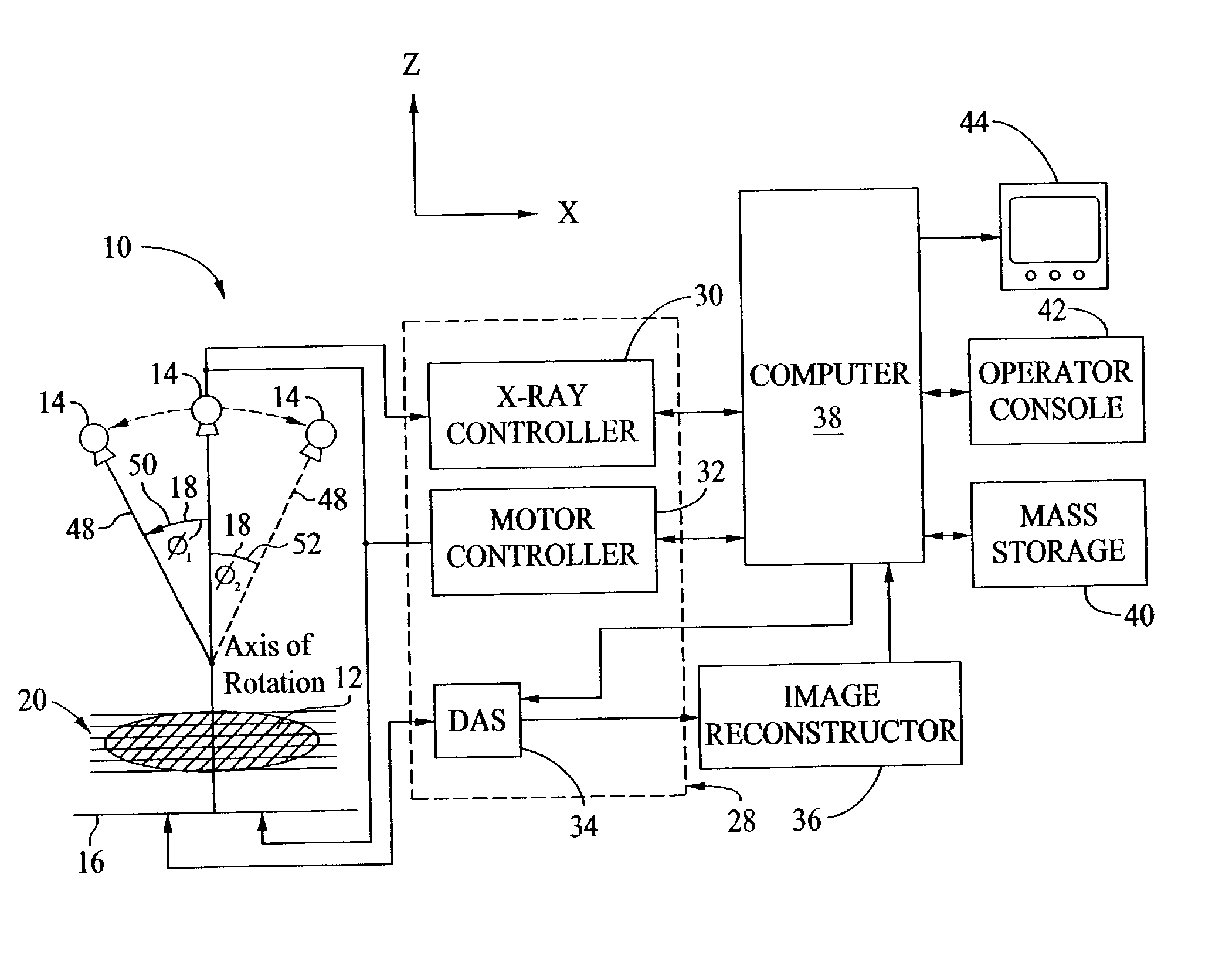
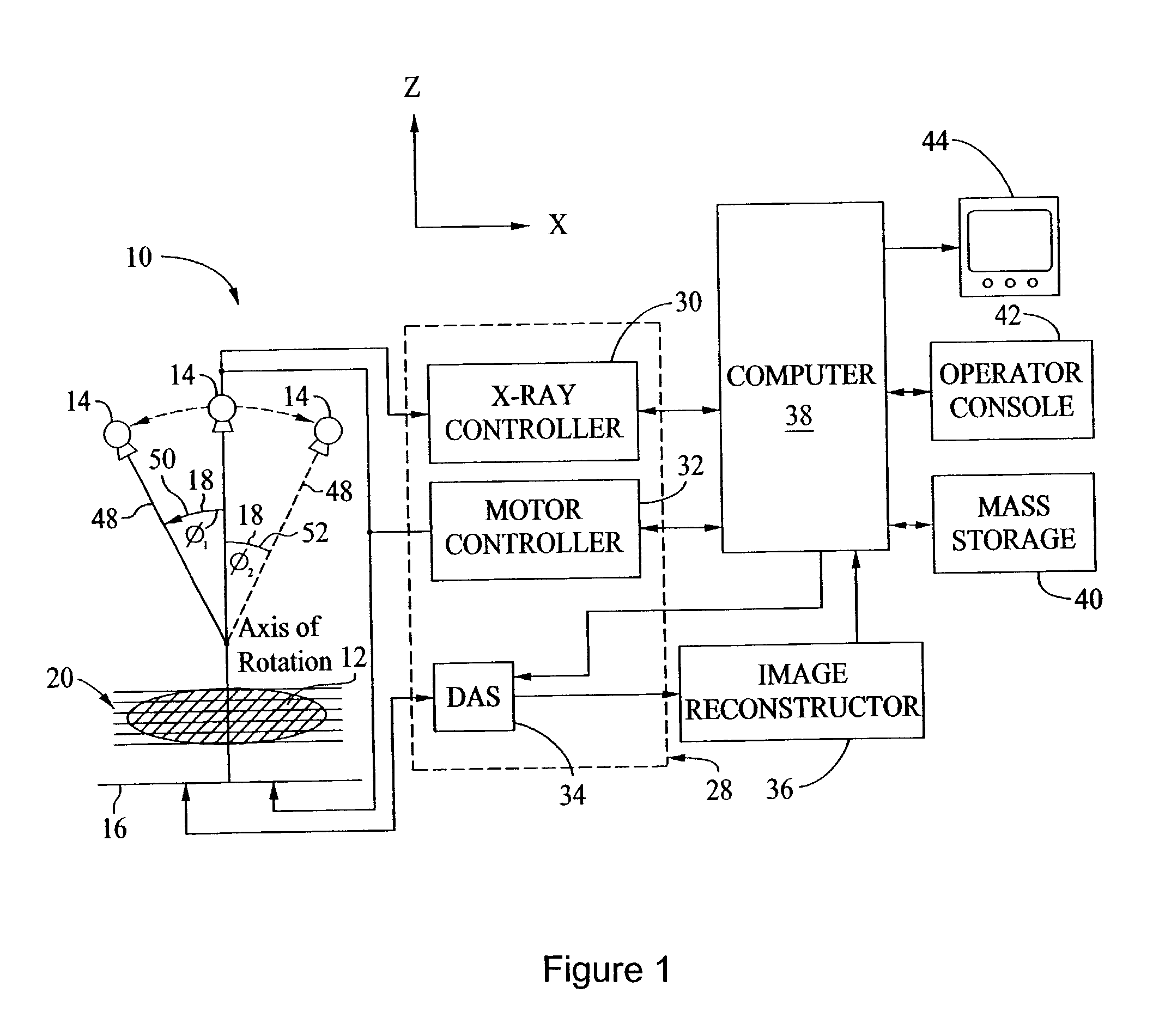
[0016]Referring to FIG. 1 and in an exemplary embodiment, a medical imaging system 10 generates a three-dimensional dataset representative of an imaged object 12, such as a patient's breast 12 in mammographic tomosynthesis. System 10 includes a radiation source 14, such as an x-ray source 14, and at least one detector array 16 for collecting views from a plurality of projection angles 18. Specifically, and in one embodiment, system 10 includes a radiation source 14 which projects a cone-shaped beam of x-rays which pass through object 12 and impinge on detector array 16. The views obtained at each angle 18 can be used to reconstruct a plurality of slices, i.e., images representative of structures located in planes 20 parallel to detector 16. Detector array 16 is fabricated in a panel configuration having a plurality of pixels (not shown) arranged in rows and columns so that an image is generated for an entire object of interest such as breast 12. In one embodiment, detector array 16 ...


PUM


Abstract

A method for facilitating an enhancement of a visibility of an object in an x-ray image includes generating an x-ray image including at least one object, generating an estimate of a background surrounding the at least one object, subtracting the background estimate from the x-ray image to generate an estimate of pixel intensities due the object, mapping the estimate of pixel intensities due to the object, and combining the mapped estimate of pixel intensities due to the object and the x-ray image to generate an enhanced image.

Description

STATEMENT REGARDING FEDERALLY SPONSORED RESEARCH & DEVELOPMENT[0001]The government may have rights in this invention pursuant to Subcontract 22287 issued from the Office of Naval Research / Henry M. Jackson Foundation.BACKGROUND OF THE INVENTION[0002]This invention relates generally to an imaging system, and more particularly, to methods and apparatus for enhancing an image using a medical imaging system.[0003]Detecting microcalcifications is important because they are often the only pathology indicators in mammography revealing cancer on a mammogram at a stage when the disease is still curable. For example, microcalcifications are currently used to detect over 50% of non-palpable disease on mammograms. However, some microcalcifications are relatively small and can be virtually imperceptible to the human eye even after the display settings for the image have been adjusted to improve visualization of the pathology.BRIEF DESCRIPTION OF THE INVENTION[0004]In one aspect, a method for faci...

Claims


Application Information

Patent Timeline
IPC IPC(8): G06K9/00A61B6/00G06F19/00G06K9/40G06T5/00G06T5/40G06T5/50H04N5/32
CPCG06T5/008G06T5/40G06T7/0012G06T2207/10112G06T2207/10116G06T2207/20012G06T2207/20144G06T2207/30068G06T2207/30096G06T7/194
Inventor KAUFHOLD, JOHN PATRICK
Owner GENERAL ELECTRIC CO
Why Eureka
Try Eureka