Method for automated analysis of digital chest radiographs

a technology of digital chest radiographs and automated analysis, applied in image analysis, image enhancement, instruments, etc., can solve the problems of reducing the performance of low-level image processing, difficult to automatically and accurately detect lung regions, and poor quality, so as to improve the display quality of radiographs, improve the reliability of the system, and improve the accuracy and reliability of boundary detection.

Active Publication Date: 2007-05-22
CARESTREAM HEALTH INC
View PDF5 Cites 32 Cited by
  • Summary
  • Abstract
  • Description
  • Claims
  • Application Information

AI Technical Summary

Benefits of technology

[0014]1. Chest radiographs are normalized based on the anatomic regions, which not only improves the display quality of radiographs, but also makes the system robust;
[0015]2. The algorithm incorporates both high-level analysis and low-level image processing, which enables the system to deal with radiographs with abnormal anatomy, noise and poor quality;
[0016]3. The shape-based deformable lung model is tolerant of shape and positional variances as well as image disturbances.
[0018]5. The employed edge information combines difference order derivative and different direction edge information, which makes the boundary detection more accurate and reliable.

Problems solved by technology

These approaches pose problems when processing images of abnormal anatomy, or images with excessive noise and poor quality, because the abnormal anatomic structures or noise often confuse the segmentation routines.
Although the latter methods demonstrate improved performance, to automatically and accurately detect lung regions is still a difficult problem.
The latter structures cause the lung boundaries to appear ambiguous, which greatly reduces the performance of low-level image processing.

Method used


Image

  • Method for automated analysis of digital chest radiographs
  • Method for automated analysis of digital chest radiographs
  • Method for automated analysis of digital chest radiographs

Examples


Embodiment Construction

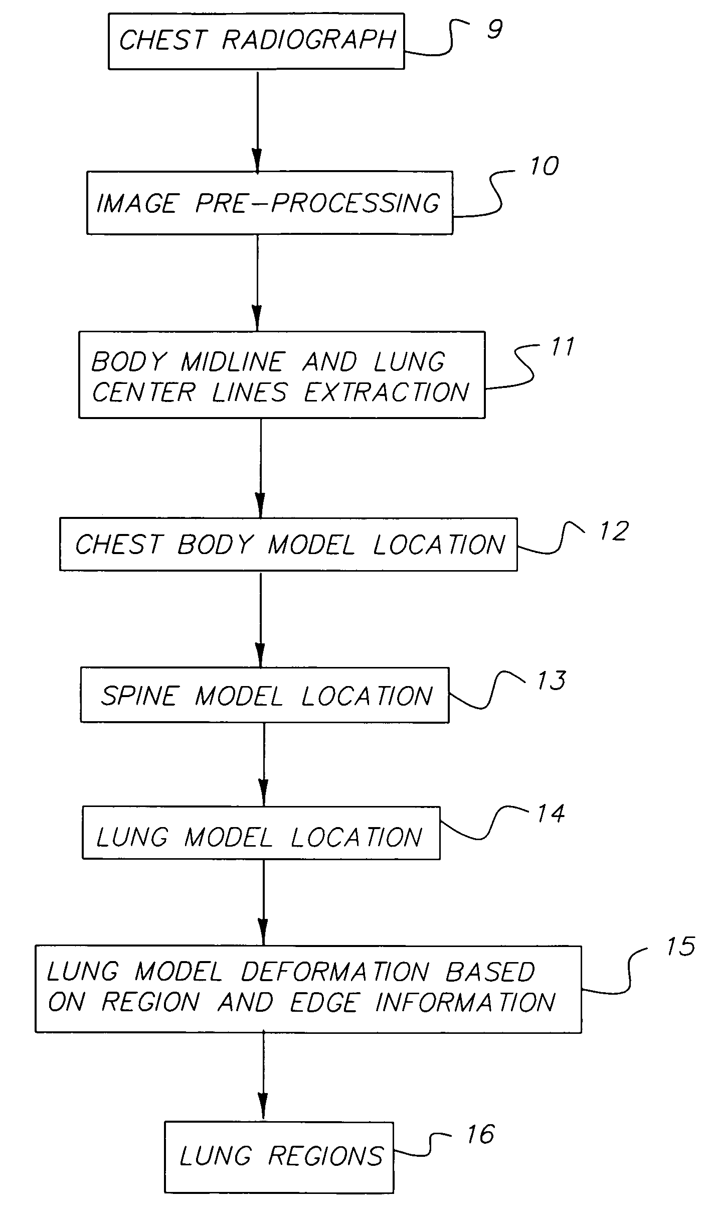
[0036]The present invention relates in general to the processing of chest radiographic images. FIG. 17 is a block diagram of a radiographic system incorporating the present invention. As shown a radiographic image, such as a chest radiographic image is acquired by an image acquisition system 1600. Image acquisition system 1600 can include one of the following: (1) a conventional radiographic film / screen system in which a body part (chest) of a patient is exposed to x-radiation from an x-ray source and a radiographic image is formed in the radiographic image is formed in the radiographic film. The film is developed and digitized to produce a digital radiographic image. (2) A computed radiography system in which the radiographic image of the patient's body part is formed in a storage phosphor plate. The storage phosphor plate is scanned to produce a digital radiographic image. The storage phosphor plate is erased and reused. (3) A direct digital radiography system in which the radiogr...


PUM


Abstract

A method for automatically segmenting lung regions in a chest radiographic image comprising; providing an input digital chest radiograph image; preprocessing the input digital radiographic image; extracting the chest body midline and lung centerlines from the preprocessed image. Locating one-by-one, the chest body model, the spine model and the two lung models in the image based on the extracted chest body midline and two lung centerlines; and detecting the lung contours by deforming the lung shape models to converge to the true lung boundaries as a function of the extracting and locating image processing.

Description

FIELD OF THE INVENTION[0001]The present invention relates to techniques for automated analysis of digital chest radiographs, and more specifically to a method for automated detection of lung regions in digital chest radiographs.BACKGROUND OF THE INVENTION[0002]Identification of lung regions in chest radiographs is an important pre-processing step for most types of computer analysis of digital chest radiographs, such as interstitial disease, pneumothorax, cardiomegaly and pulmonary nodules. A considerable amount of work in recent literature has addressed this topic, and various image processing methods have been applied. These methods can be basically classified into two categories. One is feature-based pixel classification and the other is ruled-based reasoning. In pixel classification systems, each pixel in the image is represented by a set of features, such as density, histogram, entropy, and gradients etc, and is classified into a region type based on the output of Neural Network...

Claims


Application Information

Patent Timeline
Patent Type & Authority Patents(United States)
IPC IPC(8): G06K9/00G06K9/34G06K9/46G06T5/00G06T7/00G06V10/28
CPCG06K9/38G06T7/0012G06T7/0083G06T7/0089G06T2207/10116G06T2207/20008G06T2207/30061G06T7/12G06T7/149G06V10/28
Inventor LUO, HUIFOOS, DAVID H.
Owner CARESTREAM HEALTH INC
Why Eureka
Try Eureka