
X-ray mammography/tomosynthesis of patient's breast

a breast and x-ray technology, applied in tomosynthesis, medical science, diagnostics, etc., can solve the problems of patient's discomfort or inability to sleep, patient's startled, and consequent motion artifacts in the image, so as to increase discomfort and the likelihood of motion artifacts

Active Publication Date: 2007-07-17
HOLOGIC INC
View PDF2 Cites 212 Cited by
  • Summary
  • Abstract
  • Description
  • Claims
  • Application Information

AI Technical Summary

Problems solved by technology

It has been discovered that the motion of the x-ray source in this approach, at least for positions that include the CC position, may raise concerns of patient comfort, patient safety and patient motion artifacts.
For example, if a scan that includes the CC position starts when the x-ray source is away from and moves toward the patient's shoulder, this typically would require the patient to cock her head away at the start of the scan and during positioning of the breast, which may be uncomfortable or not even possible for some patients.
In addition, motion of the patient's body in the course of cocking the head away and possible moving the head during the scan away from an uncomfortable position, may introduce motion of her breast relative to the x-ray imager and consequent motion artifacts in the image.
If the starting position is away from the patient's head, so that the x-ray source moves toward the patient's head during the scan, and a shield is used to position her head outside the x-ray source path, the patient may be startled by the approach of the x-ray source and move, with consequent possibility of motion artifacts in the image.
If there is no shield in such a scan, the patient may have to cock her head rapidly during the scan to avoid contact, with increased discomfort and likelihood of motion artifacts, or else the patient would require her head to remain cocked throughout the scan so as to avoid contact as the source approaches the end of the scan.
An additional challenge relates to using the same equipment for both tomosynthesis imaging and mammogram imaging.
If the mammogram data acquisition is done separately from the scan for tomosynthesis data acquisition, e.g., before or after the scan for tomosynthesis data, the time to position the equipment for mammogram data acquisition and separately for tomosynthesis data acquisition may unduly increase the examination time.
Yet another challenge arises if an anti-scatter grid is used for mammogram data acquisition but not for tomosynthesis data acquisition.
Yet another challenge concerns the use of a patient shield.
One problem with this approach is that the housing will be rather large.
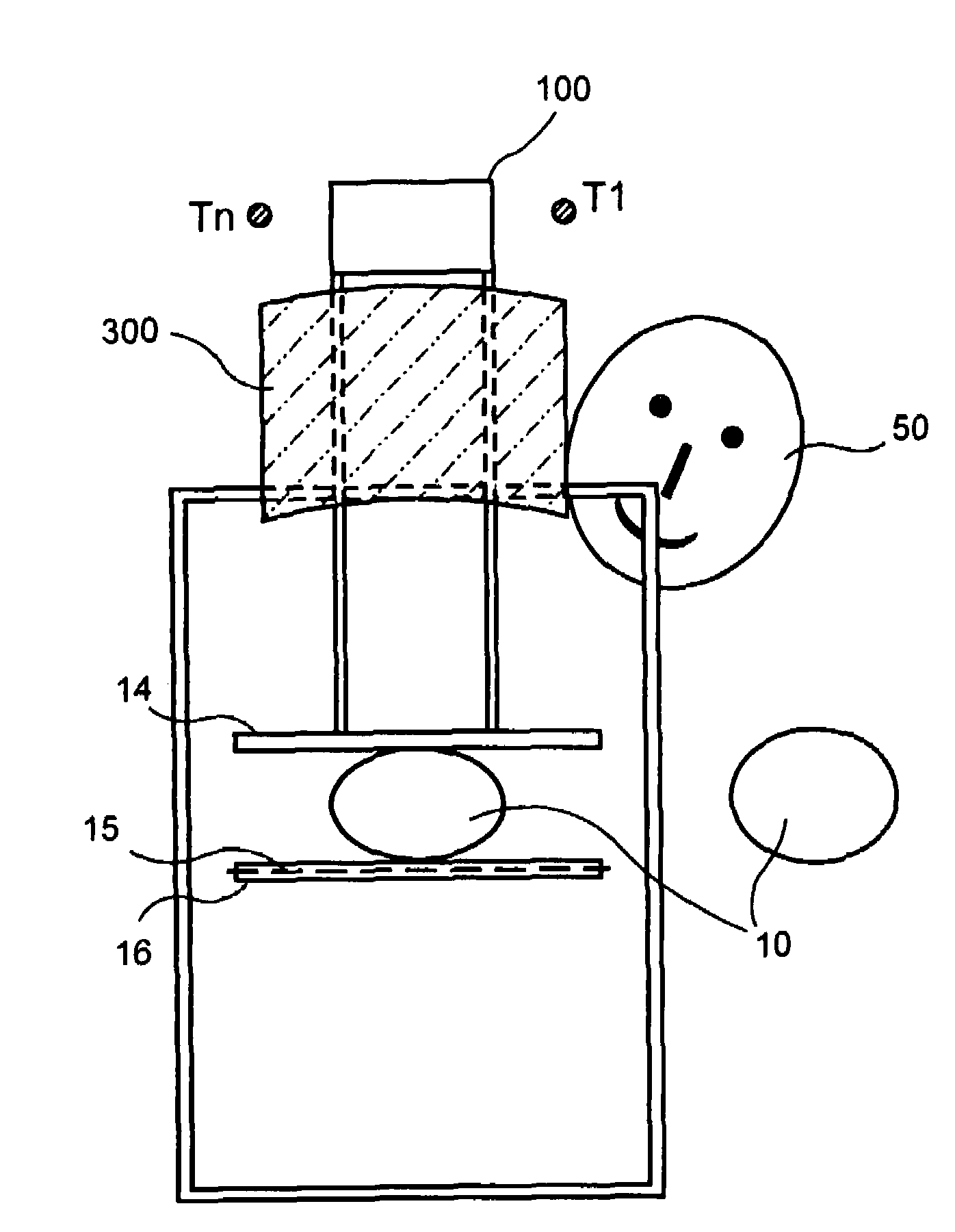
If there is a separate shield in front of the tube, or between the tube's scanning path and the patient's head, then known solutions present problems in requiring the patient's head to cock to one side to avoid pressing against the shield, and additionally the shield can prevent convenient access to the patient's breast and the breast compression hardware, presenting difficulties for the mammographic technologist when positioning the patient.

Method used


Image

  • X-ray mammography/tomosynthesis of patient's breast
  • X-ray mammography/tomosynthesis of patient's breast
  • X-ray mammography/tomosynthesis of patient's breast

Examples


Embodiment Construction

[0026]In describing preferred embodiments illustrated in the drawings, specific terminology is employed for the sake of clarity. However, the disclosure of this patent specification is not intended to be limited to the specific terminology so selected and it is to be understood that each specific element includes all technical equivalents that operate in a similar manner.

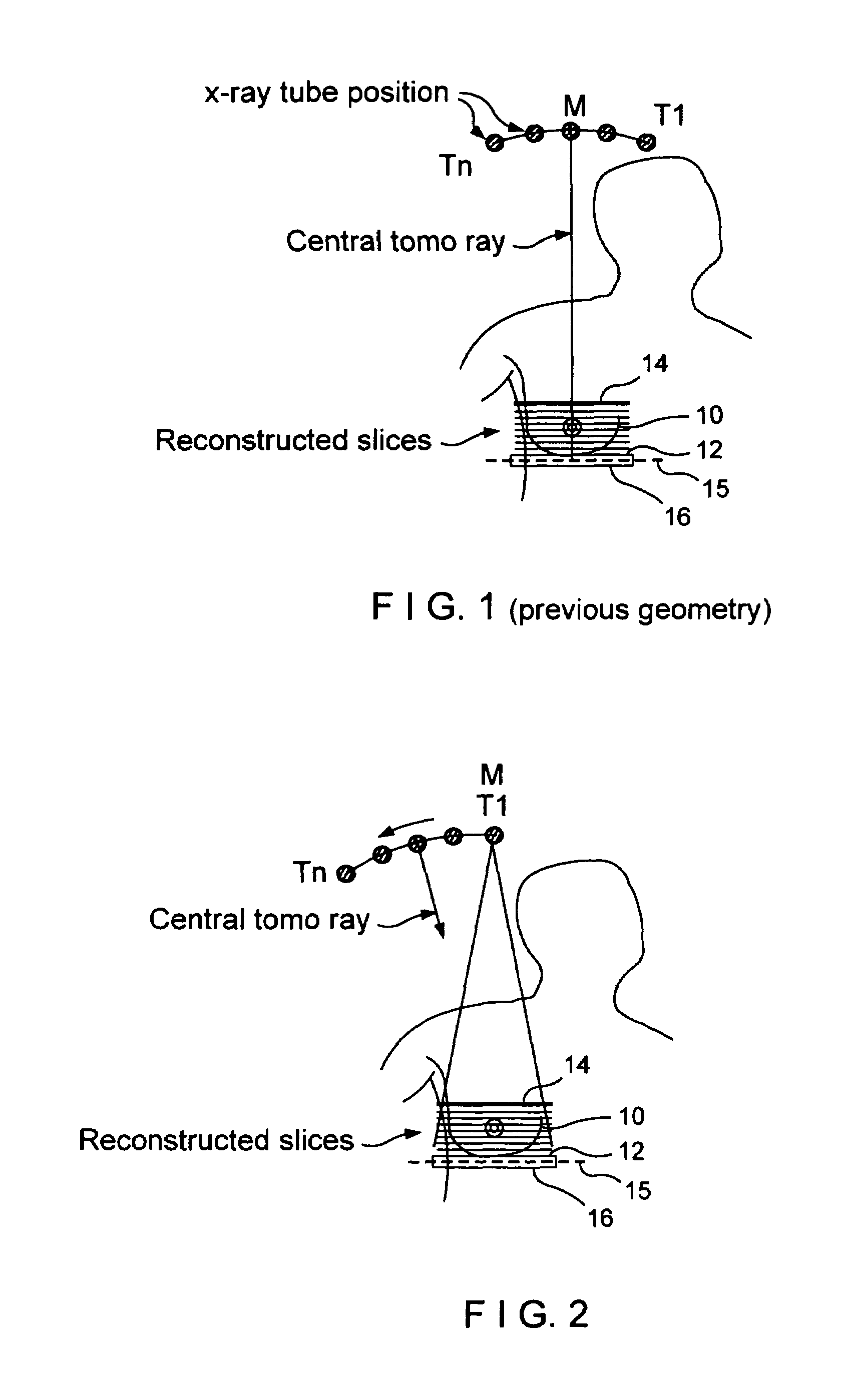
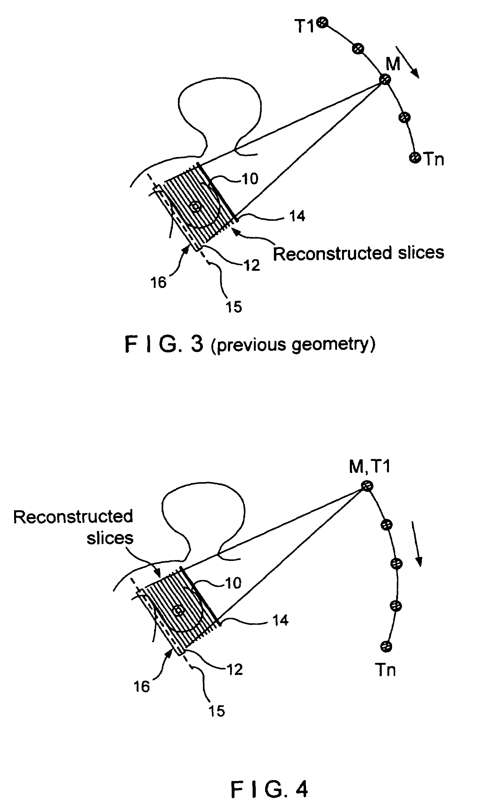
[0027]FIGS. 1 and 2 illustrate scanning geometry differences between a previously proposed system (FIG. 1) and a new system disclosed in this patent specification (FIG. 2). As illustrated in FIG. 1, a patient's right breast 10 is immobilized on a breast platform 12 with the use of a compression paddle 14, in a CC imaging position. An x-ray source such as an x-ray tube with collimation for the x-ray beam moves relative to breast 10 along a trajectory defined by points T1–Tn, for example through an arc of ±15° relative to a normal to breast platform 12, which normal coincides with the central x-ray (labeled central to...


PUM


Abstract

A breast x-ray system and method using tomosynthesis imaging in which the x-ray source generally moves away from the patient's head. The system may include an operation mode in which it additionally takes mammogram image data.

Description

FIELD[0001]This patent specification is in the field of x-ray imaging of a patient's breast and pertains more specifically to obtaining mammograms and / or tomosynthesis images using a geometry in which the x-ray source moves over a trajectory that is generally to one side of a normal to the breast platform, and to improved shielding of the patient's head and to image acquisition, processing and display improvements.BACKGROUND[0002]Breast cancer and other breast lesions continue to be a significant threat to women's health. X-ray mammography currently is the most widely used tool for early detection and diagnosis, and is the modality approved by the U.S. Food and Drug Administration to screen for breast cancer in women who do not show symptoms of breast disease. A typical x-ray mammography system compresses and immobilizes a patient's breast on a breast platform positioned between an x-ray source and an x-ray imager, and takes a projection x-ray image (called here a conventional mammo...

Claims


Application Information

Patent Timeline
Patent Type & Authority Patents(United States)
IPC IPC(8): A61B6/04
CPCA61B6/025A61B6/4417A61B6/502A61B6/4028A61B6/4429A61B6/102A61B6/583
Inventor JING, ZHENXUENIKLASON, LORENSTEIN, JAYSHAW, IANDEFREITAS, KENFARBIZIO, TOMRUTH, CHRISTOPHERREN, BAORUISMITH, ANDREW
Owner HOLOGIC INC
Learn More