Enhanced optical coherence tomography for anatomical mapping

an anatomical mapping and optical coherence tomography technology, applied in the field of coherent waveform based imaging, can solve problems such as loss of depth information, and achieve the effect of reducing total information content and enhancing anatomical mapping

Active Publication Date: 2007-11-27
UNIV OF MIAMI
View PDF15 Cites 272 Cited by
  • Summary
  • Abstract
  • Description
  • Claims
  • Application Information

AI Technical Summary

Problems solved by technology

Specifically, the squaring and summing over the spectra results in a loss of all depth information.

Method used


Image

  • Enhanced optical coherence tomography for anatomical mapping
  • Enhanced optical coherence tomography for anatomical mapping
  • Enhanced optical coherence tomography for anatomical mapping

Examples


Embodiment Construction

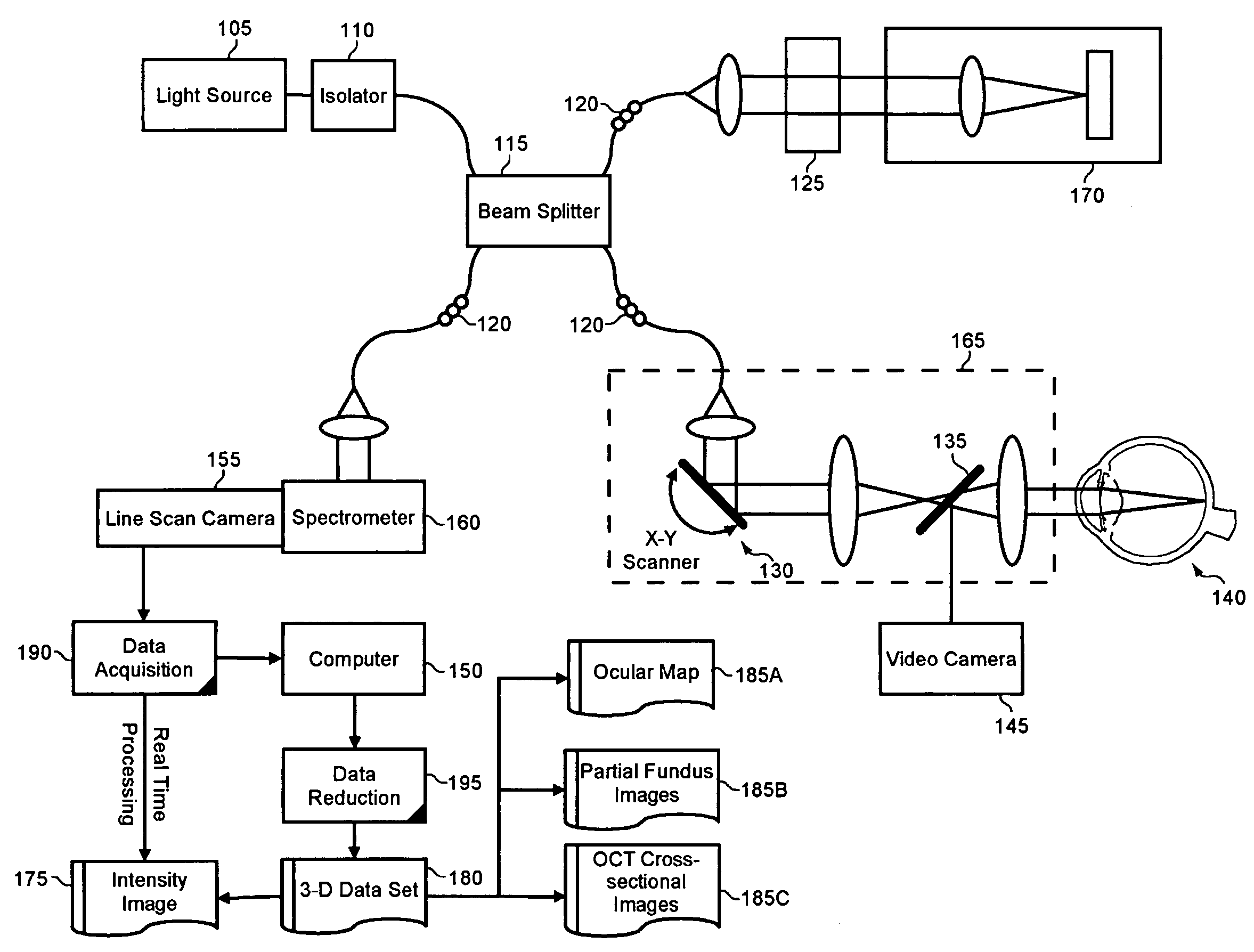
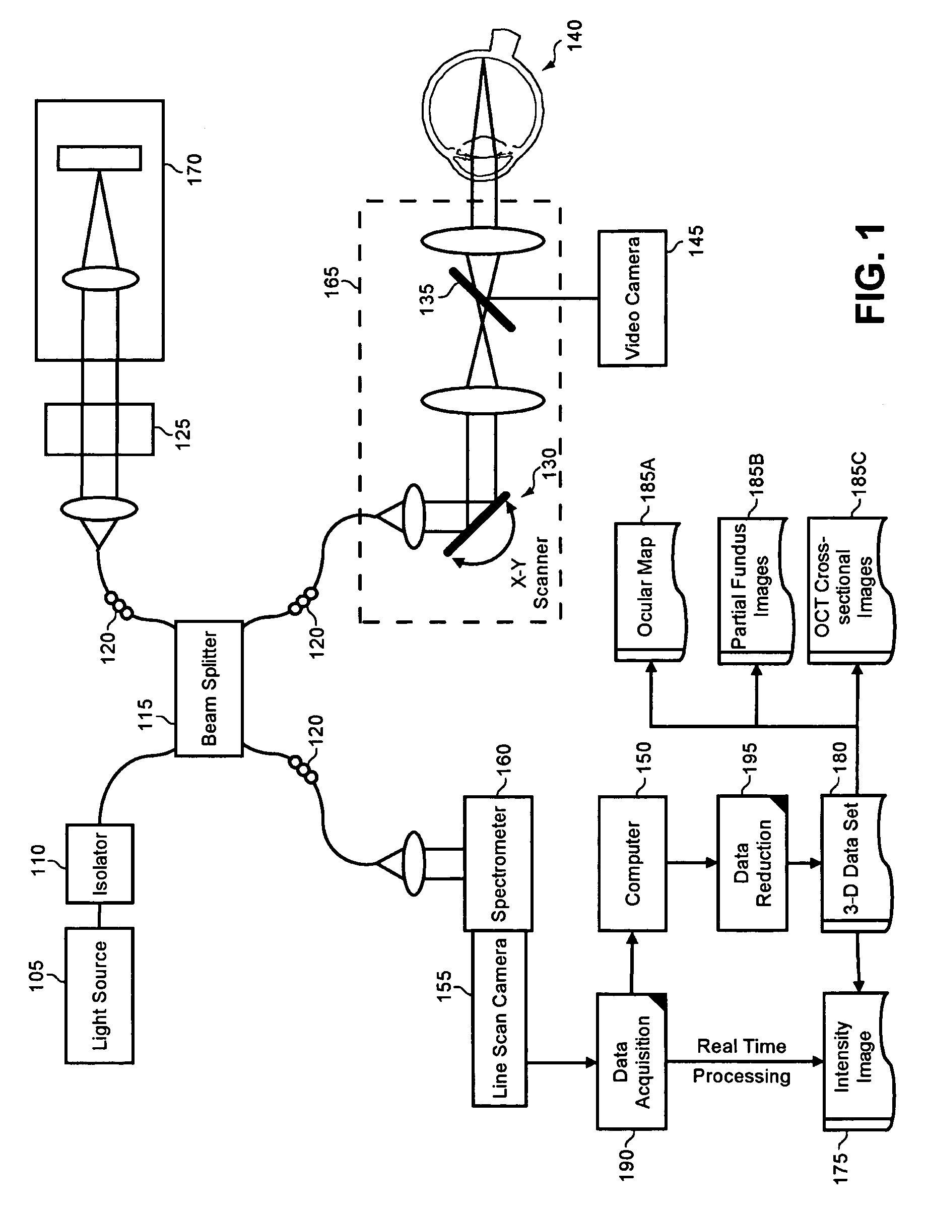
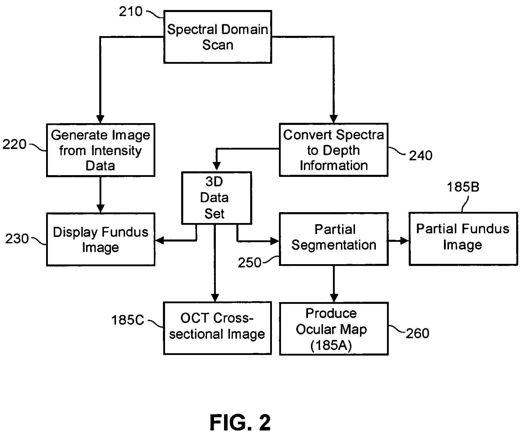
[0017]The present invention is a system, method and apparatus for anatomical mapping through the production of a 3-D data set, for instance through imagery produced utilizing OCT. In accordance with the present invention, a fundus intensity image can be acquired from a spectral domain scanning of light back-reflected from an eye. The 3-D data set can be reduced to generate an ocular mapping, including edema mappings and thickness mappings. Optionally, a partial fundus intensity image can be produced from the spectral domain scanning of the eye to generate an en face view of the retinal structure of the eye without first requiring a full segmentation of the 3-D data set.

[0018]In further illustration, FIG. 1 is a schematic illustration of an OCT imaging system configured for anatomical mapping in accordance with the present invention. As shown in FIG. 1, a low coherent light source 105 can be provided. The low coherent light source 105 can be a super-luminescent diode, such as a super...


PUM


Abstract

A system, method and apparatus for anatomical mapping utilizing optical coherence tomography. In the present invention, 3-dimensional fundus intensity imagery can be acquired from a scanning of light back-reflected from an eye. The scanning can include spectral domain scanning, as an example. A fundus intensity image can be acquired in real-time. The 3-dimensional data set can be reduced to generate an anatomical mapping, such as an edema mapping and a thickness mapping. Optionally, a partial fundus intensity image can be produced from the scanning of the eye to generate an en face view of the retinal structure of the eye without first requiring a full segmentation of the 3-D data set. Advantageously, the system, method and apparatus of the present invention can provide quantitative three-dimensional information about the spatial location and extent of macular edema and other pathologies. This three-dimensional information can be used to determine the need for treatment, monitor the effectiveness of treatment and identify the return of fluid that may signal the need for re-treatment.

Description

PRIORITY[0001]This application claims the benefit of the filing date under 35 U.S.C. § 119(e) of U.S. Provisional Patent Application Ser. No. 60 / 632,387, filed on Dec. 2, 2004, which is hereby incorporated by reference in its entirety.BACKGROUND OF THE INVENTION[0002]1. Technical Field[0003]The present invention relates to coherent waveform based imaging, and more particularly to an optical coherence tomography (OCT) imaging system.[0004]2. Statement of the Related Art[0005]Many imaging systems utilize coherent waveforms to obtain information regarding target objects of interest. Examples include OCT, ultrasound diagnostics, and synthetic aperture radar. OCT is a low-coherence interferometer-based noninvasive medical imaging modality that can provide high-resolution sectional images of biological tissues (see for example, U.S. Pat. Nos. 5,321,501, 5,459,570, Huang, D. et al. (1991). “Optical coherence tomography.”Science 254(5035): 1178-81). Since first introduced, OCT has been used...

Claims


Application Information

Patent Timeline
Patent Type & Authority Patents(United States)
IPC IPC(8): G01B9/02
CPCA61B3/102G01B9/02089G01B9/02091G01B9/02044
Inventor KNIGHTON, ROBERT W.JIAO, SHULIANGGREGORI, GIOVANNIPULIAFITO, CARMEN A.
Owner UNIV OF MIAMI
Why Eureka
Try Eureka