Diagnosis of lysosomal storage disorders using saposins and other markers

a technology of lysosomal storage and saposin, which is applied in the direction of material testing goods, biochemistry apparatus and processes, instruments, etc., can solve the problems of increasing the size and number of lysosomes within the cells, increasing the accumulation of substrates, etc., and achieves positive treatment outcomes

Inactive Publication Date: 2008-05-27
WOMENS & CHILDRENS HOSPITAL
View PDF6 Cites 4 Cited by
  • Summary
  • Abstract
  • Description
  • Claims
  • Application Information

AI Technical Summary

Benefits of technology

[0015]The invention also provides methods of monitoring a patient with galactosialidosis, MPS IVA or Pompe's disease. Such methods entail determining a baseline level of α glucosidase in a tissue sample from the patient with the disorder before treatment with the agent. The baseline level is then compared with a level of the α-glucosidase in a tissue sample from the patient after treatment with the agent. An increase relative to the baseline indicates a positive treatment outcome.

Problems solved by technology

The deficiency leads to the accumulation of substrates, normally degraded within the lysosome, and an increase in size and number of lysosomes within the cells.

Method used


Image

  • Diagnosis of lysosomal storage disorders using saposins and other markers
  • Diagnosis of lysosomal storage disorders using saposins and other markers
  • Diagnosis of lysosomal storage disorders using saposins and other markers

Examples


examples

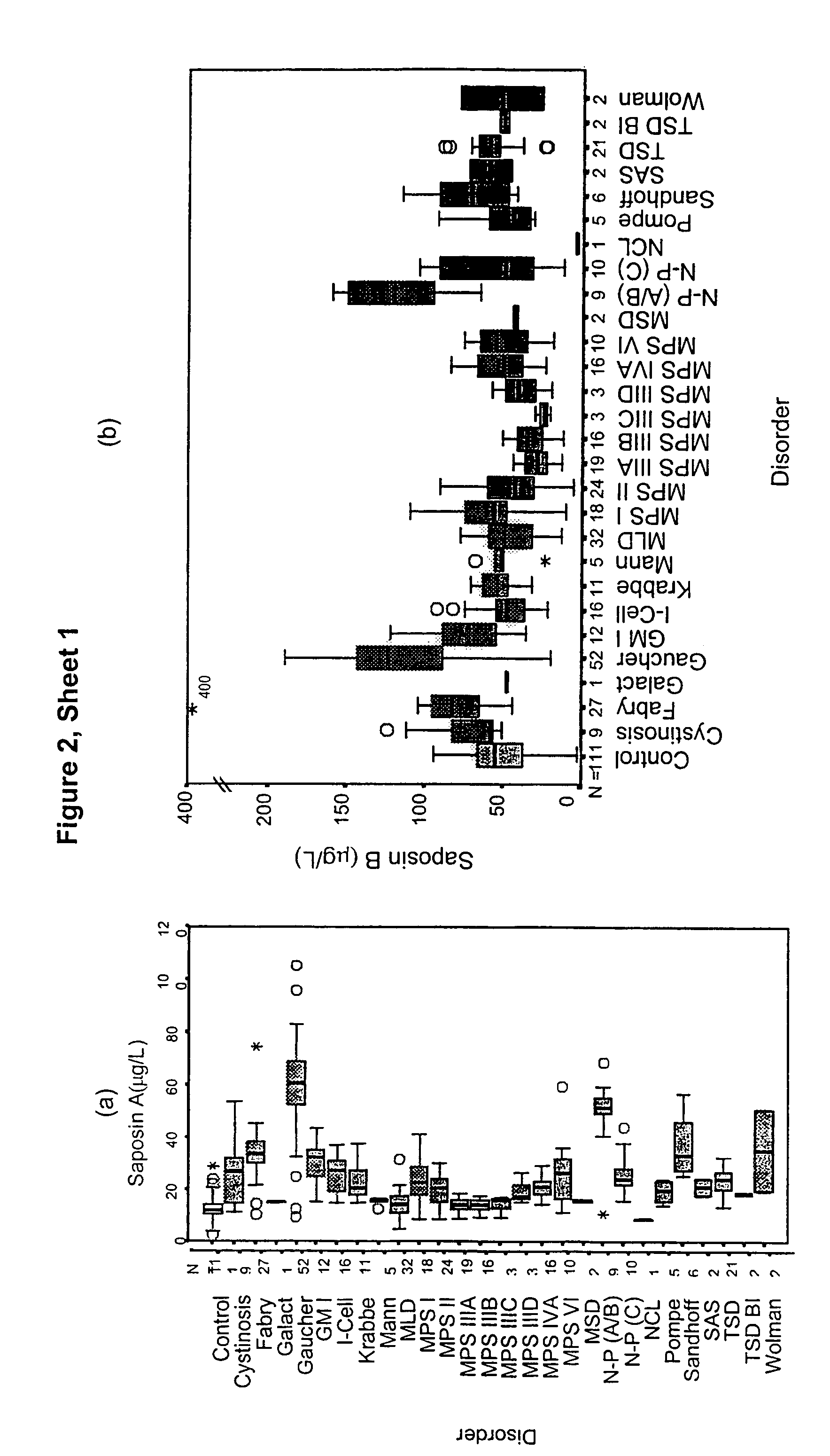
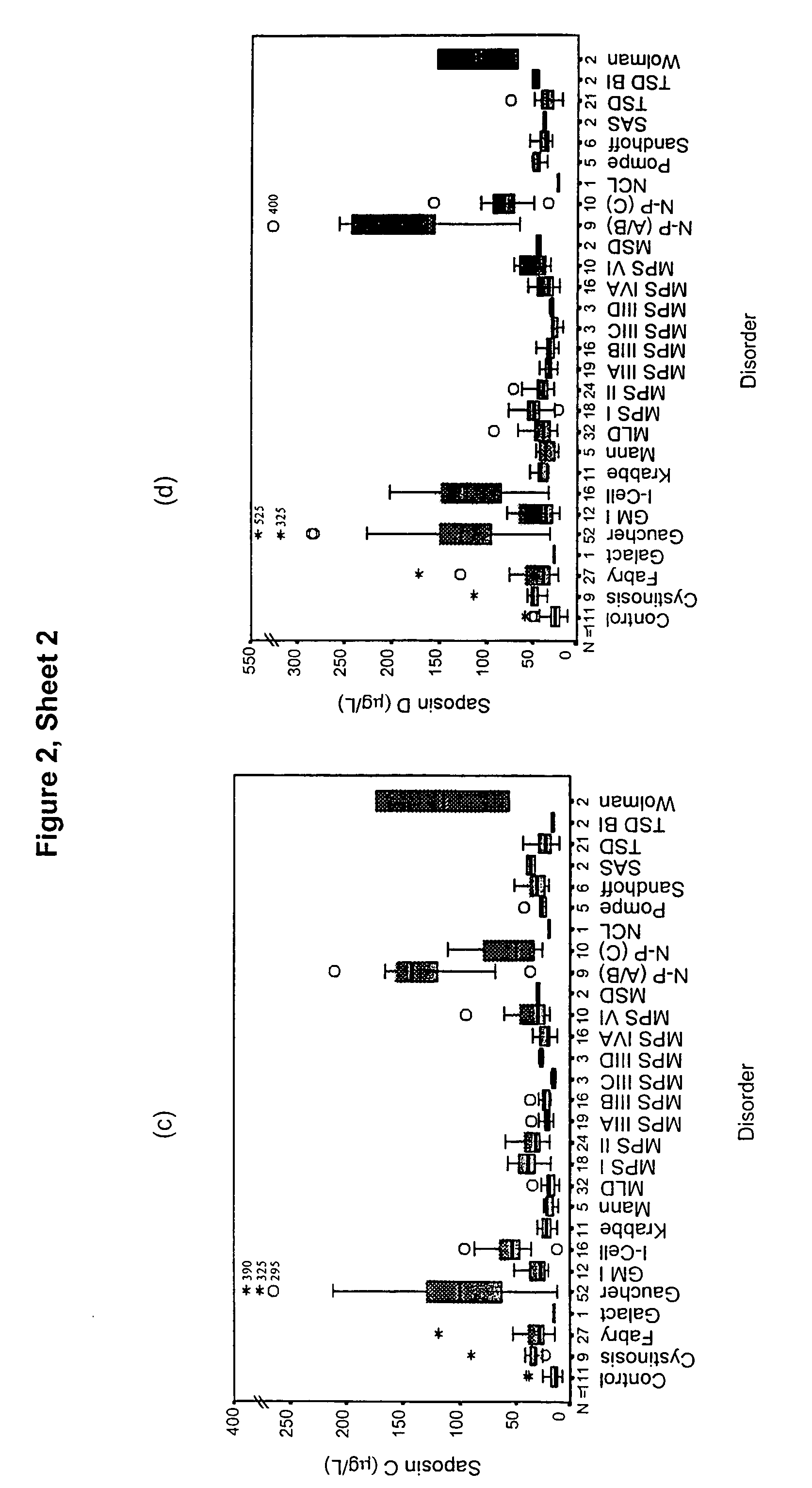
1. Saposins as a Marker for Lysosomal Storage Disease

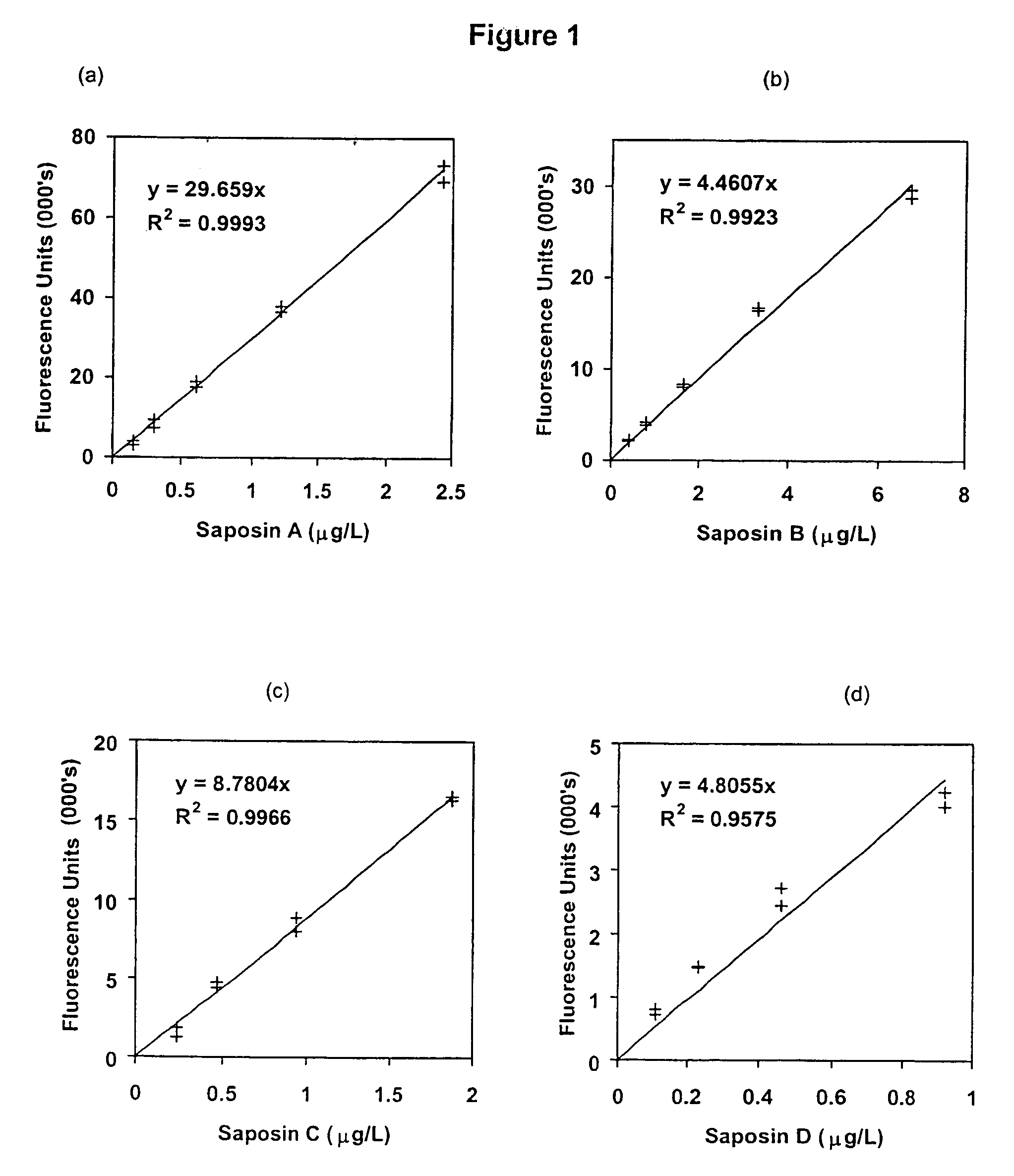
[0066]This example shows the suitability of saposins A, B, C and D as a screening marker for LSD by determining the levels of these proteins in plasma samples taken from unaffected and LSD-affected individuals.

[0067]Materials and Methods

[0068]Patient Samples

[0069]Plasma samples used were from samples submitted to the National Referral Laboratory (Women's and Children's Hospital, Adelaide, Australia) for LSD screening and processing for routine biochemistry. Whole blood samples for fractionation studies were obtained from healthy volunteers within the laboratory.

[0070]Polyclonal Antibodies

[0071]Anti-saposins A, B, C and D polyclonal antibodies were produced and characterised as previously described [15]. Each antibody was purified on a 2-mL Hitrap™ Protein G column (Pharmacia Biotech, Uppsala, Sweden) and quantified by absorbance at 280 nm (absorbance=1.4 for 1.0 g / L).

[0072]Europium Labelling of Polyclonal Antibodies

[0073]Purified ...

example 2

[0098]Plasma samples have been assayed for α-glucosidase protein by an immunoquantification assay using the same technology as described for the saposin study. Results are expressed as a box plot in FIG. 3. Results are also presented in Table 5 showing the median level in each disorder group, and the % of patients with an α-glucosidase level elevated above the 95th centile of the control population. Overall 55% of patients showed an elevation of α-glucosidase. All Pompe patients showed a level of α-glucosidase below the 5th centile of the control population.

[0099]Therefore, α-glucosidase is a useful marker for the detection of a range of LSD, by detecting either an elevation or a decrease in the level of this protein in plasma serum or whole blood.

[0100]

TABLE 5α-glucosidase levels in plasma from control and LSD affected individualsPERCENTILESCountMinMaxMeanStd Errelevateda0525507595control801.922.310.4.553.87.410.112.320.6acid lipase264.887.376.111.310076.1——cystinosis813.557.225.65...

example 3

[0101]The experiment shown in FIG. 2 was repeated for some of the samples using a combination of monoclonal and polyclonal antibodies to saposin C. Microliter plates were coated with the monoclonal antibody 7B2 (4 mg / L, 16 h, 4° C.), washed then incubated with plasma samples (2 μL) in assay buffer (6 h, 4° C.). The plate was washed again, then incubated with solution-phase europium labelled antibodies as indicated at 500 μg / mL (16 h, 4° C.). The plate was washed and developed with enhancement buffer (200 μL / well) and the fluorescence read. Concentrations of saposin C were calculated based on calibration curves using recombinant saposin C.

[0102]

Detection AntibodiesPlasma SamplePoly(EU)13A1(Eu)23A1(Eu) / Poly(Eu)Control(C2)4.507.8Control(C3)6.3011.3Gaucher(614)460.541Gaucher(1500)37.54.476MLD(451)4.42636MLD(552)9.439461Anti-saposin C polyclonal antibody2Anti-saposin C monoclonal antibody

[0103]The 7B2 antibody behaved similarly to polyclonal sera in the capture step. However, the 3A1 ant...


PUM

PropertyMeasurementUnit
concentrationsaaaaaaaaaa

Abstract

The invention provides methods of diagnosing or monitoring lysosomal storage disorders based on detecting levels of saposins, LAMPs and/or α-glucosidase in patient sample. Elevated levels of saposins and/or LAMPs are indicative of a disorder. Elevated levels of α glucosidase are indicative of some types of lysosomal storage disorders and decreased levels of α glucosidase are indicative of other types of lysosomal storage disorder. In some methods, the profile of elevation of different saposins, LAMPs and α glucosidase allows distinction between different types of lysosomal storage disorder.

Description

CROSS-REFERENCE TO RELATED APPLICATIONS[0001]The present application derives priority from U.S. Ser. No. 60 / 124,864, filed Mar. 17, 1999 and GB 99 08190.3, filed Apr. 9, 1999, both of which are incorporated by reference in their entirety for all purposes.BACKGROUND[0002]Lysosomal storage disorder (LSDs) are a large family of genetic disorders that can lead to the manifestation of severe clinical symptoms. Although the frequency of each individual disease is relatively rare, the collective incidence of all lysosomal storage disorders is about 1 in 7000 newborns. This is greater than that of other diseases, such as phenylketonuria, for which newborn screening methods are available. Each lysosomal storage disorder results from a deficiency in a lysosomal enzyme, transporter or protein involved in lysosomal biogenesis or function [1]. The deficiency leads to the accumulation of substrates, normally degraded within the lysosome, and an increase in size and number of lysosomes within the ...

Claims


Application Information

Patent Timeline
Patent Type & Authority Patents(United States)
IPC IPC(8): C12Q1/00
CPCG01N33/68G01N2405/08G01N2800/044
Inventor MEIKLE, PETER JOHNHOPWOOD, JOHN JOSEPHWINCHESTER, BRYAN GORDON
Owner WOMENS & CHILDRENS HOSPITAL
Why Eureka
Try Eureka