

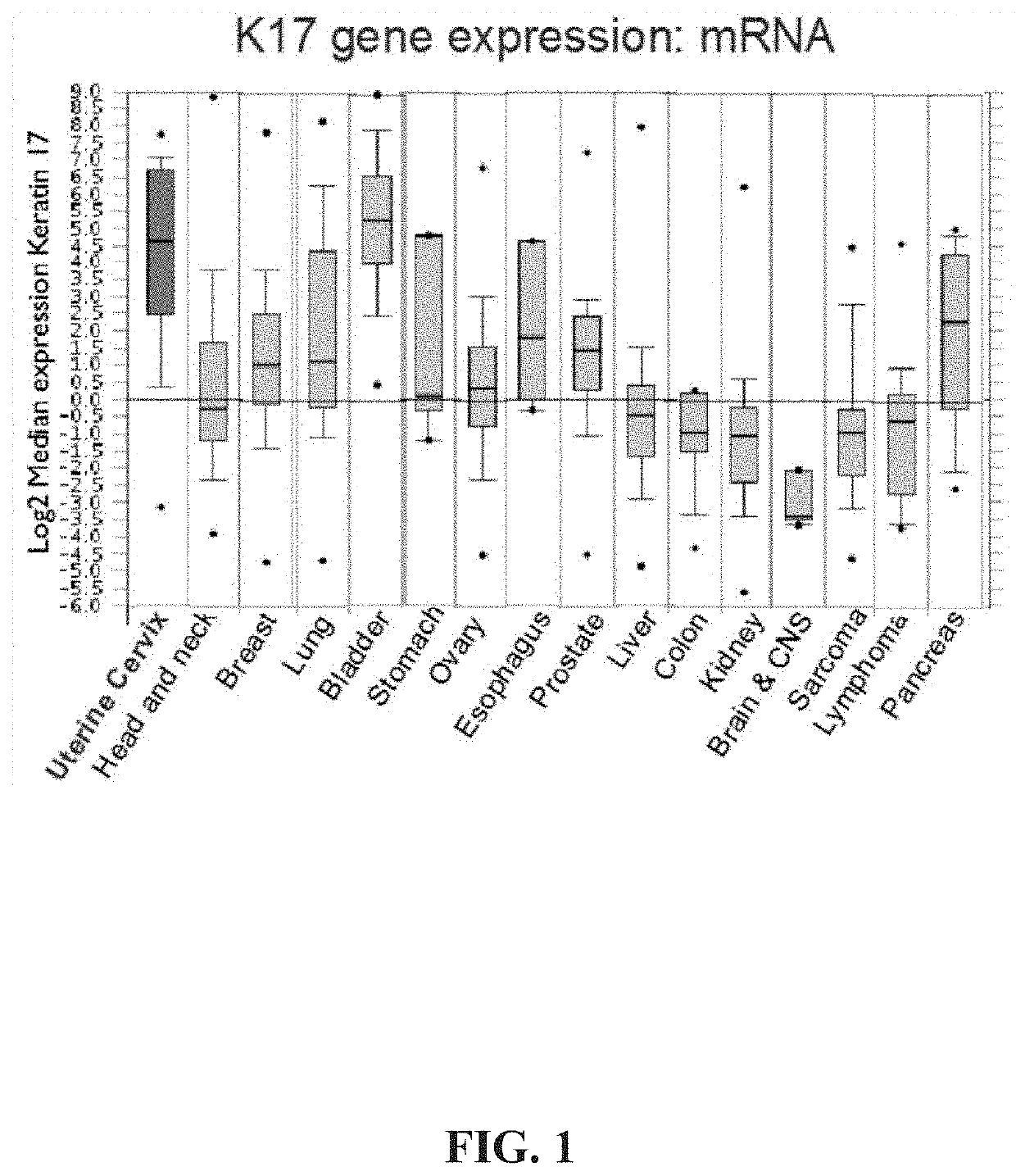
Keratin 17 as a biomarker for bladder cancer
a biomarker and bladder cancer technology, applied in the field of keratin 17 as a biomarker for bladder cancer, can solve the problems of ineffective detection of low-grade tumors of the bladder mucosa by urovysion®, and a much more bleak outcome, so as to reduce the amount of false negative readings, improve diagnosis, and improve treatment outcomes
- Summary
- Abstract
- Description
- Claims
- Application Information
AI Technical Summary
Benefits of technology
Problems solved by technology
Method used
Image

Examples
example 1
and Methods
[0058]The studies carried out included the analysis of 84 formalin-fixed paraffin-embedded surgical tissue blocks (Table 1). All surgical tissue blocks were obtained from subjects (patients) suspicious for having bladder cancer. These study cases comprised the following diagnostic categories: benign urothelium, (n=12), papillary urothelial neoplasm of low malignant potential (PUNLMP) (n=9), low-grade papillary urothelial carcinoma. (n=23), high-grade papillary urothelial carcinoma (n=14), transitional / urothelial carcinoma (n=26). In certain cases, a tissue block was selected following histologic review by a pathologist (DCM) of hematoxylin-eosin stained sections from either transurethral resection (TURBT), bladder biopsy or cystectomy specimens to confirm that diagnostic tissue as originally reported, was adequately represented in the remaining tissue blocks. Cases that had insufficient residual tissue or for which diagnostic tissue could not be preserved for future clini...
example 2
7 Detection in Immunohistochemical Stained Cells as Diagnostic Method for Bladder Cancer
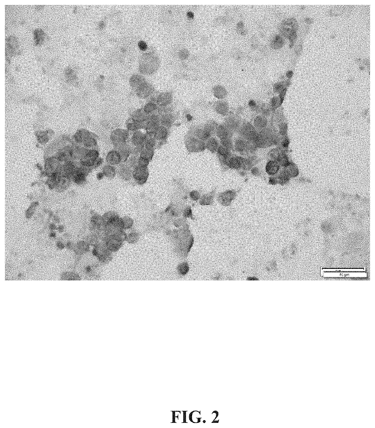
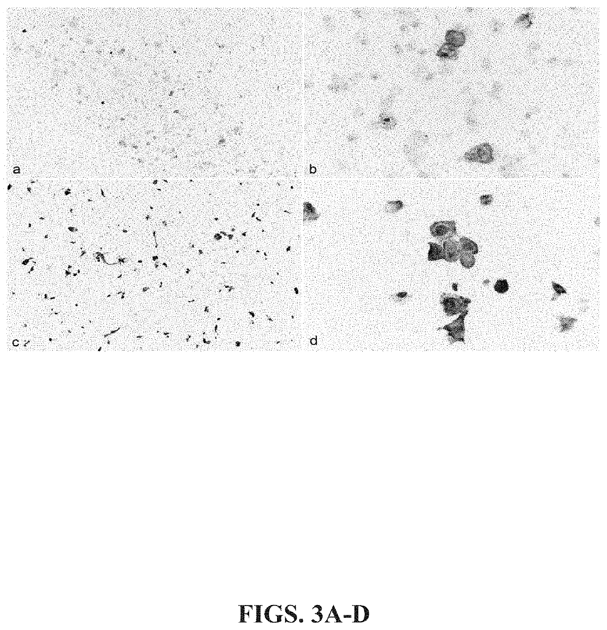
[0067]To determine the diagnostic values of K17 in one or more diagnostic categories of early stage bladder cancer, immunohistochemical staining (Table 1) was performed for K17 on tissue of archived bladder cancer patient tissue samples from four diagnostic categories: benign bladder mucosa (benign), papillary urothelial neoplasia of low malignant potential (PUNLMP), low-grade papillary urothelial carcinoma (LG), high grade papillary urothelial carcinoma (HG) and urothelial carcinoma.
[0068]K17 staining was only faintly detectable (mean PathSQ score of 2.08) in benign bladder mucosa, but was highly expressed in all diagnostic categories of bladder cancer examined. For example, Table 1 shows that mean PathSQ scores ranged from 12.3831954 to 45.1781993 for HG and PUNLMP samples, respectively (Table 1), whereas the mean PathSQ score of control (non-cancerous) samples was approximately 2. Further, FIG...
example 3
7 Detection in Cells Through Automated Device Based Detection Methods
[0071]In addition to immunostaining of urine cytology samples, K17 positive cells in urine cytology samples is detectable by flow cytometry and microfluidic device systems capable of detecting fluorescently tagged proteins. Here, labeled cancer cells known to be positive for KI 7 (HeLa, FIG. 8A) and K17-negative, control cells (C33, FIG. 8B) were isolated, fluorescently labeled with K17 antibodies and K17 expression was analyzed on a microfluidic detection device, as set forth above. As seen in FIGS. 8A-8B, there are significantly more K17-labeled cells in Hela (90.4% K17-positive) compared to control, C33 cells (35.1% K17-positive). Taken together, these data show that detection of K17 positive cells can be efficiently achieved using a microfluidic device coupled with K17 immunostaining.
PUM
| Property | Measurement | Unit |
|---|---|---|
| OD | aaaaa | aaaaa |
| temperature | aaaaa | aaaaa |
| detectable wavelength | aaaaa | aaaaa |
Abstract
Description
Claims
Application Information