Hybrid imaging method to monitor medical device delivery and patient support for use in the method

a medical device and hybrid imaging technology, applied in the field of medical imaging, can solve the problems of limiting the ability to use even real-time imaging data, the original targeting coordinates are no longer correct, and the needle may deviate from the desired path and deflect on the route to the target, so as to facilitate any treatment or diagnostic procedure, the effect of less invasiveness to the patient, and faster speed

Active Publication Date: 2008-05-27
INVIVO CORP
View PDF32 Cites 153 Cited by
  • Summary
  • Abstract
  • Description
  • Claims
  • Application Information

AI Technical Summary

Benefits of technology

[0009]The present technology uses 3D imaging data obtained by conventional non-invasive imaging techniques, particularly MRI (magnetic resonance imaging), US (ultrasound), positron emission tomography (PET), computerized tomography (CT), or other three-dimensional imaging system. The technology discloses a number of imaging and interventional functions required for complete breast-MRI patient management, including screening for breast cancer, determination of tumor extent and multi-focality of previously diagnosed cancer, and diagnosis of suspicious lesions. Further applications of the technology include MR-guided positioning of wires and marking devices in the breast to facilitate any treatment or diagnostic procedure, such as those including but not limited to surgical excision / biopsy, MR-guided core biopsy for lesion diagnosis without surgery, and MR-guided and monitored minimally invasive therapy to destroy diseased tissue. Multi-modality MR / US breast imaging disclosed in the descriptions of the present technology enables a more effective means of interventional device positioning (more accurate, faster, less invasive to the patient), a means of tissue diagnosis without biopsy (through ultrasound (referred to herein as “US examination”), and a means of monitoring tissue ablation boundaries when performing minimally invasive therapies.
[0010]While the method of the present technology was specifically optimized for breast cancer management, it will be understood by those of ordinary skill in the art that the techniques and apparatus of this technology can be easily adapted to various other body parts and pathologies.
[0014]A third aspect of the present technology is to provide a transport stretcher or gurney to aid patient access to the interventional area, the apparatus to include a bridged interventional gap, IV (intravenous) poles which accompany the patient during the entire procedure, a headrest which accommodates the patient's arms and permits a view out of the magnet, and mirrors and lighting to help better position the patient.

Problems solved by technology

In cases where tissue stiffness variations are extreme, the needle may deviate from the desired path and deflect on-route to the target tissue.
Similarly, the needle may distort the tissue itself and thereby move the target tissue to a new location, such that the original targeting coordinates are no longer correct.
Further limitations of current systems include the fact that needle position is often determined by reference to its artifact generated in the MR images.
In many situations this is appropriate; however, when targeting small lesions (i.e. <7 mm), the needle artifact (often 5-9 mm) may obscure the target, limiting the ability to use even real-time imaging data, as from MRI, to validate needle / target position.
However, these “open” systems are not always available and are often of suboptimal field strength, which can result in reduced image quality.
While this approach usefully addresses the issues of tissue / needle deflection, it also removes the normally close interaction between the radiologist and patient, which may lead to high levels of patient anxiety.

Method used


Image

  • Hybrid imaging method to monitor medical device delivery and patient support for use in the method
  • Hybrid imaging method to monitor medical device delivery and patient support for use in the method
  • Hybrid imaging method to monitor medical device delivery and patient support for use in the method

Examples


example 1

MR-Guided Wire Localization

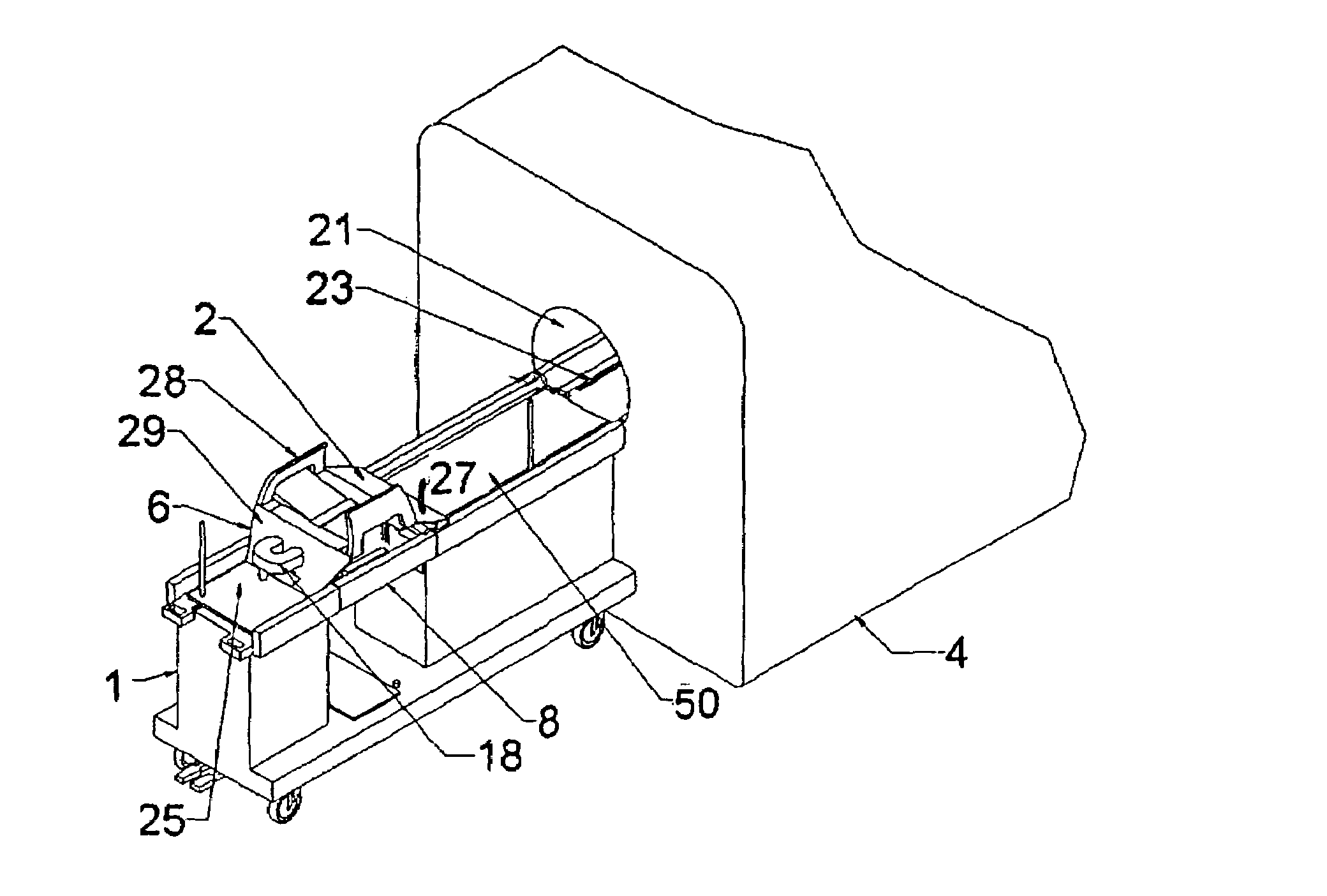
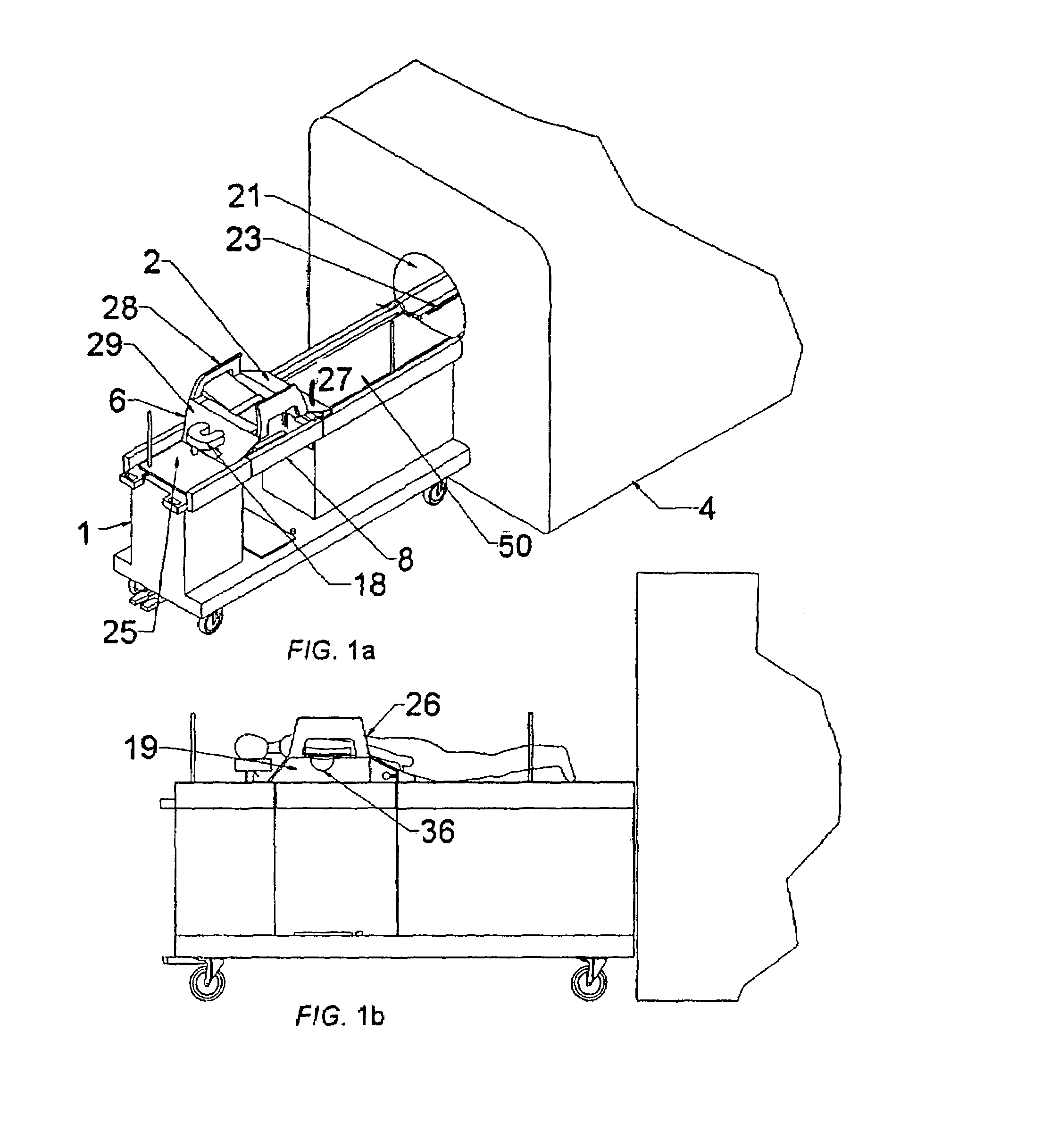
[0117]The use of device guidance techniques to deliver a localization wire or marker to guide surgical intervention is illustrated in the flowchart shown in FIG. 25. The patient is first positioned on the patient support apparatus. The breast of interest is then compressed between two fenestrated compression plates with attached fiducial markers, which are attached in turn to the compression plate locking supports. These fenestrated plates are sterile and can be introduced while the patient is in the prone position. These plates can be moved in the anterior-posterior direction to positions near the chest wall to enable full interventional access to the breast. MR imaging coils are attached to these compression plates. MR imaging is then used to identify the lesion and the fiducial markers. Using this information, the appropriate fenestrated plate aperture and needle guide plug hole are determined in order to position the needle as closely as possible to th...

example 2

MR-Guided Angulated Breast Biopsy

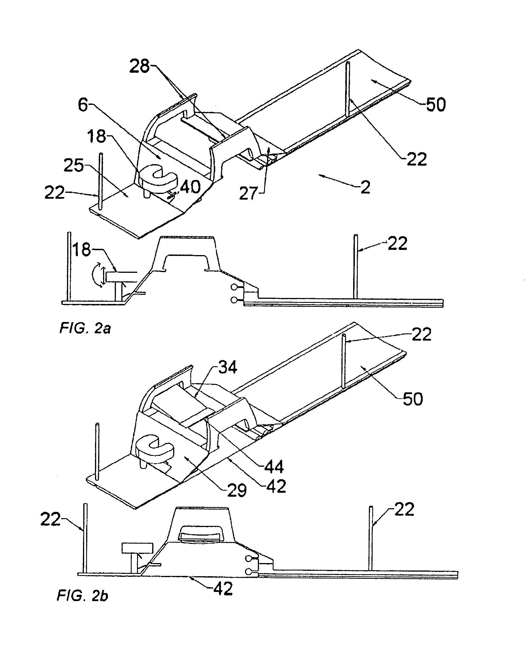
[0119]According to the invention, a core biopsy needle may be delivered to the breast on an oblique trajectory as illustrated in the flowchart shown in FIG. 26. A needle guide plug having straight (medial-lateral) holes of a larger diameter than the biopsy needle, or angulating guide plugs may be used in cases. Patient positioning with fenestrated plates and MR imaging is performed in an identical manner as described under MR-guided wire localization, with the option of implementing the various compression plates indicated in the diagram. Calculation of the needle trajectory is done using compound angles to define the needle trajectory and a goniometer is used to set the orientation of the guide plug. In cases where access to some targets is limited by the fenestration access, the fenestrated plate's position may be altered without moving the breast.

[0120]Introducer needle insertion is preceded by producing an incision in the skin at the center of th...

example 3

MR-Guided Marker Placement

[0121]The concept of MR-guided biopsy presented in Example 2 can easily be extended to placement of small position markers in the breast, according to the invention. This may or may not be done in conjunction with MR-guided biopsy, or done in conjunction with MR-guided needle localization. This application would take advantage of the fact that a large tissue segment can be accessed through a single incision point (repositioning of the fenestrated plate to provide better access may be required). As shown in FIG. 15, a linear tumor 320 can be delineated by way of a set of markers 326 These markers 326 may be identified after the procedure defining the maximal extent of the tumor. Markers such as endovascualar occlusion coils, surgical clips, or any of the devices as disclosed by Foester et al. in U.S. patent application Ser. No. 2002 / 0193815 A1 may be used. Furthermore, radiotherapy implantable seeds, or local chemotherapy delivery devices may also be distrib...


PUM


Abstract

This invention discloses a method and apparatus to deliver medical devices to targeted locations within human tissues using imaging data. The method enables the target location to be obtained from one imaging system, followed by the use of a second imaging system to verify the final position of the device. In particular, the invention discloses a method based on the initial identification of tissue targets using MR imaging, followed by the use of ultrasound imaging to verify and monitor accurate needle positioning. The invention can be used for acquiring biopsy samples to determine the grade and stage of cancer in various tissues including the brain, breast, abdomen, spine, liver, and kidney. The method is also useful for delivery of markers to a specific site to facilitate surgical removal of diseased tissue, or for the targeted delivery of applicators that destroy diseased tissues in-situ.

Description

RELATED APPLICATION DATA[0001]This application claims priority from U.S. Provisional Patent Application Ser. No. 60 / 506,784, filed Sep. 30, 2003.BACKGROUND OF THE INVENTION[0002]1. Field of the Invention[0003]This invention relates to the field of medical imaging and particularly to a hybrid imaging method and apparatus used to monitor and optimize the placement of interventional medical devices in human tissues.[0004]2. Background of the Art[0005]A number of techniques, methodologies, apparatus and systems have been proposed to improve the accuracy of instrumentality placement such as needle or catheter placement into tissue based on measurements from 3D imaging formats. These imaging formats (such as Magnetic Resonance Imaging, sonographs (ultrasound), fluoroscopy, X-ray, and the like) locate the needle entry device in relation to treatment- or therapy-targeted tissue, such as MR-detected target tissue. These imaging formats generate imaging data that are used to determine the app...

Claims


Application Information

Patent Timeline
Patent Type & Authority Patents(United States)
IPC IPC(8): A61B5/05A47B13/00A47B7/00A61B5/055A61B6/00A61B6/04A61B8/00A61B8/08A61B10/00A61B17/34A61B19/00A61G1/04G01S7/52
CPCA61B8/0825A61B19/20A61B8/0841A61B8/406A61B8/4416A61B8/5238A61B19/201A61B19/203G01S7/52049A61B5/0555A61B10/0233A61B2017/3411A61B2017/3413A61B2019/205A61B2019/5236A61B2019/5276A61B2019/5408A61B8/0833A61B8/4245A61B90/10A61B90/17A61B90/11A61B90/14A61B2090/374A61B2090/378A61B2090/3908A61B5/055
Inventor PIRON, CAMERON ANTHONYLUGINBUHL, CHRISTOPHER ALEXANDERPLEWES, DONALD B.
Owner INVIVO CORP
Why Eureka
Try Eureka