
Methods and apparatus for model-based detection of structure in view data

a technology of view data and model data, applied in the field of model-based techniques for detecting structure in view data, can solve the problems of affecting the resulting image resolution, reconstructed image data has less resolution than view data, and loss of resolution, etc., to obscure and/or eliminate the detail of the reconstructed imag

Active Publication Date: 2009-02-17
BROWN UNIVERSITY
View PDF22 Cites 41 Cited by
  • Summary
  • Abstract
  • Description
  • Claims
  • Application Information

AI Technical Summary

Benefits of technology

[0024]Another embodiment according to the present invention includes a method of processing view data of structure obtained from an X-ray scanning device capable of scanning, in situ, at least a portion of a human's anatomy, the method comprising an act of detecting at least one dimension of a structure, wherein the at least one dimension is less than 500 microns.
[0025]Another embodiment according to the present invention includes a method of processing view data of structure obtained from a micro-computer tomographic (microCT) X-ray scanning device, the method comprising an act detecting at le...

Problems solved by technology

This averaging blurs the image and affects the resulting image resolution.
The result is that the reconstructed image data has less resolution than the view data from which it was generated.
This loss of resolution may obscure and / or eliminate detail in the reconstructed image.
However, this process may be difficult and time consuming.
For example, it may be difficult to assess 3D biological structure by attempting to follow 2D structure through a series of stacked 2D images.
In particular, it may be perceptually difficult and time consuming to understand how 2D structure is related to 3D structure as it appears, changes in size and shape, and / or disappears in successive 2D image slices.
To further frustrate this process, when anatomical structure of interest is small, the structure may be difficult to discern or absent altogether in the reconstructed image.
However, segmentation on reconstructed images has proven difficult and Applicant has appreciated that it may be ineffective in detecting small structures whose features have been obscured or eliminated during image reconstruction.
Reconstructed images of view data obtained from conventional X-ray scanning devices may be limited to a resolution of approximately 500 microns.
As a result, conventional imaging techniques may be unable to image structure having dimensions smaller than 500 microns.
That is, variation in the density distribution of these small structures cannot be resolved by conventional image reconstruction.
However, microCT cannot image large objects such as a human patient and therefore is unavailable for in situ imaging of the human anatomy.
Applicant has appreciated that when the structure being modeled is complex and includes a number of deformable parameters, the combinatorial problem of configuring the model may become intractable.
That is, as the number of parameters over which the model is allowed to vary increases, the number of possible configurations of the model tends to explode.
In addition, Applicant has appreciated that with no guidance on how to initially configure the model, a poorly chosen initial hypothesis may cause a subsequent optimization scheme to converge to an undesirable local minimum.
As a result, the selected model configuration may poorly reflect the actual structure that was scanned.

Method used


Image

  • Methods and apparatus for model-based detection of structure in view data
  • Methods and apparatus for model-based detection of structure in view data
  • Methods and apparatus for model-based detection of structure in view data

Examples


Embodiment Construction

[0047]As discussed above, segmentation of reconstructed images is often difficult and is limited to information describing structure at the reduced resolution resulting from the image reconstruction process. Structure at or below this resolution, though present in the view data, may be unavailable to detection and segmentation algorithms that operate on reconstructed image data. Conventional model based techniques that seek to avoid image reconstruction have been frustrated by the combinatorial complexity of fitting a model configuration to the observed view data.

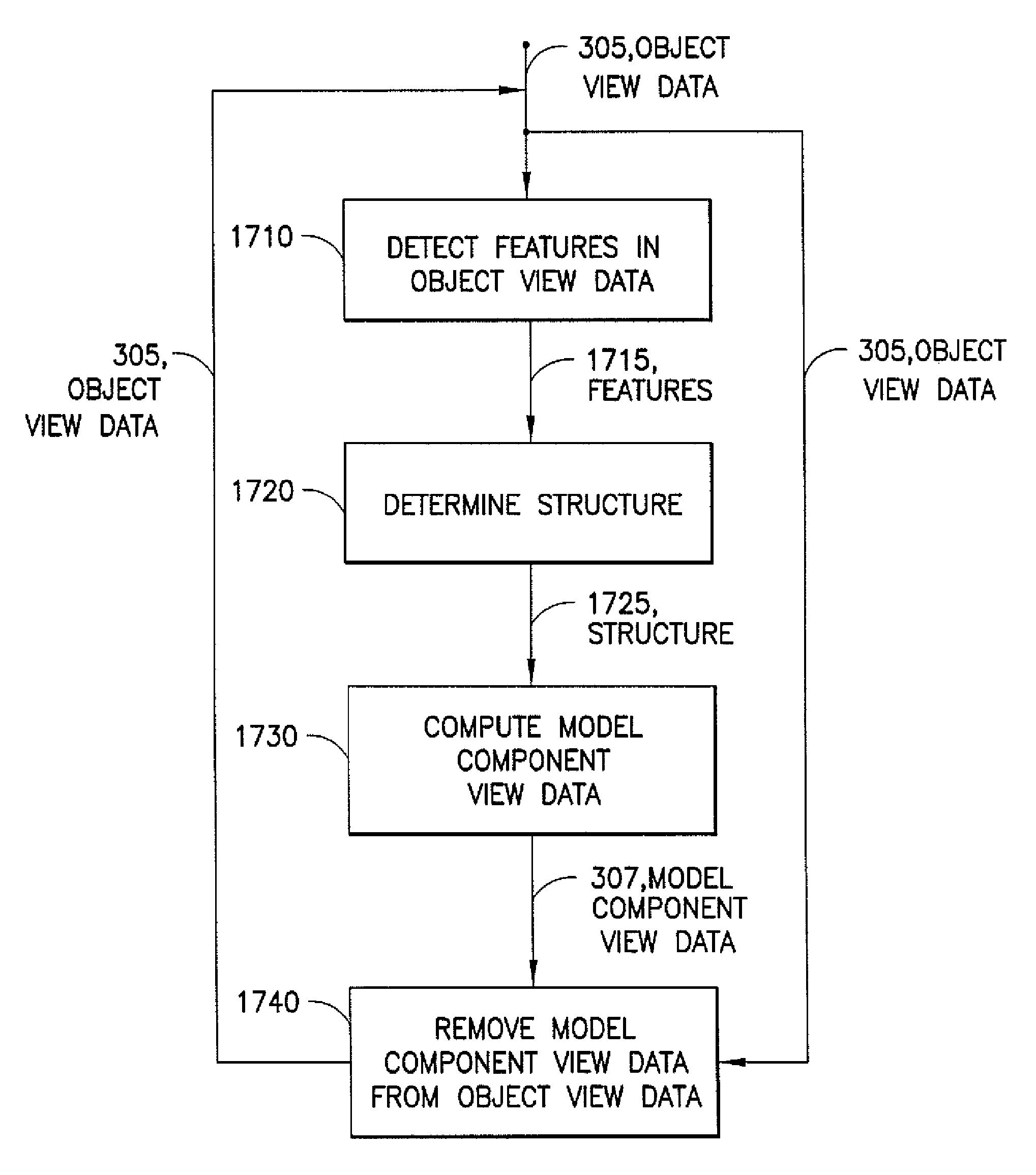
[0048]In one embodiment according to the present invention, a model is generated to describe structure to be detected in view data obtained from scanning the structure. The view data may be processed to detect one or more features in the view data characteristic of the modeled structure and employed to determine a value of one or more parameters of a configuration of the model, i.e., information in the view data may be used...


PUM


Abstract

In one aspect, a method and apparatus for determining a value for at least one parameter of a configuration of a model associated with structure of which view data has been obtained including detecting at least one feature in the view data, and determining the value for the at least one parameter of the configuration of the model based at least in part on the at least one feature. In another aspect, a method and apparatus for detecting at least one blood vessel from object view data obtained from a scan of the at least one blood vessel including generating a model of the at least one blood vessel, the model having a plurality of parameters describing a model configuration, determining a hypothesis for the model configuration based, at least in part, on at least one feature detected in the object view data, and updating the model configuration according to a comparison with the object view data to arrive at a final model configuration, so that the final model configuration represents the at least one blood vessel.

Description

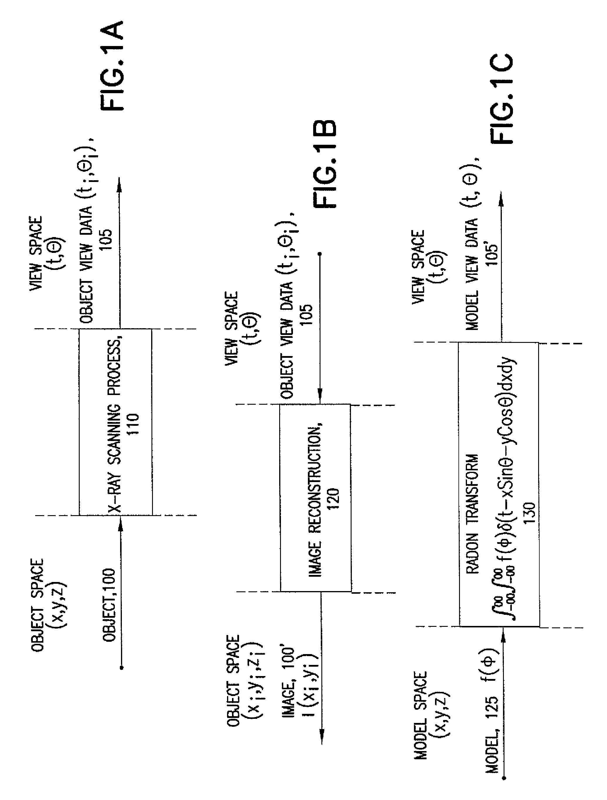
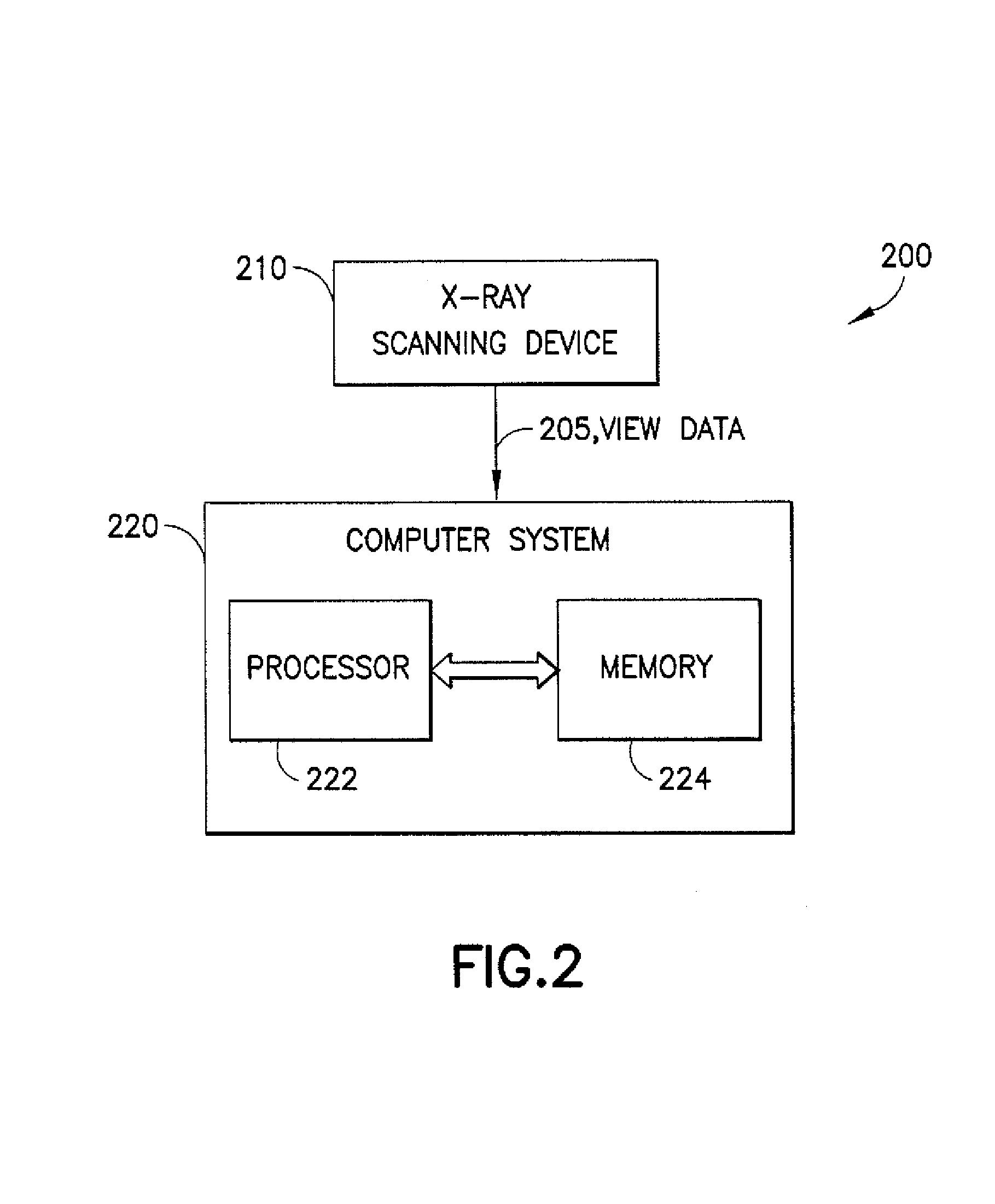
RELATED APPLICATIONS[0001]This application claims the benefit under 35 U.S.C. §119(e) of U.S. Provisional Application No. 60 / 479,114, filed Jun. 17, 2003, entitled “MODEL-BASED TOMOGRAPHIC RECONSTRUCTION OF VESSEL NETWORKS,” by Mundy et al., which is incorporated by reference herein in its entirety.FIELD OF THE INVENTION[0002]The present invention relates to imaging and more particularly to model-based techniques for detecting structure in view data, for example, in view data obtained from an X-ray scanning device.BACKGROUND OF THE INVENTION[0003]X-ray imaging technology provides a non-invasive technique of visualizing the internal structure of an object of interest by exposing the object to high energy electromagnetic radiation (i.e., X-rays). X-rays emitted from a radiation source pass through the object and are absorbed at varying levels by the internal structures of the object. As a result, X-ray radiation exiting the object is attenuated according to the various absorption char...

Claims


Application Information

Patent Timeline
IPC IPC(8): G06K9/00G01N23/046A61B6/03A61B6/12A61B34/10G01N23/044G06T5/00G06T7/00G06T11/00G06T17/10
CPCA61B6/032A61B6/12A61B6/466A61B6/504G06T7/0012G06T7/0083G06T17/10G06T2207/10081G06T2207/30101G06T7/12A61B6/5217G06T7/0014G06T2207/10116
Inventor MUNDY, JOSEPH L.KIMIA, BENJAMIN
Owner BROWN UNIVERSITY
Learn More