
Multidimensional bioelectrical tissue analyzer

a bioelectrical tissue and analyzer technology, applied in the field of diagnostics of medical pathologies, can solve the problems of affecting the widespread use of these existing technologies, presenting certain health risks to patients, and each of these existing technologies has drawbacks

Inactive Publication Date: 2009-03-03
DELPHINUS MEDICAL TECH
View PDF31 Cites 185 Cited by
  • Summary
  • Abstract
  • Description
  • Claims
  • Application Information

AI Technical Summary

Benefits of technology

[0056]It is thus a first advantage of the invention that a method and apparatus are provided for the detection and characterization of medical pathologies using bioimpedance measurements that make use of the complex impedance, including resistive, capacitive, and inductive components.
[0057]It is a second advantage of the invention that a method and apparatus are provided that use a current waveform, rather than a voltage waveform, to stimulate electrical currents within an object under study in order to detect and characterize medical pathologies with the use of bioimpedance measurements.
[0058]It is a third advantage of the invention that a method and apparatus are provided for the detection and characterization of medical pathologies that make use of a continuum model of epithelial tissue to analyze bioimpedance measurements.
[0059]It is a fourth advantage of the invention that a method and apparatus are provided for the detection and characterization of medical pathologies that make use of a model of epithelial tissue that contains nonlinearities in order to analyze bioimpedance measurements.
[0060]It is a fifth advantage of the invention that a method and apparatus are provided for the detection and characterization of medical pathologies that use electrodes positioned on gloves to collect bioimpedance data. This is advantageous because it permits a practitioner to palpate tissues as part of an evaluation and to perform intraoperative evaluations.
[0061]It is a sixth advantage of the invention that a method and apparatus are provided for the automatic detection and characterization of medical pathologies using a trained evaluation system to analyze bioimpedance measurements.

Problems solved by technology

Each of these existing technologies has drawbacks, however.
For example, x-rays, mammography, and CT scans all use ionizing radiation and therefore present certain health risks to a patient, such as cell mutations.
Also, both CT scans and MRI involve procedures that are relatively expensive, thereby hampering their widespread use.
Mammography is particularly uncomfortable for the patient since it requires that the breast being studied be compressed to allow more uniform tissue density, better x-ray penetration, and tissue stabilization.
The cost and availability of functional imaging techniques such as SPECT and PET has yet to be sufficiently reliably documented to justify their application in routine procedures.
The lack of functional, or tissue characterization, imaging makes standard imaging non-specific.
Lack of specificity causes further testing and increases costs.
One area that has received limited attention involves the measurement of the electromagnetic field of living organisms; methods and devices that use such measurements are relatively crude and exploit only a portion of the data that can be collected.
Other local effects, such as distortions in biologically closed circuits, may also occur.
Also, the tumor may not respond significantly to growth factors, while the enzymes and growth factors produced may significantly affect normal cells surrounding the tumor.
Most previous methods for using potentials measured at the surface of a living organism are predicated on an overly simplistic hypothesis.
One disadvantage is that these measurements are affected by factors irrelevant to the diagnosis and which make such a diagnosis more uncertain.
Unfortunately, potentially discriminant digital data is thus converted to an analog image for operator interpretation and discrimination.
Even more problematic is that there is a significant dependency on the skill of the operator to recognize and identify image artifacts during scanning.
Rather than digital separation and potential elimination, artifacts from underlying ribs and other structures thus detract from an operator's evaluation.
Because of this limitation, the principles on which the device is based are not applicable for analysis of other systems.
Furthermore, the device is not well suited to an increase in the number of potential current pathways since even such an increase would not improve the analysis.
As a representation of living tissue, such models are rather crude and their actual usefulness is correspondingly limited.
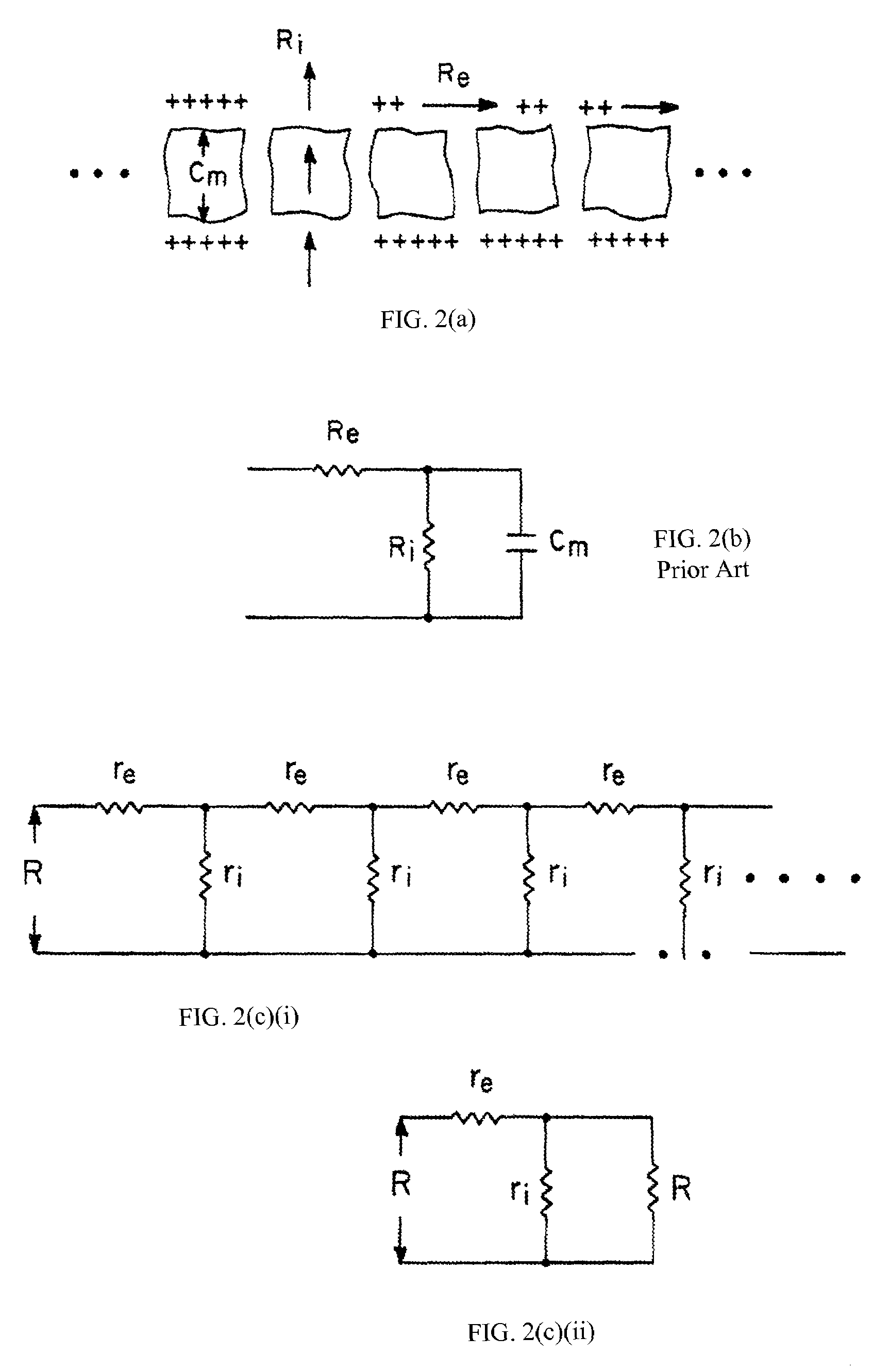
The lumped-element models have a number of limitations that are either inherent or have been introduced as a result of simplifying assumptions.
No serious attempt has been made to account for the random orientations of the epithelia and the resulting complex current paths associated with in vivo measurements.
A further weakness with lumped-element models is that only the capacitive reactance has been considered.

Method used


Image

  • Multidimensional bioelectrical tissue analyzer
  • Multidimensional bioelectrical tissue analyzer
  • Multidimensional bioelectrical tissue analyzer

Examples


first embodiment

[0120]There are three distinct modes in which the acoustic subsystem of the invention is used. In each of these embodiments, an array of sensitive microphones is placed on the body at positions that a clinician considers to be most appropriate for the disorder under study. In a first embodiment, these microphones measure acoustic variations associated with the process under study, such as the heartbeat, blood flow through arteries and veins, respiration, neuromuscular activity, and the gastrointestinal tract. These passive acoustic data are preferably digitized at a rate of at least 30 kHz, and even higher rates are preferred if warranted by specific high-frequency abnormalities. The fast Fourier transform of the digitized acoustic data is calculated and compared with the resulting power spectra to that of healthy functional tissue. These data provide detailed functional data about heart health, and about air and fluid transport in organs that may become afflicted by any pathology. ...

second embodiment

[0121]In a second embodiment, a monochromatic acoustic wave at a preset frequency is excited by a loud speaker in contact with the body at a position determined by the clinician. The array of microphones detects only that part of the total acoustic response that is at the drive frequency, and at a constant phase relationship to the drive. The resulting magnitude of the sound observed at each microphone using this synchronous detection technique is used to infer the acoustic impedance magnitude and phase angle at that position in the array. This acoustic impedance is modulated by a slower (much slower compared to the period of the acoustic wave) biological process, such as the heartbeat, respiration, gastrointestinal activity, etc. These measurements provide far more detailed information and detect far more pervasive patterns specific to the pathology than do simple potential sensing, as in systems such as electrocardiography (EKG), electroencephalography (EEG), electromyography (EMG...

third embodiment

[0122]In a third embodiment, the microphone array is taken out of the synchronous detection mode, and set to sense frequencies that are detuned from the drive frequency. This detection technique provides information about the nonlinear acoustic character of the tissue as a function of drive amplitude, and also as a direct sensor of acoustic Doppler-effect detuning associated with fluid flow or mass motion in the body.

6.9. Exemplary Applications

[0123]While the invention has been described above with particular reference to the detection and characterization of cancer within a human or animal body, it is not so limited. Several examples of the application of the invention to both oncologic and other applications are described below.

[0124]6.9.1. Oncologic

[0125]In several embodiments, the invention is used in the diagnosis of oncologic disease. While devices in the prior art are able to distinguish benign from proliferative (i.e. premalignant and malignant) epithelium, the more sensitiv...


PUM

PropertyMeasurementUnit
oscillation frequencyaaaaaaaaaa
specific gravityaaaaaaaaaa

Abstract

A method and apparatus that use complex impedance measurements of tissue in human or animal bodies for the detection and characterization of medical pathologies is disclosed. An analysis of the complex impedance measurements is performed by a trained evaluation system that uses a nonlinear continuum model to analyze the resistive, capacitive, and inductive measurements collected from a plurality of sensing electrodes. The analysis of the impedance measurements results in the construction of a multidimensional space that defines the tissue characteristics, which the trained evaluation system uses to detect and characterize pathologies. The method and apparatus are sufficiently general to be applied to various types of human and animal tissues for the analysis of various types of medical pathologies.

Description

[0001]This application is a nonprovisional of and claims the benefit of U.S. Appl. No. 60 / 185,413, filed on Feb. 28, 2000, the disclosure of which is herein incorporated by reference in its entirety for all purposes.1. FIELD OF THE INVENTION[0002]This invention relates to the diagnosis of medical pathologies in tissues and internal organs of a human or animal body. More particularly, the invention relates to the diagnosis of oncologic pathologies in tissues or internal organs of a human or animal body and to the diagnosis of other pathologies in the cardiac, pulmonary, neurologic, and skeletal systems using bioelectrical impedance measurements.2. BACKGROUND OF THE INVENTION2.1. Introduction[0003]There is an ongoing need in medicine for methods and apparatuses that can accurately detect and characterize medical pathologies within tissues and internal organs in a human or animal body. There is currently a particular need for a versatile method and apparatus to provide safe, reliable, ...

Claims


Application Information

Patent Timeline
Patent Type & Authority Patents(United States)
IPC IPC(8): A61B5/05A61B5/053G01N27/02G01N27/04G01N27/24
CPCA61B5/053A61B5/4312A61B5/4869A61B5/6806G01N33/48728
Inventor LITTRUP, PETERDUNCAN, ROBERT
Owner DELPHINUS MEDICAL TECH
Learn More