[0004]Tomosynthesis as used in the systems and methods disclosed in this patent specification typically involves acquiring a plurality of tomosynthesis projection images Tp at respective angles relative to the breast, and reconstructing therefrom a plurality of tomosynthesis reconstructed images Tr representative of breast slices that have selective thicknesses. Proper display techniques are desirable to make the presentation of Tp and / or Tr images (collectively referred to here as T images) more effective and efficient for review by health professionals. When tomosynthesis projection images Tp are acquired along with conventional 2D mammograms Mp, improved display methods are desirable that facilitate the display of both T and Mp images. Effective display approaches also are desirable when tomosynthesis images Tp and / or Tr that are acquired at one time need to be compared to mammograms Mp and / or to tomosynthesis images Tp and / or Tr acquired at a different time. Effective displays also are desirable when only Tr and / or Tp images are being displayed. Another display issue relates to Computer Aided Detection (CAD) methods that use computer analysis of images to identify locations and possibly other characteristics of suspected abnormalities. CAD marks currently are placed on or are otherwise associated with mammogram images Mp, but it may be useful to place them at the appropriate location on Tr and / or Tp images o to otherwise associate them with Tr / Tp images. Conversely, it may be desirable to obtain CAD marks by processing Tp and / or Tr images, and place them at appropriate locations on Mp images. Here the notation Mp refers to a conventional mammogram, which is a two-dimensional projection image of a breast; the term Mp encompasses both a digital image as acquired by a flat panel detector or another imaging device and the image after conventional processing to prepare it for display to a health professional or for storage, e.g. in the PACS system of a hospital or another institution. Tp refers to an image that is similarly two-dimensional but is taken at a respective tomosynthesis angle between the breast and the origin of the imaging X-rays (typically the focal spot of an X-ray tube), and also encompasses the image as acquired as well as the image after being processed for display or for some other use. Tr refers to an image that is reconstructed from images Tp, for example in the manner described in said earlier-filed patent applications, and represents a slice of the breast as it would appear in a projection X-ray image of that slice at any desired angle, not only at an angle used for Tp or Mp images. The terms Tp, Tr and Mp also encompasses information, in whatever form, that is sufficient to describe such an image for display, further processing, or storage. The images Mp, Tp and Tr typically are in digital form before being displayed, and are defined by information identifying properties of each pixel in a two-dimensional array of pixels. The pixel values typically relate to respective measured or estimated or computed responses to X-rays of corresponding volumes in the breast (voxels or columns of tissue).
[0015]Images of different types and from different sources can be displayed in desirable size and resolution. For example, an image can be displayed in (1) Fit To View Port mode, in which the size of the displayed image size is maximized such that the entire imaged breast tissue is visible, (2) True Size mode, in which a display pixel on the screen corresponds to a pixel of the image, or (3) Right Size mode, in which the size of a displayed image is adjusted so that it matches that of another image that is concurrently displayed or with which the displayed image is or can be toggled. For example, if two images of the same breast are taken and are not the same size or do not have the same special resolution, provisions are made to selectively zoom in or zoom out one of them, or zoom both, such that they appear to be the same size on the screen when they are concurrently displayed or the user toggles between them, to facilitate comparison or to otherwise facilitate detection / diagnosis. Known interpolation / extrapolation and weighting techniques can be used in such re-sizing, and known image processing technology can be used to make other characteristics of the displayed images similar in a way that facilitates detection / diagnosis.
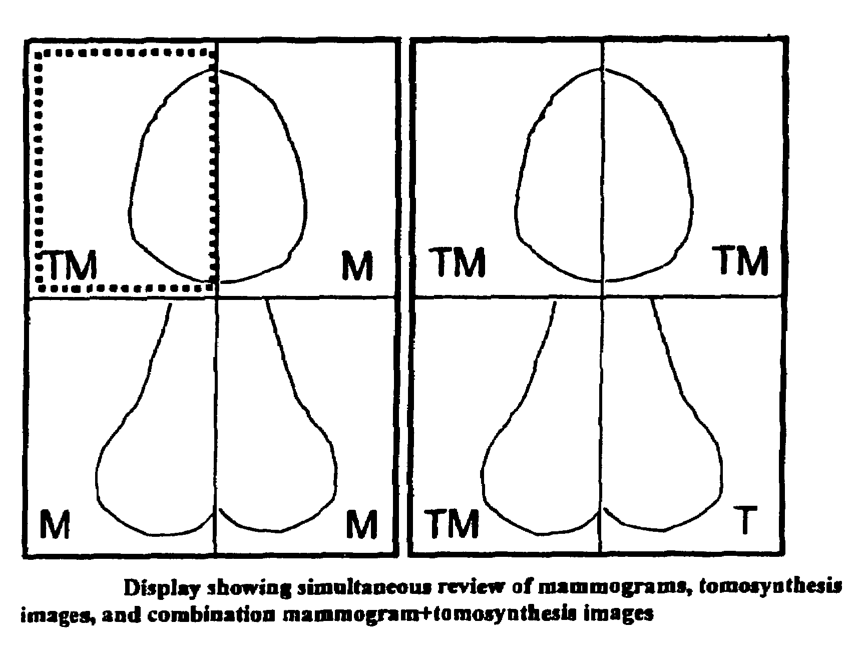
[0017]When CAD information is available that is associated with any of the images that can be displayed, linking can be provided such that CAD information derived from one of the types of displayed images can be automatically associated with corresponding locations on another displayed image. For example, if CAD information has been derived on the basis of an Mp image and the Mp image is displayed at the same time as, or is toggled with, Tr images, provisions are made to selectively display CAD information associated with the appropriate locations on Tr images. Alternatively, if CAD information is derived on the basis of Tr and / or Tp images, it can be automatically selectively displayed in association with an Mp image that is displayed at the same time or is toggled with the Tr / Tp images. In one example, an Mp image with CAD marks thereon remain displayed on the screen while Tr images of the same breast are scrolled or shown in cine mode on the screen, to facilitate identification and / or assessment of suspected abnormalities in the Tr images.

