Axial length measurement of the eye, although simple in concept is difficult in practice because the eye cannot be held stationary during the exam.
Furthermore, the eye is pliable and excessive pressure applied to the eye during the measurement process will compress the eye and create inaccurate results.
Any one of these three variables, if not controlled, can confound the measurement process and introduce major errors during lens calculation for cataract surgery.
Despite excellent technician skill, axial length measurement has been confounded by these variables since its inception.
A central problem discovered when utilizing and evaluating prior art for axial length ultrasound measurement is that each solution typically focuses on minimizing one or two potentially confounding factors, leaving the other variables to chance.
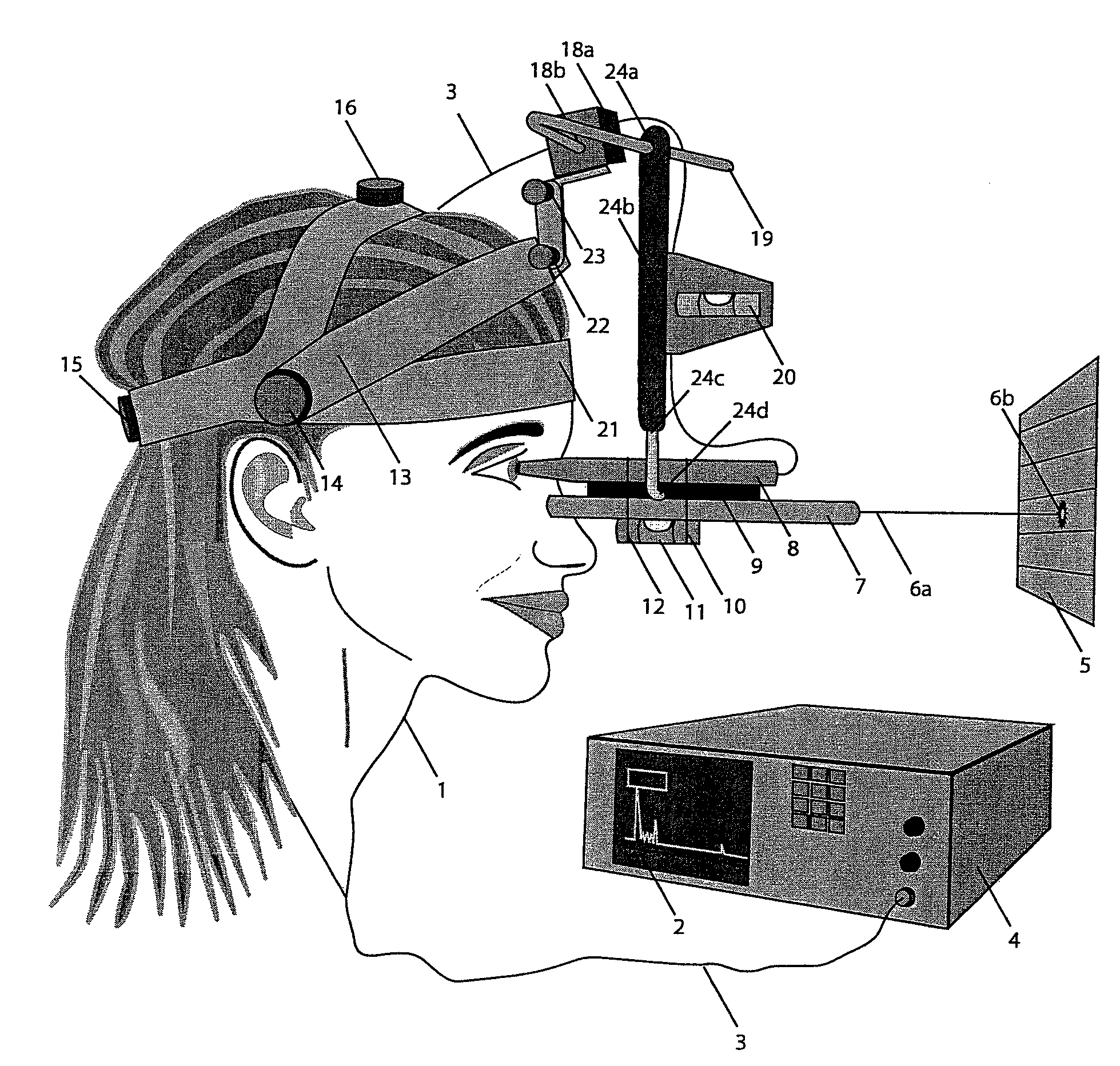
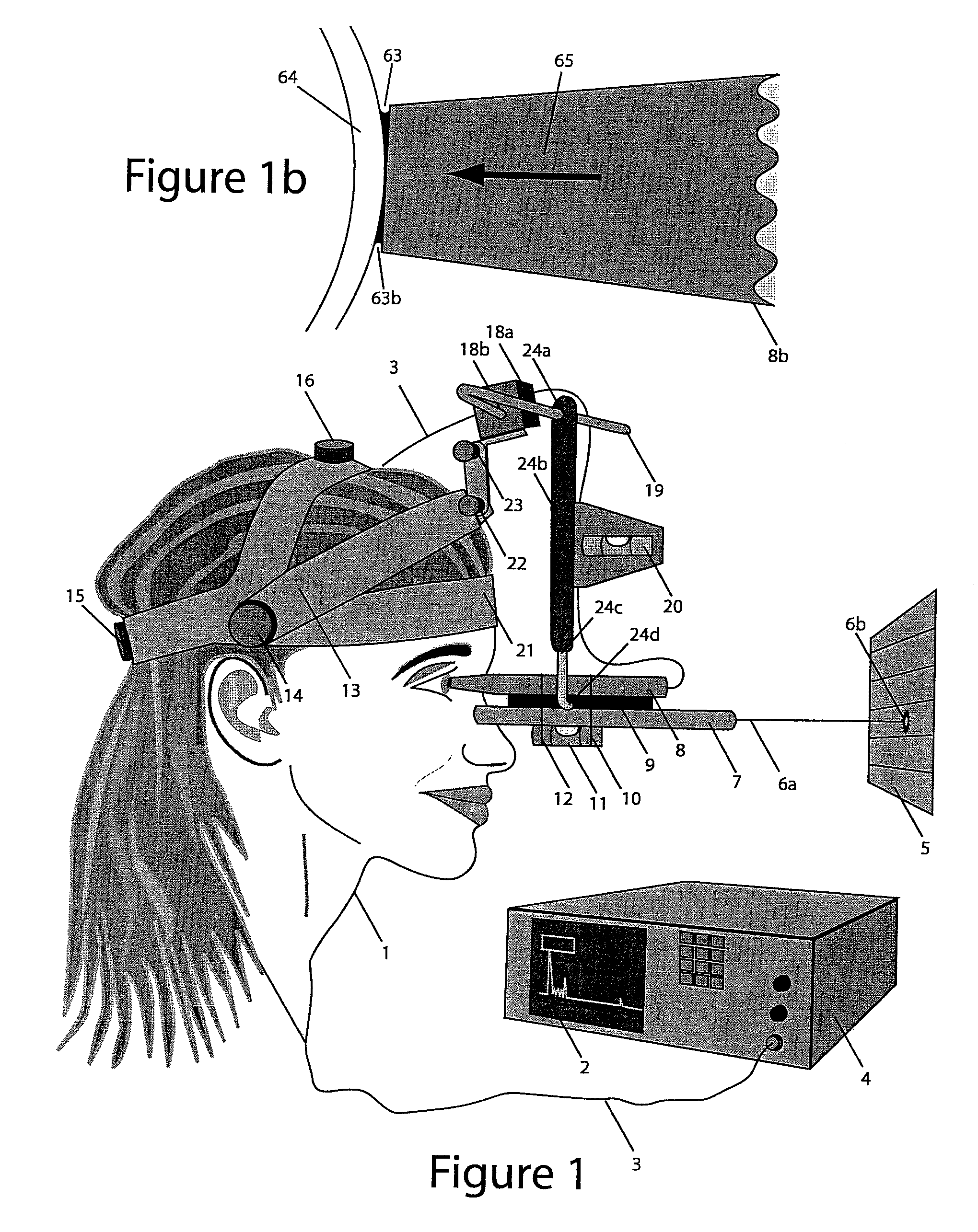
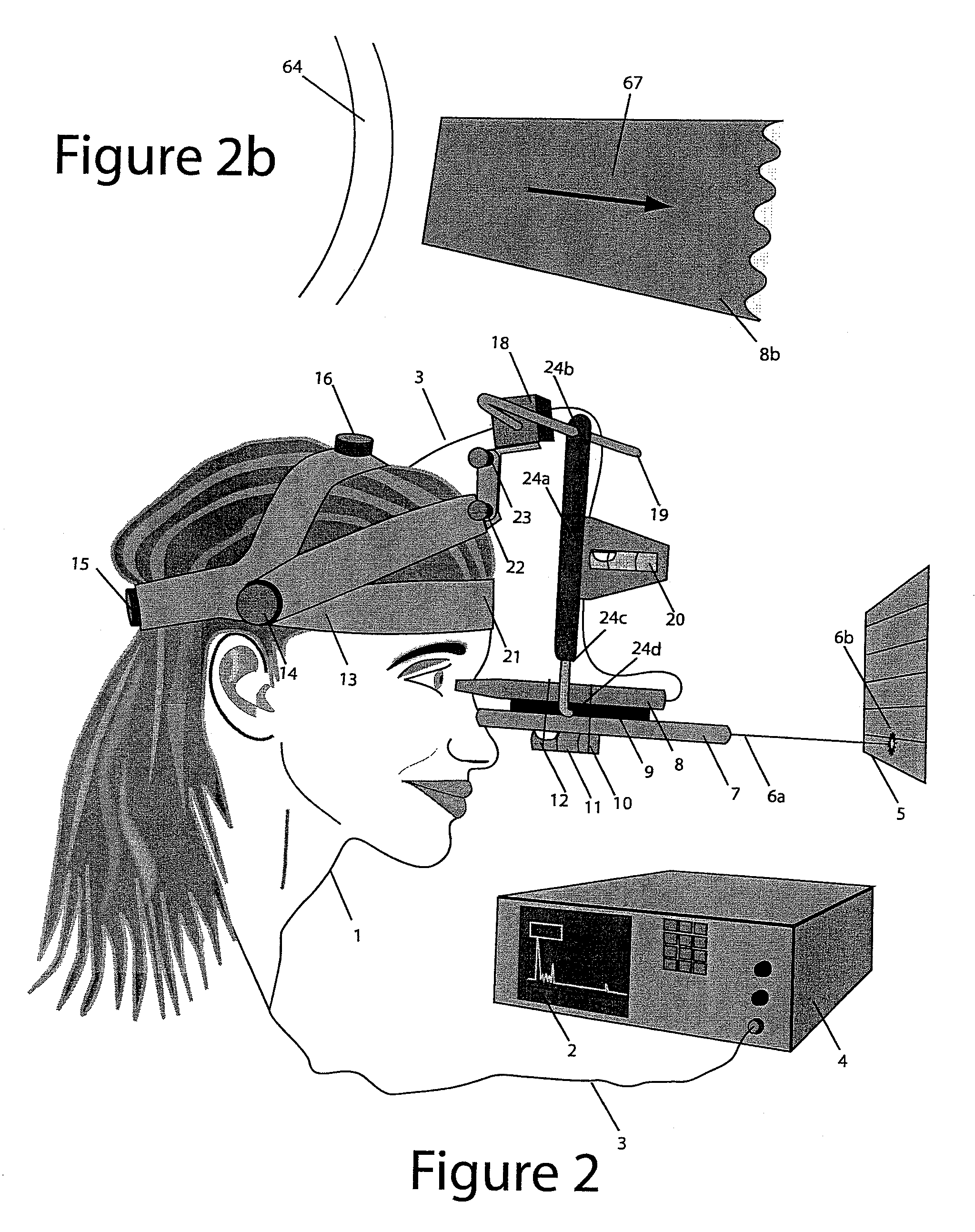
1) Misalignment of the Probe during measurement: Misalignment of the probe during ultrasound measurement causes gross errors in axial length measurement as a result of off-center and / or off-axis positioning.
Either of these errors will lead to incorrect calculations of the lens power used for cataract surgery.
These internal fixation lights and the process of using them have limitations in their ability to assure proper alignment during ultrasound measurement.
The fundamental limitation to this process is that the internal fixation point in the prior art is too large to adequately limit movement of the eye.
As a result, patients cannot assist in maintaining axial alignment during measurement.
Another fundamental problem is that the complex movements of the eye are extremely difficult to follow using a hand-held probe.
The first limitation of the internal fixation light provided in the prior art, U.S. Pat. No. 4,934,370 to Campbell, is the large size of the fixation light.
This invention improves upon the previous process, where no fixation target was employed; however, the internal fixation system has limitations because of its large size.
Because of the large apparent size of a 1 mm fixation spot after it contacts the cornea, the current standard in the industry provides a rough guide for fixation, but does not precisely assure proper alignment of the ultrasound probe with the visual axis.
The large spot size combined with the inherent difficulty of aligning the probe along the visual axis of a “live” eye leads to confounding errors during measurement.
Visually aligning the probe along the visual axis while holding the probe stationary against the eye with the correct amount of pressure is quite difficult.
Either of these positioning errors will give inaccurate readings.
Off Center Problems: The first aspect of achieving the correct ultrasound probe alignment is locating and maintaining the correct contact point between the tip of the probe and the topographical center of the cornea.
Extrapolating the pupil's center from this location can be quite difficult.
Typically, the patient's eye is wandering about throughout the measurement process because the fixation spot inside the prior art probe is too large to confine the movement of the eye as stated above.
Since the cornea is a sphere, any off-center positioning of the probe tip will give an erroneously short measurement.
In fact, short measurements are the most commonly encountered error seen in ultrasound axial measurements, in part because of the difficultly of visually maintaining the proper orientation of the probe on the moving cornea.
Off Axis Problems: The second aspect of achieving the correct ultrasound probe alignment is maintaining the proper relationship between the tip and the tail of the ultrasound probe.
Patients typically do not give feedback during this procedure, so there is no objective way of knowing if the ultrasound probe is correctly aligned.
Therefore, proper axial alignment cannot be objectively confirmed.
Axial alignment of the ultrasound probe, also known as the tip to tail alignment, is quite complex as it relates to a moving eye.
This extremely complex movement is very difficult to perform in a “live” eye that has the ability to move at a much higher rate of speed than the hand that is trying to follow it.
In addition to the fine movements of the eye, the patient's head is also unstable and tends to move during the procedure.
Finally, most patients tend to have an avoidance response and their entire body moves away from the technician during the course of the measuring process thereby altering critical landmarks and making it quite difficult to maintain proper alignment.
Due to these limitations, the internal fixation spot presented in the prior art is not the ideal way to maintain fixation during axial length measurement of the eye with ultrasound.
It is no surprise that difficulty abounds in attempting to provide consistently accurate ultrasound readings.
This apparatus works relatively well in practice; however, there are two limitations to the proposed mechanism.
The first limitation is that the device must be held in the technician's hand and the problems of “off-center” and “off-axis” alignment discussed above become a confounding problem.
The device can be connected to a slit lamp for more precise control, but there is still no inherent way to assure alignment with the visual axis.
The second limitation is that the resistance applied by the spring to the tip of the probe is fixed at a certain amount.
This means that if the intra-ocular pressure is less than this amount, the probe will compress the eye and cause short readings.
This probe dampens excessive pressure in the plane directly to and away from the eye, but any tangential movement on the surface of the eye moves the inner barrel of the spring loaded probe against the side wall of the outer sleeve and may decrease the ease with which the two sleeves slide past one another causing the resistance applied to the tip of the device to increase, thereby compressing the cornea.
It does not address the compression of the cornea, nor does it address the axial alignment issue.
This approach addresses the compression issue, since the intervening water bath prevents contact between the probe and the eye; however, it does not address the axial alignment issue.
Furthermore, the water bath technique takes much longer to perform, involves laying the patient in a supine position, using a lid speculum and requires the use of a coupling agent (water bath) which can be messy.
Due to limitations listed above, neither the slit lamp technique nor the water bath technique fully address all three limitations of ultrasound measurement of the eye, which include, 1) proper alignment of the ultrasound probe along the visual axis, 2) a mechanism of gently positioning the ultrasound probe against the eye during measurement with the correct and constant pressure applied to the eye by the ultrasound probe tip, and 3) stationary positioning of the ultrasound probe relative to the patient's head and eye during the measurement process.
