Methods and apparatus for processing medical images

Inactive Publication Date: 2010-01-12
KENT RIDGE DIGITAL LABS
View PDF8 Cites 27 Cited by
  • Summary
  • Abstract
  • Description
  • Claims
  • Application Information

AI Technical Summary

Benefits of technology

[0011]A system combining various aspects of the invention may be fast, almost fully automatic, and need not require any hardware accelerator. This is because the registration technique makes use of anatomical knowledge to narrow the search within the brain space, and permits the use of simple analysis operations.

Problems solved by technology

However, to our best knowledge, there is no commercial application of brain atlas in neuroradiology yet.
Several non-linear warping approaches have also been proposed [1], but they are time consuming, highly dependent on starting points and at present far from clinical applications yet.

Method used


Image

  • Methods and apparatus for processing medical images
  • Methods and apparatus for processing medical images
  • Methods and apparatus for processing medical images

Examples


Embodiment Construction

1. Introduction

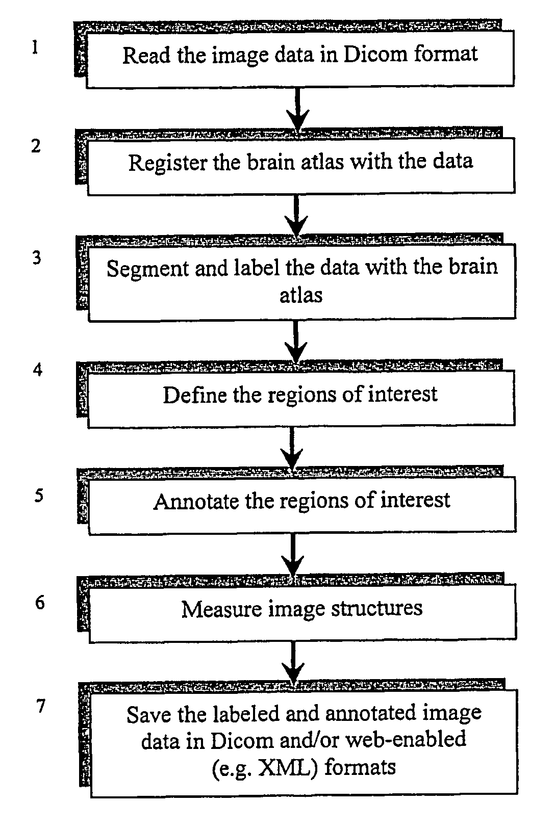
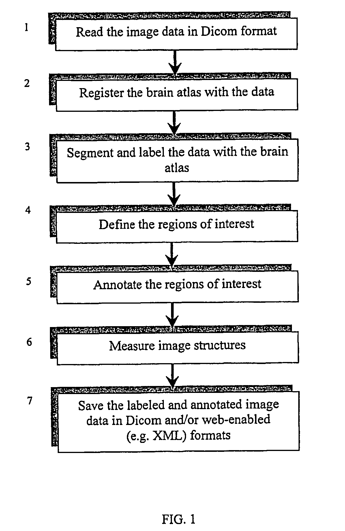
[0038]FIG. 1 illustrates in conceptual terms a method according to the present invention.

[0039]The patient specific image data are read in Dicom format (step 1), which is a standard format in diagnostic imaging and Dicom readers are commonly available. Then in step 2, the brain atlas is registered with the data (a fast, automatic method to do it is given below), so the neuroradiologist can segment and label the data by means of the atlas (step 3). In particular, the region(s) of interest can be defined on the images (step 4) and annotated (step 5), and segmentation and labeling can be done within these regions of interests. In addition, the image structures can optionally be measured (step 6). In step 7, the atlas-enhanced data can be stored for further processing by other medical professionals, such as neurologists and neurosurgeons. In particular, the atlas-enhanced data can be stored in Dicom format and / or in any web-enabled format such as XML. These steps are desc...


PUM


Abstract

A system for analysing a brain image compares the image with a brain atlas, labels the image accordingly, and annotating the regions of interest and / or other structures. This atlas-enhanced data is written to a file (or more than one file) in the Dicom format or any web-enabled format such as SGML or XML format. The image used may be produced by any medical imaging modality. A fast algorithm is proposed for a landmark-based piecewise linear mapping of one volumetric image into another volumetric image. Furthermore, a new set of brain landmarks are proposed, and algorithms for the automatic identification of these landmarks are formulated for three orientations, axial, coronal, and sagittal.

Description

FIELD OF INVENTION[0001]The present invention relates to methods and apparatus for manipulating medical images of brains. In particular it relates to systems for using a brain atlas to annotate a medical image, and saving the resultant image for subsequent utilisation.BACKGROUND OF THE INVENTION[0002]Modern medical imaging technologies offer unprecedented views into human body. Our ability to generate images is much greater than the ability to understand and interpret them. Deformable body models along with warping techniques provide means for analysis of medical images and deformable brain atlases, in particular, are useful for analysis of brain scans. The purpose of the present invention is to facilitate the interpretation of brain scans by neuroradiologists and to provide means to transfer this knowledge to other medical professionals such as neurologists and neurosurgeons.[0003]The present inventor has developed an electronic brain atlas database [3]. There is a strong commercia...

Claims


Application Information

Patent Timeline
IPC IPC(8): G06K9/00G06T7/00
CPCG06T7/0012G06T7/0028G06T7/0038G06T2207/30016G06T7/33G06T7/38
Inventor NOWINSKI, WIESLAW L.THIRUNAVUUKARASUU, ARUMUGAM
Owner KENT RIDGE DIGITAL LABS
Why Eureka
Try Eureka