Methods and systems for automated detection and analysis of lesion on magnetic resonance images

a magnetic resonance image and automatic detection technology, applied in the field of digital image processing, can solve the problems of obscuring the tumor, affecting the detection accuracy of the image, and the common cause of cancer deaths

Inactive Publication Date: 2010-07-13
CARESTREAM HEALTH INC
View PDF10 Cites 16 Cited by
  • Summary
  • Abstract
  • Description
  • Claims
  • Application Information

AI Technical Summary

Problems solved by technology

Breast cancer is a common cancer of women and a common cause of cancer deaths.
However, mammography has been considered by some to have limitations in its ability to detect cancer, with one reported sensitivity being estimated at 80-85%.
This limitation may result from the obscuration of the tumor by superimposed fibroglandular tissue.
Advances have been made in the field of breast MRI, however, there is no well-defined standard or optimal imaging technique for performing contrast-enhanced breast MRI.
In addition, there are no standardized interpretation criteria and no unified definition of what constitutes clinically important contrast enhancement.
It should be recognized that if the patient moves even slightly between the two scans, detail information in the images may be distorted, thus resulting a loss of information due to a misregistration of the two scans acquired at the different times. Image registration may be required to reduce artifacts due to patient movement.
The limitation of the above approaches is that the interpretation based on temporal analysis of contrast enhancement only.

Method used


Image

  • Methods and systems for automated detection and analysis of lesion on magnetic resonance images
  • Methods and systems for automated detection and analysis of lesion on magnetic resonance images
  • Methods and systems for automated detection and analysis of lesion on magnetic resonance images

Examples


Embodiment Construction

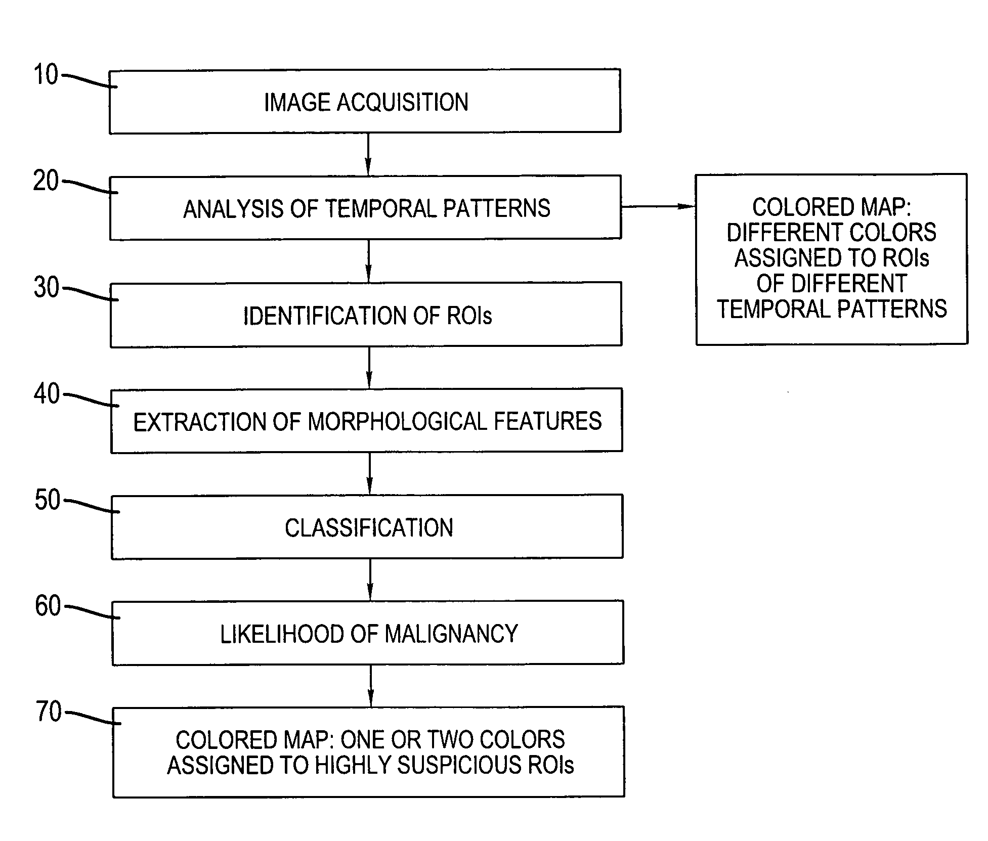
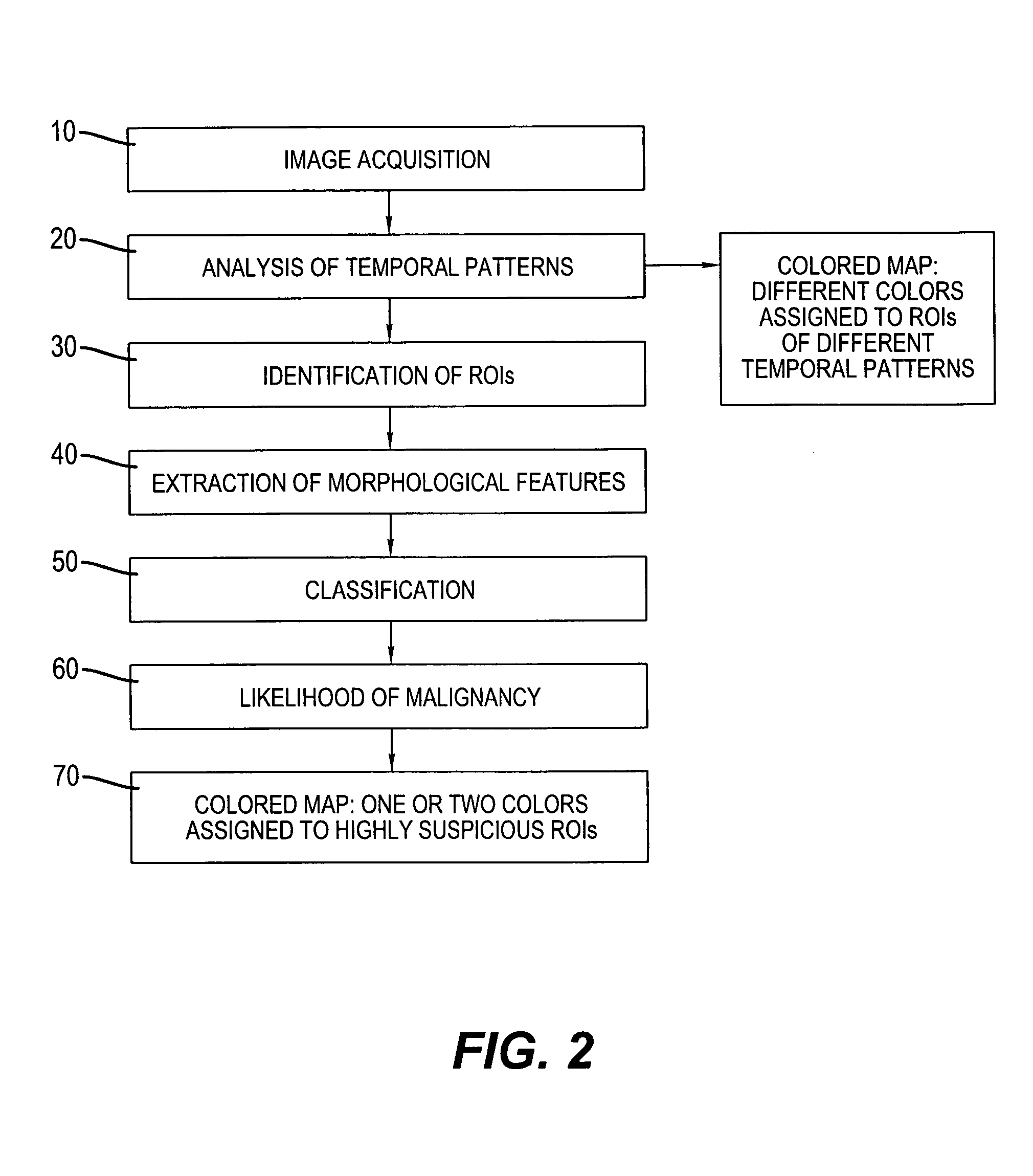
[0033]The following is a detailed description of the preferred embodiments of the invention, reference being made to the drawings in which the same reference numerals identify the same elements of structure in each of the several figures.

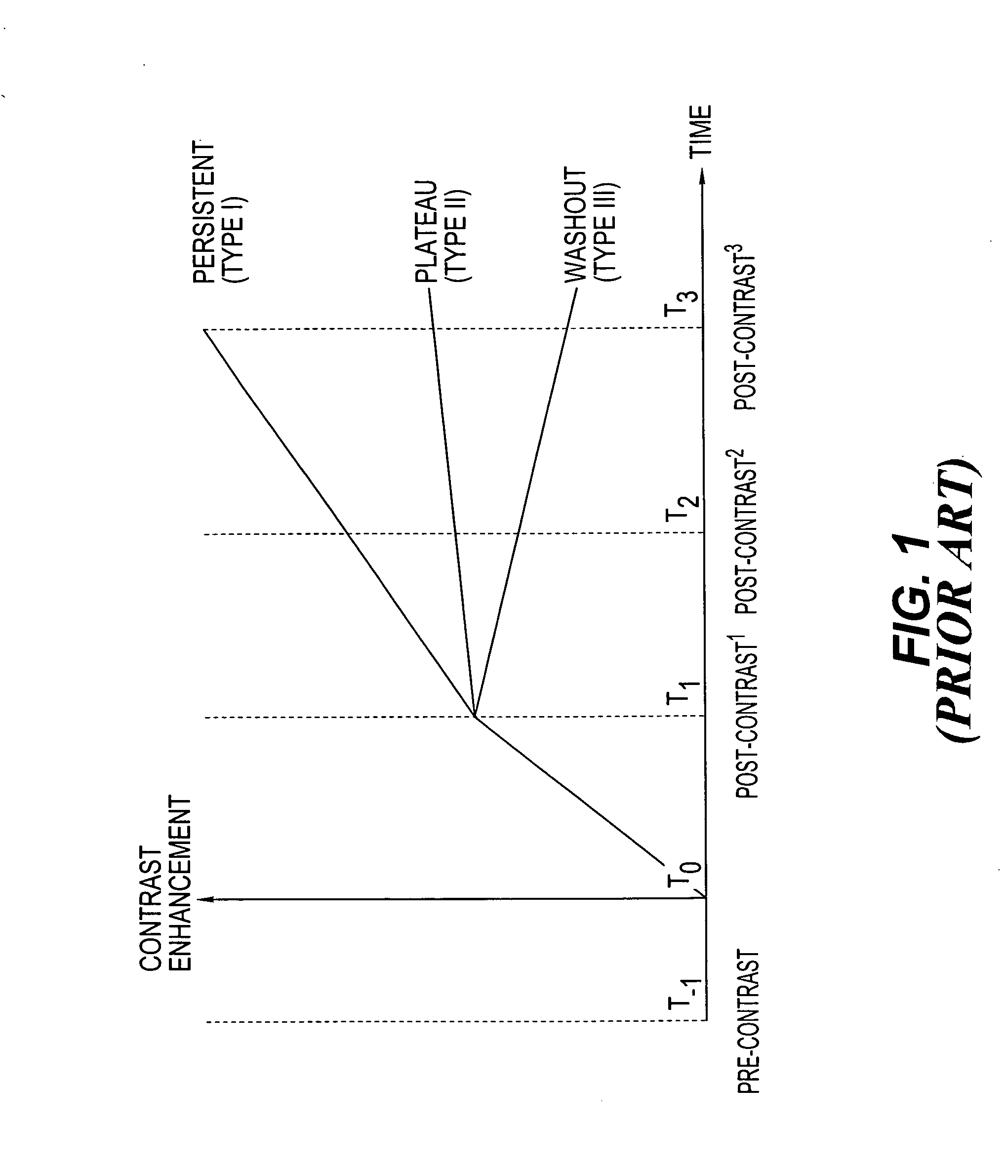
[0034]FIG. 2 shows a flow chart or diagram generally illustrating an automated method for the detection and characterization of lesions in MR images in accordance with the present invention. Generally, 3-dimensional (3D) MR images of the same breast are acquired over a period of time (step 10). During acquisition, at least one scan is acquired prior to injection of a contrast agent (pre-contrast) and at least two scans are acquired after injection (post-contrast). As shown in FIG. 1 for illustrative, exemplary purposes, pre-contrast injection is at time T-1; contrast injection is at time T0; and three post contrasts are acquired at times T1, T2, T3. The acquired 3D images comprise a volume (data set) and are presented in digital format.

[0035]It is n...


PUM


Abstract

A method for detecting and analyzing candidate lesions in a magnetic resonance image of a breast. The method includes the steps of: accessing a plurality of temporal magnetic resonance images of the breast; identifying candidate lesions by performing a temporal pattern analysis of the plurality of images to produce temporal features based on an uptake phase and a washout phase; performing a morphological operation on the candidate lesions to produce morphological features; and classifying the candidate lesions using the morphological features and temporal features to produce classified candidate lesions.

Description

CROSS REFERENCE TO RELATED APPLICATIONS[0001]Reference is made to, and priority is claimed from, provisional application U.S. Ser. No. 60 / 675,589, entitled “METHODS AND SYSTEMS FOR AUTOMATED DETECTION AND ANALYSIS OF LESION ON MAGNETIC RESONANCE IMAGES”, filed on Apr. 28, 2005, in the names of Huo et al, and assigned to the assignee of this application.FIELD OF THE INVENTION[0002]The present invention relates to digital image processing. More particularly, the invention is directed to a method for medical image analysis, such as to detect and diagnose breast cancer using contrast enhanced MRI images.BACKGROUND OF THE INVENTION[0003]Breast cancer is a common cancer of women and a common cause of cancer deaths. Mammography is an imaging modality which has provided some effectiveness in the early detection of clinically occult breast cancer, and is viewed by some to be a primary imaging modality for breast cancer screening.[0004]However, mammography has been considered by some to have ...

Claims


Application Information

Patent Timeline
Patent Type & Authority Patents(United States)
IPC IPC(8): G06K9/00A61B5/00
CPCG06T7/0012G06T7/60G06T2207/10096G06T2207/20036G06T2207/30068
Inventor HUO, ZHIMINWANG, HUILIU, XU
Owner CARESTREAM HEALTH INC
Why Eureka
Try Eureka