

Methods of using regenerative cells in the treatment of musculoskeletal disorders
a technology of musculoskeletal disorders and regenerative cells, applied in the field of regenerative cells, can solve the problems of substantial social economic problems, non-surgical methods for treating musculoskeletal injuries, and musculoskeletal disorders
- Summary
- Abstract
- Description
- Claims
- Application Information
AI Technical Summary
Problems solved by technology
Method used
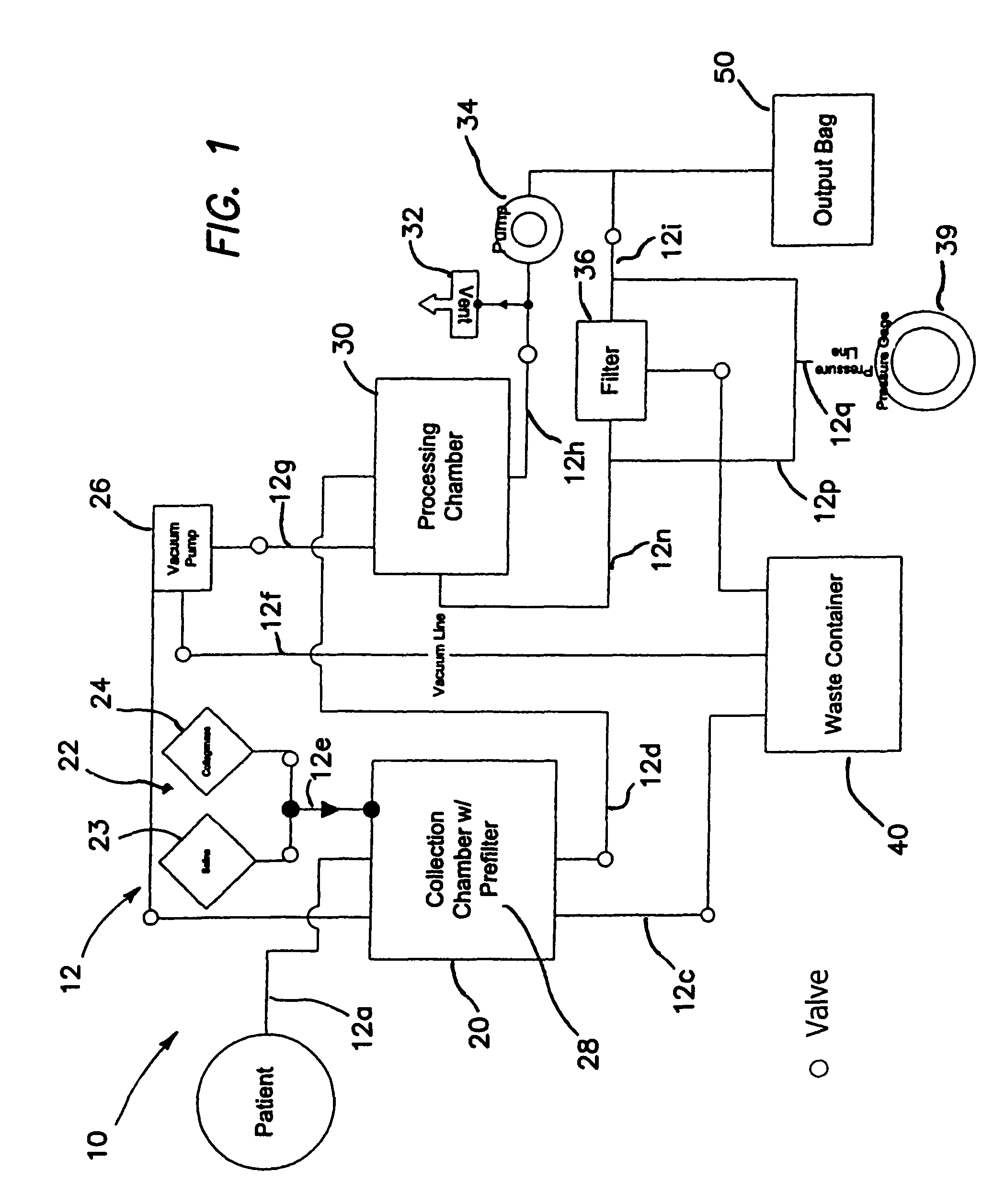
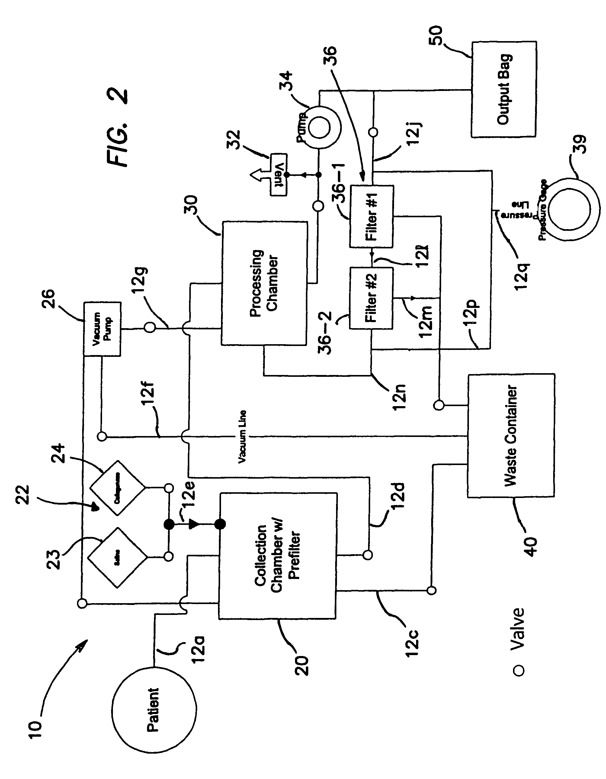
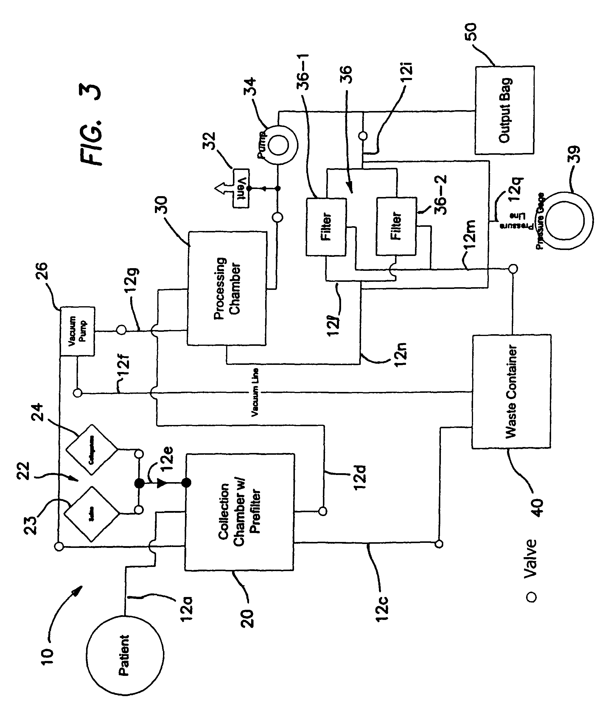
Image

Examples
example 1
Regenerative Cells Form Cartilage, Bone and Skeletal Muscle In Vitro
Materials and Methods:
Regenerative Cell Preparation
[0180]Human regenerative cells were harvested by enzymatic digestion as described in herein. Isolated regenerative cells were then separated into CD34 (+) and CD34 (−) populations using immuno magnetic beads. Each sub-population of cells were cultured in complete medium (DMEM, 10% fetal bovine serum, 5% horse serum, and 1% antibiotic, antimycotic solution) at multiple densities in multi-well culture plates. At 3 weeks, CD34 (−) clones were selected from these cultures and subcultured for approximately 2 months in one of the following culture mediums: complete medium, osteogenic medium, or chondrogenic medium.
Analysis:
[0181]Differentiation of clonal cells towards cartilage or bone was determined by staining with alcian blue or alkaline phosphatase, respectively. Differentiation of clonal cells towards muscle was assessed by immunohistochemical staining of CD34 (+) an...
example 2
Human Regenerative Cells are a Rich Source of Osteoprogenitor Cells
Methods and Materials:
Liposuction:
[0184]Human adipose tissue was obtained from individuals undergoing elective lipoplasty (liposuction). In general the liposuction procedure involved creation of a 0.5 cm incision in the skin of the abdomen and / or thigh followed by infiltration of the subcutaneous space with tumescent solution (saline supplemented with lidocaine and epinephrine). After approximately 10 minutes the subcutaneous adipose tissue was aspirated through a cannula (3-5 mm diameter according to Physician preference) under machine-assisted suction. Adipose tissue, blood, and tumescent solution (lipoaspirate) was collected into a disposable container and stored at 4° C. during transport to the laboratory.
Tissue Digestion:
[0185]Adipose tissue was washed several times with warm (37° C.) saline to remove excess blood and free lipid. The tissue was then digested at 37° C. with Blendzyme collagenase (Blendzyme 1 for ...
example 3
Regenerative Cells Form Cartilage and Bone In Vivo in Non-Bony Site
Methods and Materials:
Cell Scaffolds:
PLA Scaffold Fabrication and Preparation
[0197]Porous polymer scaffolds having an approximate porosity of 90% were manufactured by a solvent-casting / particulate-leaching method. Briefly, sodium chloride was used as the leachable porogen and was sieved into particles ranging in diameter of 300-500 μm. A 3% polymer solution was made by combining 70:30 poly(L-lactide-co-DL-lactide) with chloroform. The sieved salt and polymer solution were mixed in Teflon Petri dishes to make a 9:1 weight ratio of salt to polymer and the solvent was allowed to evaporate in the hood for 2 days. The salt was leached from the scaffold by immersion in water for 3 days, with water changes every 8-12 hours, Residual solvent was removed by placing the porous polymeric sponges under low vacuum for 2 days. Scaffolds were sized for implantation by punching 8 mm diameter implants from the polymer sheets. Scaffol...
PUM
| Property | Measurement | Unit |
|---|---|---|
| diameter | aaaaa | aaaaa |
| diameter | aaaaa | aaaaa |
| length | aaaaa | aaaaa |
Abstract
Description
Claims
Application Information