Method and apparatus for three-dimensional interactive tools for semi-automatic segmentation and editing of image objects

a semi-automatic, image object technology, applied in the field of three-dimensional interactive tools, can solve the problems of time-consuming and laborious manual segmentation process, inability to detect internal defects in objects, and complex anatomical segmentation process,

Active Publication Date: 2010-08-10
GENERAL ELECTRIC CO
View PDF8 Cites 9 Cited by
  • Summary
  • Abstract
  • Description
  • Claims
  • Application Information

AI Technical Summary

Problems solved by technology

For instance, internal defects in an object may be detected.
Anatomical segmentation, however, is a complex problem.
Manual segmentation is a tedious, time consuming process.
Fully automatic segmentation, although ideal, currently does not yield acceptable results.
The live wire algorithm, however, may only be used in the two dimensional space and hence, this technique is a slice-by-slice segmentation, where the drawn contour on one slice becomes the initial contour on the next slice and this initial contour can be deformed.
The difficulty in this strategy is to define the required extent of displacement.
Since some medical objects may assume several forms, it is very hard to choose the correct model for the actual medial images or the degree of deformation allowable.
In addition, once an object has been segmented, a user still has the problem of manually editing the object.
Present editing tools are time consuming and difficult to use, making post-segmentation editing a disproportionately long and difficult task.

Method used


Image

  • Method and apparatus for three-dimensional interactive tools for semi-automatic segmentation and editing of image objects
  • Method and apparatus for three-dimensional interactive tools for semi-automatic segmentation and editing of image objects
  • Method and apparatus for three-dimensional interactive tools for semi-automatic segmentation and editing of image objects

Examples


Embodiment Construction

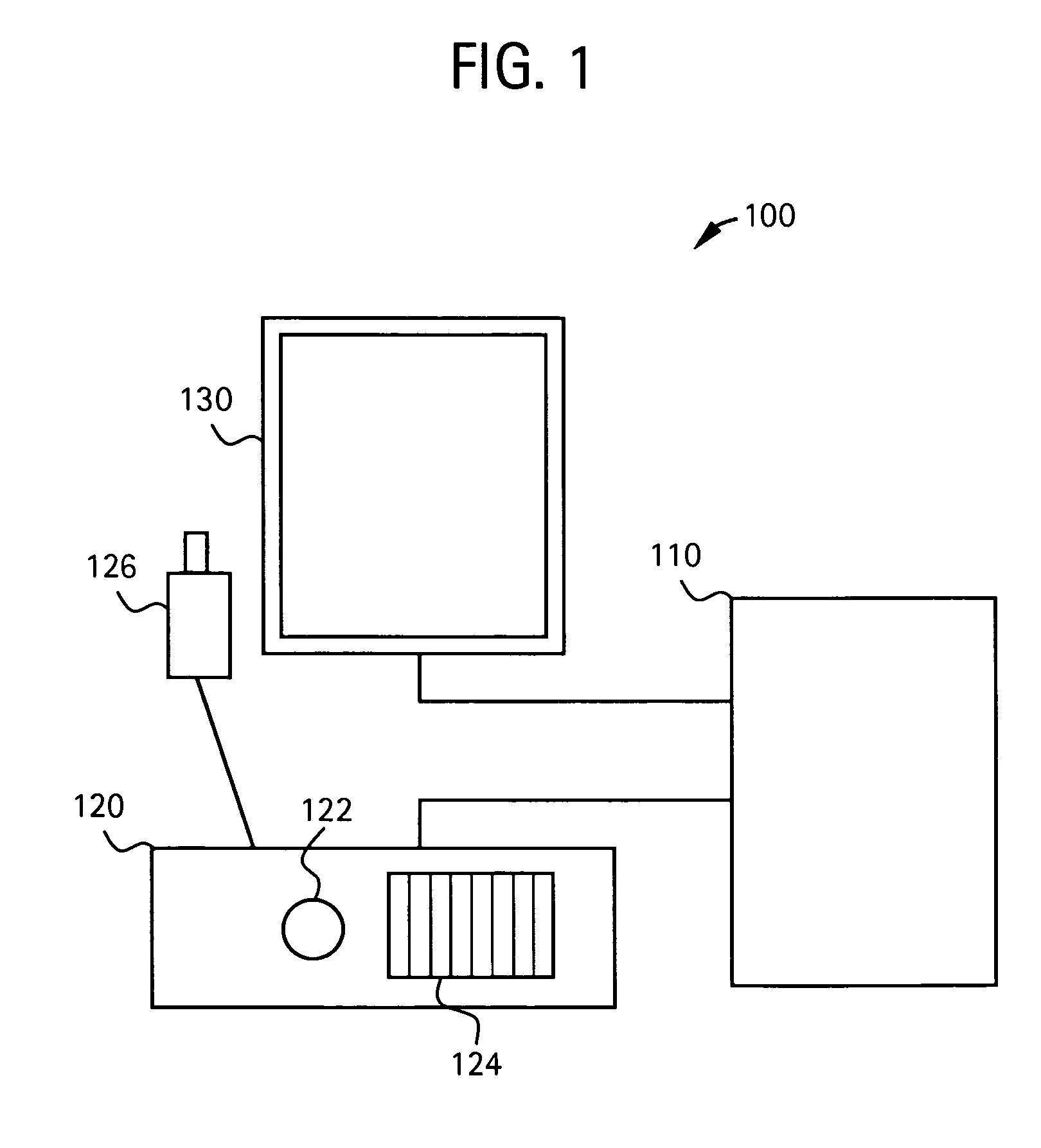
[0028]FIG. 1 illustrates a system 100 for controlling the display and segmentation of medical images. The system 100 includes a computer unit 110. The computer unit 110 may be any equipment or software that permits electronic medical images, such as x-rays, ultrasound, CT, MRI, EBT, MR, or nuclear medicine for example, to be electronically acquired, stored, or transmitted for viewing and operation. The computer unit 110 may be connected to other devices as part of an electronic network.

[0029]The system 100 also includes an input unit 120. The input unit 120 may be a console having a track ball 122 and keyboard 124. The input unit 120 may also have a tracing pen 126. Other input devices may be used to receive input from a user as part of the input unit 120. For example a microphone may be used to receive verbal input from a user. The tracing pen 126 may communicate with the input unit 120 through a wire. The tracing pen 126 may also communicate with the input unit 120 in a wireless f...


PUM


Abstract

A system and method for segmenting and editing anatomical objects from medical images is disclosed. The system may be a medical diagnostic imaging system. A computer unit may execute computer software for segmenting anatomical objects from medical images. The computer software may extract an anatomical object from planar curves. Additionally, the computer software may correct the shape of an existing three-dimensional anatomical object from planar curves. The planar curves may be orthogonal to each other. A user may contour of an anatomical object on a plurality of slices, such as an axial slice a sagittal slice, a coronal slice, or some combination thereof. The contour may be drawn using a tracing pen on a display unit. The display unit may receive touch screen input from the tracing pen. The display unit may display the three-dimensional segmented anatomical object.

Description

RELATED APPLICATIONS[0001][Not Applicable]FEDERALLY SPONSORED RESEARCH OR DEVELOPMENT[0002][Not Applicable]MICROFICHE / COPYRIGHT REFERENCE[0003][Not Applicable]BACKGROUND OF THE INVENTION[0004]The present invention generally relates to a system and method for improved medical imaging. Particularly, the present invention relates to a more efficient system and method for segmenting anatomical objects and correcting the segmentation of anatomical objects.[0005]Medical diagnostic imaging systems encompass a variety of imaging modalities, such as x-ray systems, computerized tomography (CT) systems, ultrasound systems, electron beam tomography (EBT) systems, magnetic resonance (MR) systems, and the like. Medical diagnostic imaging systems generate images of an object, such as a patient, for example, through exposure to an energy source, such as x-rays passing through a patient, for example. The generated images may be used for many purposes. For instance, internal defects in an object may ...

Claims


Application Information

Patent Timeline
Patent Type & Authority Patents(United States)
IPC IPC(8): G06K9/00G06V10/26
CPCG06K9/34G06K9/4638G06K2009/366G06T2207/20101G06T2207/30004G06T7/12G06T7/149G06V10/248G06V10/26G06V10/457
Inventor FIDRICH, MARTAPAP, IMRETANACS, ATTILA
Owner GENERAL ELECTRIC CO
Why Eureka
Try Eureka