
Suture capture device

a technology of suture capture and rotator cuff, which is applied in the field of arthroscopic repair of the tom rotator cuff, can solve the problems of severe limitation, time-consuming suture capture aspect, and limited instrumentation disclosed by mulhollan et al., and achieve the effect of preventing unnecessary damage to the body tissu

Inactive Publication Date: 2011-02-01
ARTHROCARE
View PDF123 Cites 101 Cited by
  • Summary
  • Abstract
  • Description
  • Claims
  • Application Information

AI Technical Summary

Benefits of technology

The present invention is a new and improved approach to securing a mattress stitch in tissues. It involves a special instrument that combines the functions of grasping tissue and passing sutures through it. The instrument has a pair of grasping jaws that can be opened and closed to securely hold the tissue in place. The instrument is designed to be used through a trocar cannula, which allows for direct visualization during the procedure. The instrument can be retracted back through the cannula to remove it from the repair site. The invention provides a significant advance in the art of suturing soft tissues and allows for the reloading of additional sutures for placement of subsequent stitches. The instrument is designed to capture the suture material directly, making the suturing process more efficient.

Problems solved by technology

Suturing of body tissues is a time consuming aspect of most surgical procedures.
A limitation of these types of devices is that they are particularly adapted for use in open surgical procedures where there is ample room for the surgeon to manipulate the instrument.
A limitation of these designs is that they also are adapted primarily for open surgery, in that they require exposure of the tissues to be sutured in order that the tissue may be grasped or pinched between the jaws of the instrument.
This is a severe limitation in the case of endoscopic surgery.
The instrument disclosed by Mulhollan et al. is limited, in that the arc through which the needle must be driven is perpendicular to the axis of the device.
A limitation of the device is the manipulation that must be done with the snare if a suture other than a monofilament is used.
A limitation of this device is that there must be both visual and physical access to both sides of the tissue flap to be sutured.
This is a severe limitation in the instance of shoulder repair, and specifically in repair of the rotator cuff.
The instrument is somewhat limited in the case of rotator cuff repair in that an additional instrument would need to be introduced into the joint to immobilize the tissues sufficiently to allow the needle tip to penetrate the tissues to be sutured.
The major disadvantage of these kinds of staplers is that they leave metal in the body.
For some tissues this is not a problem, however in some procedures, metal staples left within the tissues can be a major hindrance to the healing process.
It is an increasingly common problem for tendons and other soft, connective tissues to tear or to detach from associated bone.
One such type of tear or detachment is a “rotator cuff” tear, causing pain and loss of ability to elevate and externally rotate the arm.
However, as mentioned, this approach places only one loop of suture in the cuff for each anchor, reducing the fundamnental strength of the repair.
The knots in the tendon can be bulky and create a painful impingement of the tendon on the bone.
None of the prior art devices are adaptable to effect the placement of a mattress stitch in grasped tissues, nor are they adaptable to place sutures precisely and controllably while making provision for needle retrieval when using endoscopic techniques.
None of the prior art devices make it possible to place a mattress stitch into, for example, the supraspinatus tendon utilizing an endoscopic approach.

Method used


Image

  • Suture capture device
  • Suture capture device
  • Suture capture device

Examples


Embodiment Construction

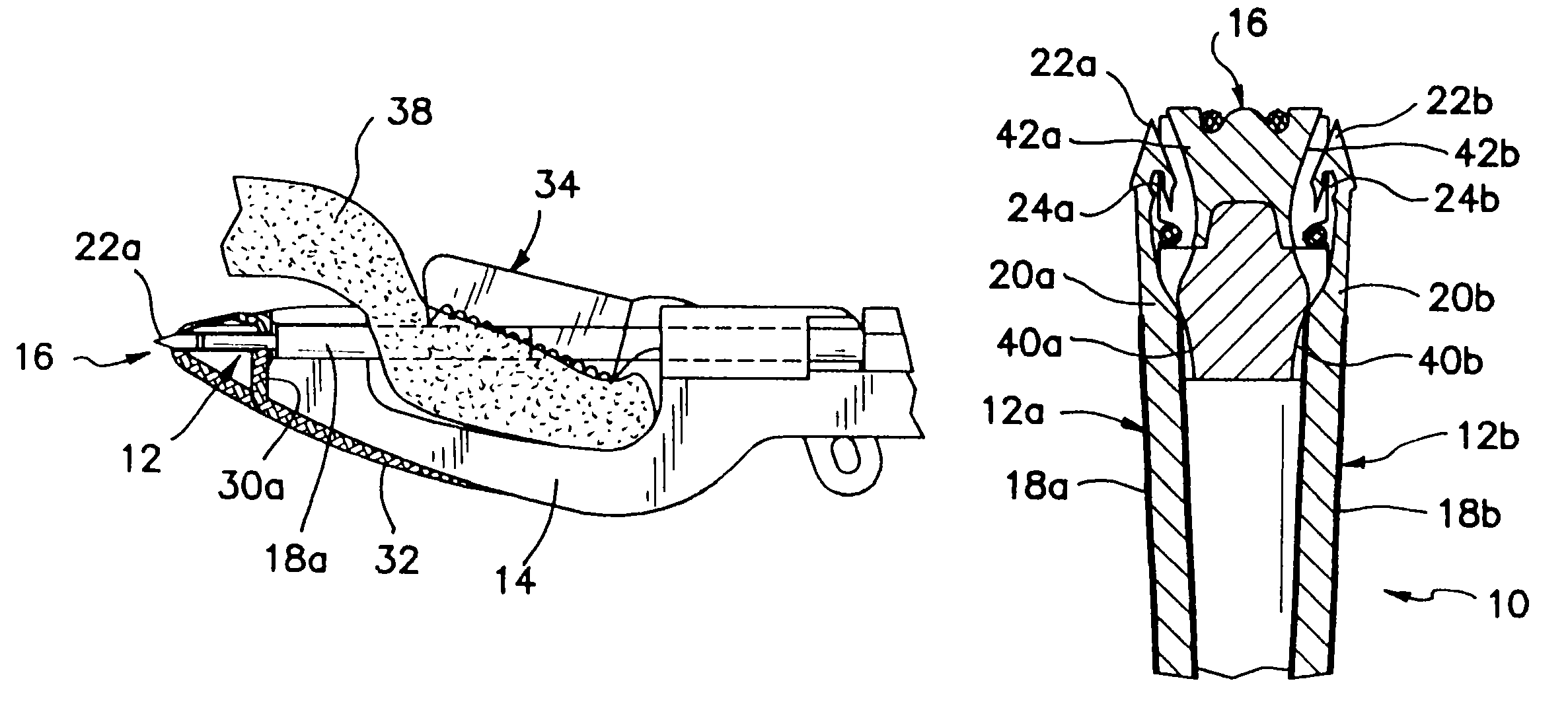
[0040]The present invention relates to a method and apparatus for the arthroscopic repair of tom tissue and bone at a surgical repair site using an inventive device which is a combination tissue grasper and suture placement device. Although the present invention is described primarily in conjunction with the repair of a tom rotator cuff, the apparatus and method could also be used in arthroscopic repair at other sites, such as the knee, elbow, hip surgery, and for other surgical techniques for securing suture in the soft tissues of the body.

[0041]A description of the basic functional elements of suture capture and retrieval, in accordance with the principles of the invention, follows.

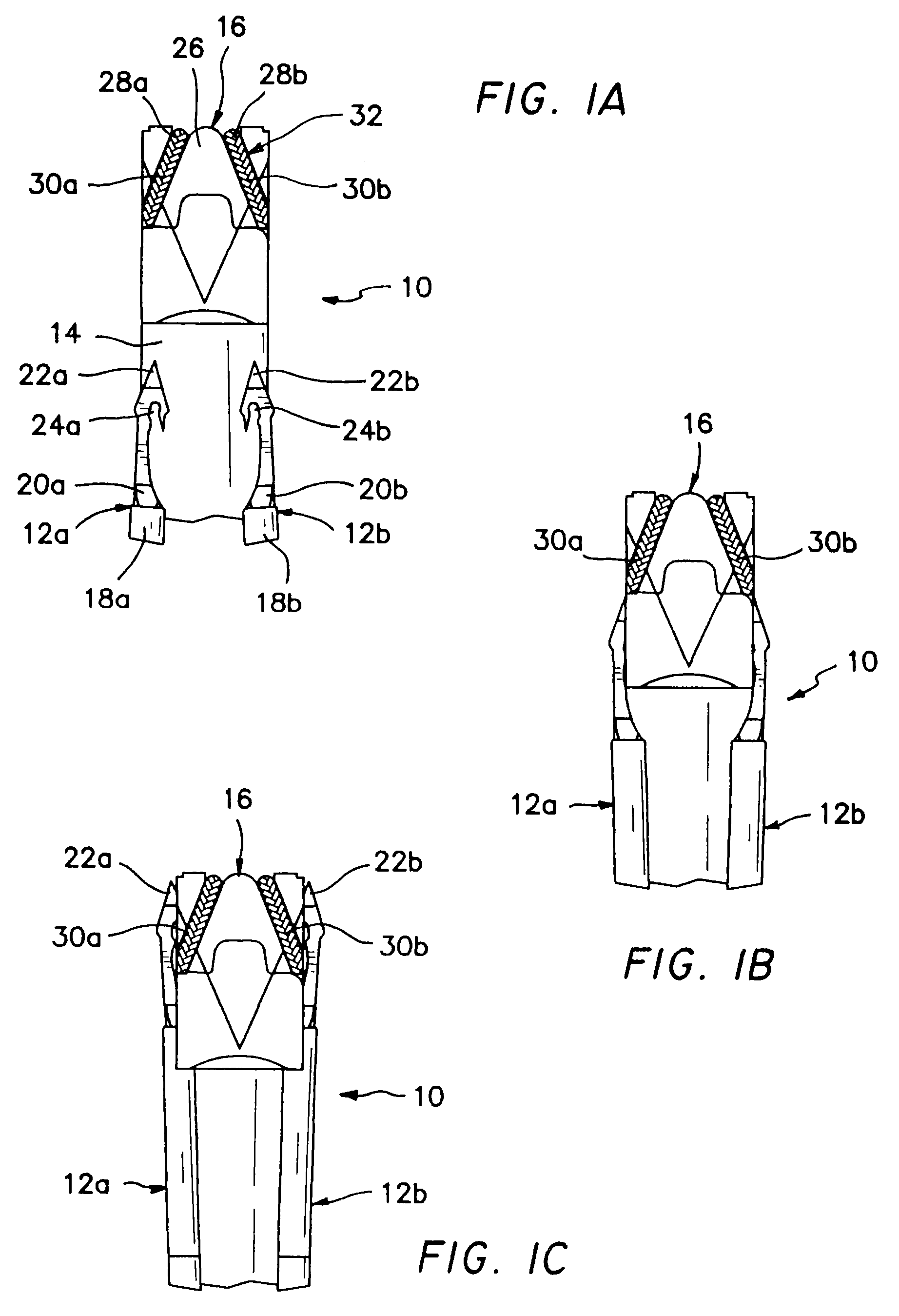
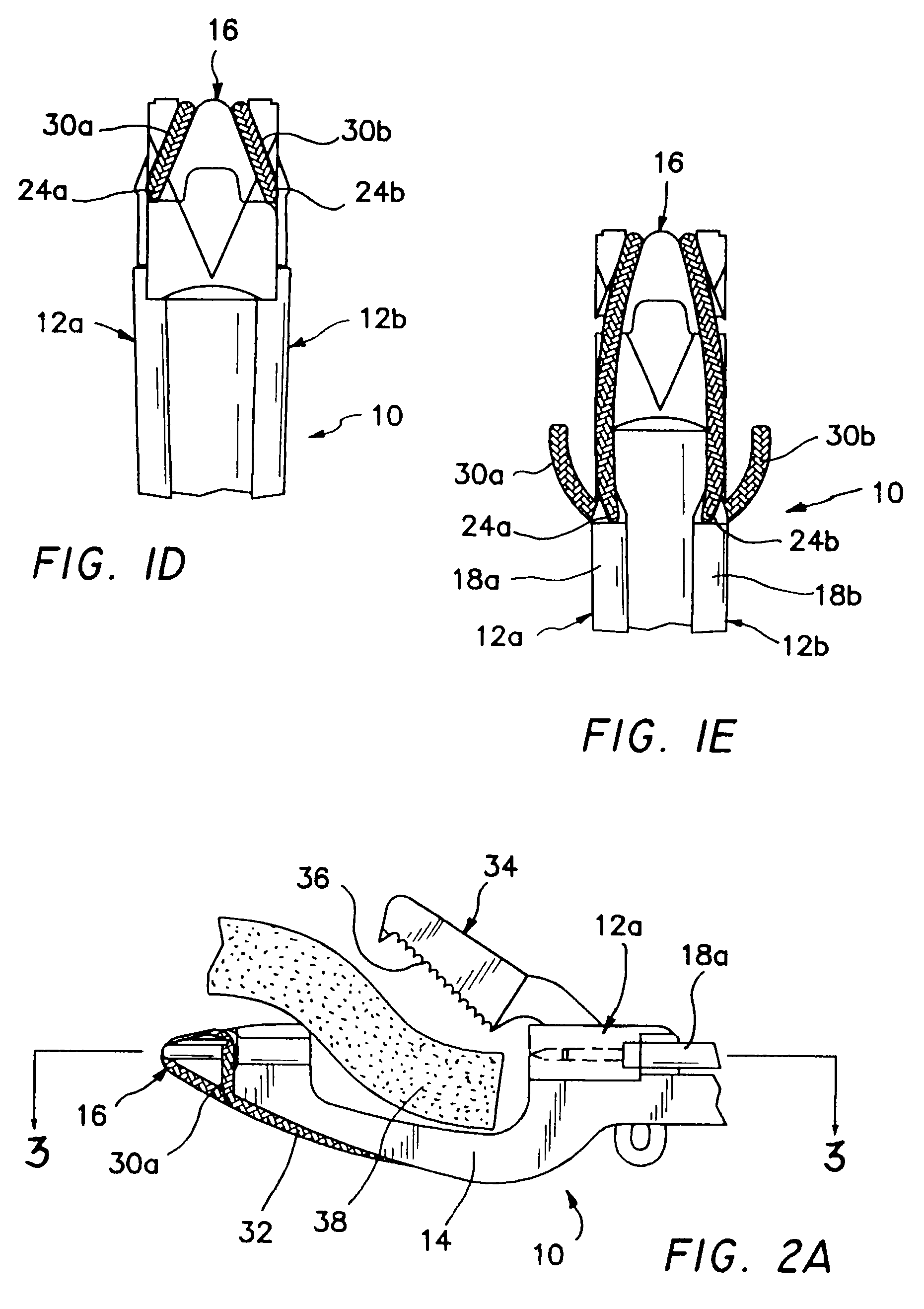
[0042]Referring to FIGS. 1A through 1E, there may be seen a plan view of the distal end of a suturing device 10 that includes a pair of needles 12a and 12b, a lower jaw 14, and a suture cartridge 16. The needles 12a, 12b further include sliding tubes or sheaths 18a and 18b, needle shafts 20a and 20b, ne...


PUM


Abstract

A suturing instrument for placing mattress stitches in soft tissue, such as a rotator cuff, includes an elongate shaft with both a stationary jaw and a movable jaw disposed at the distal end thereof. The shaft is coupled to a handle grip at the proximal end, which is configured to manipulate the jaws into open and closed positions. The jaws are configured to allow for a traumatic grasping of soft tissues. The stationary jaw may include a molded tip that holds and positions opposite ends of a single strand of suture material. The serrated upper jaw is configured to grasp and immobilize the tissue to be sutured. Hooked needles are movably positioned to pierce the grasped and immobilized tissues, and to engage and retrieve the opposite ends of the single strand of suture material. The handle is released to open the moveable jaw, whereupon the instrument may be withdrawn, trailing the suture, and leaving a mattress stitch in the grasped tissue.

Description

[0001]This application is a continuation under 35 U.S.C. 120 of U.S. patent application Ser. No. 10 / 308,732, filed on Dec. 3, 2002, now U.S. Pat. No. 6,770,084, which claims the benefit under 35 U.S.C. 119(e) of Provisional U.S. Application Ser. No. 60 / 391,676, entitled Suture Capture Device, and filed on Jun. 26, 2002, wherein both of these applications are expressly incorporated herein by reference.BACKGROUND OF THE INVENTION[0002]The present invention relates to a method and apparatus for placing sutures in soft tissue, and more particularly to methods and devices for arthroscopic repair of a tom rotator cuff.[0003]Suturing of body tissues is a time consuming aspect of most surgical procedures. Many surgical procedures are currently being performed where it is necessary to make a large opening to expose the area of, for instance, the human body that requires surgical repair. There are instruments that are becoming increasingly available that allow the viewing of certain areas of ...

Claims


Application Information

Patent Timeline
Patent Type & Authority Patents(United States)
IPC IPC(8): A61B17/04A61B17/12A61B17/06A61B17/28
CPCA61B17/0469A61B17/0482A61B17/0485A61B17/06066A61B17/29A61B2017/0472A61B2017/06009A61B2017/06019A61B2017/06042A61B2017/2926
Inventor BAIN, GREGORY H.GORDON, NORMAN S.
Owner ARTHROCARE
Learn More