
Surgical training aid apparatus

a training aid and surgical technology, applied in the field of training aids, can solve the problems of limited usefulness of current models for teaching complex surgical skills, increased disease transmission potential of trainees and mentors, and varies relative differences of human cadavers

Inactive Publication Date: 2011-04-26
SENAGORE ANTHONY +1
View PDF15 Cites 77 Cited by
  • Summary
  • Abstract
  • Description
  • Claims
  • Application Information

AI Technical Summary

Benefits of technology

"The invention is a surgical training aid apparatus that includes a housing and an anatomical replicating assembly. The anatomical replicating assembly consists of a base layer and at least one body component. The base layer has a tackiness and overlaps a portion of the top surface of the housing. The body component is positioned between the top surface of the housing and the base layer or on the base layer. The body component also has a tackiness and is releasably coupled to the base layer due to the tackiness of the two components. The apparatus may also include at least one upper layer and the body component may include a plurality of components such as a colon, ureters, artery, vein, etc. The invention also includes a method of surgical training involving cutting a layer, exposing a layer and separating them from the first layer. The technical effects of the invention include providing a realistic training aid for surgical procedures and facilitating the training of medical professionals."

Problems solved by technology

These key fact limits the usefulness of current models to teach complex surgical skills.
One problem with such an approach is that human cadavers vary relative to health, age, size, and alteration of normal anatomy by prior surgery or disease.
Moreover, a human cadaver allows for the performing of a particular procedure only once and at great expense.
Finally, current laparoscopic training requires fresh, unembalmed human cadavers which greatly increases the potential for disease transmission to trainees and mentors.
First, while body components are typically present, these body components are not realistically positioned relative to the other portions of the body, and in particular, the connection of the components to particular layers associated with the body, namely, the fusion plane between peritoneal layers and Toldt's Fascia (i.e., s specialized connective tissue layer which separates the retroperitoneal structures (muscles, bones, vascular structures, and ureters) from the intraperitoneal organs.
As such, while procedures can be discussed, the device lacks the necessary realism and realistic body component placement, such that there is no material representation of the interaction of the body components within the various layers of the body.
For example, the prior art teaching aids are quite limited in providing realistic simulation of the procedures shown in, for example Delaney, et al, “Operative Techniques in Laparoscopic Colorectal Surgery,” wherein the text is incorporated herein by reference in its entirety.

Method used


Image

  • Surgical training aid apparatus
  • Surgical training aid apparatus
  • Surgical training aid apparatus

Examples


Embodiment Construction

[0040]While this invention is susceptible of embodiment in many different forms, there is shown in the drawings and described herein in detail a specific embodiment with the understanding that the present disclosure is to be considered as an exemplification of the principles of the invention and is not intended to limit the invention to the embodiment illustrated.

[0041]It will be understood that like or analogous elements and / or components, referred to herein, may be identified throughout the drawings by like reference characters. In addition, it will be understood that the drawings are merely schematic representations of the invention, and some of the components may have been distorted from actual scale for purposes of pictorial clarity.

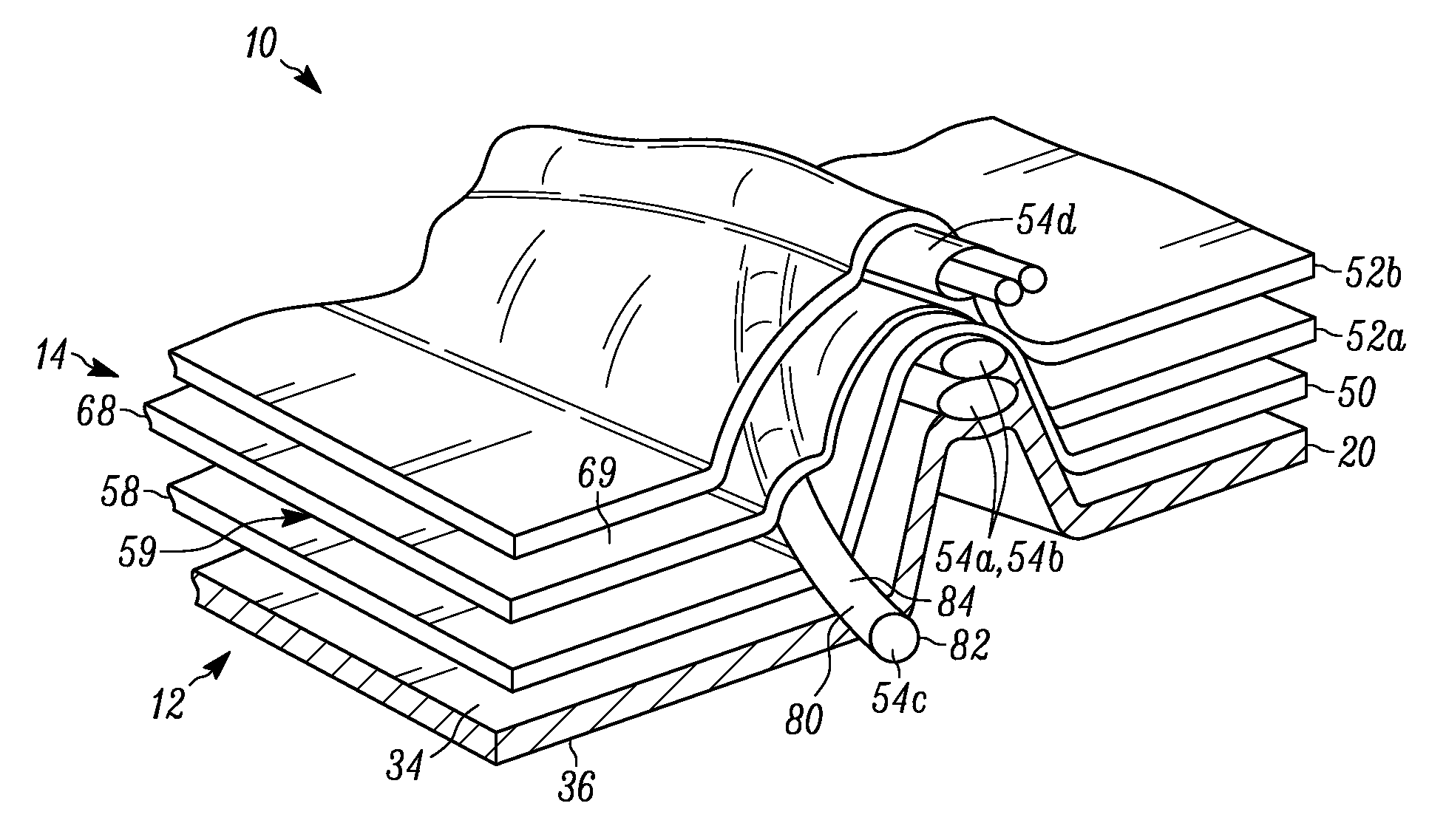
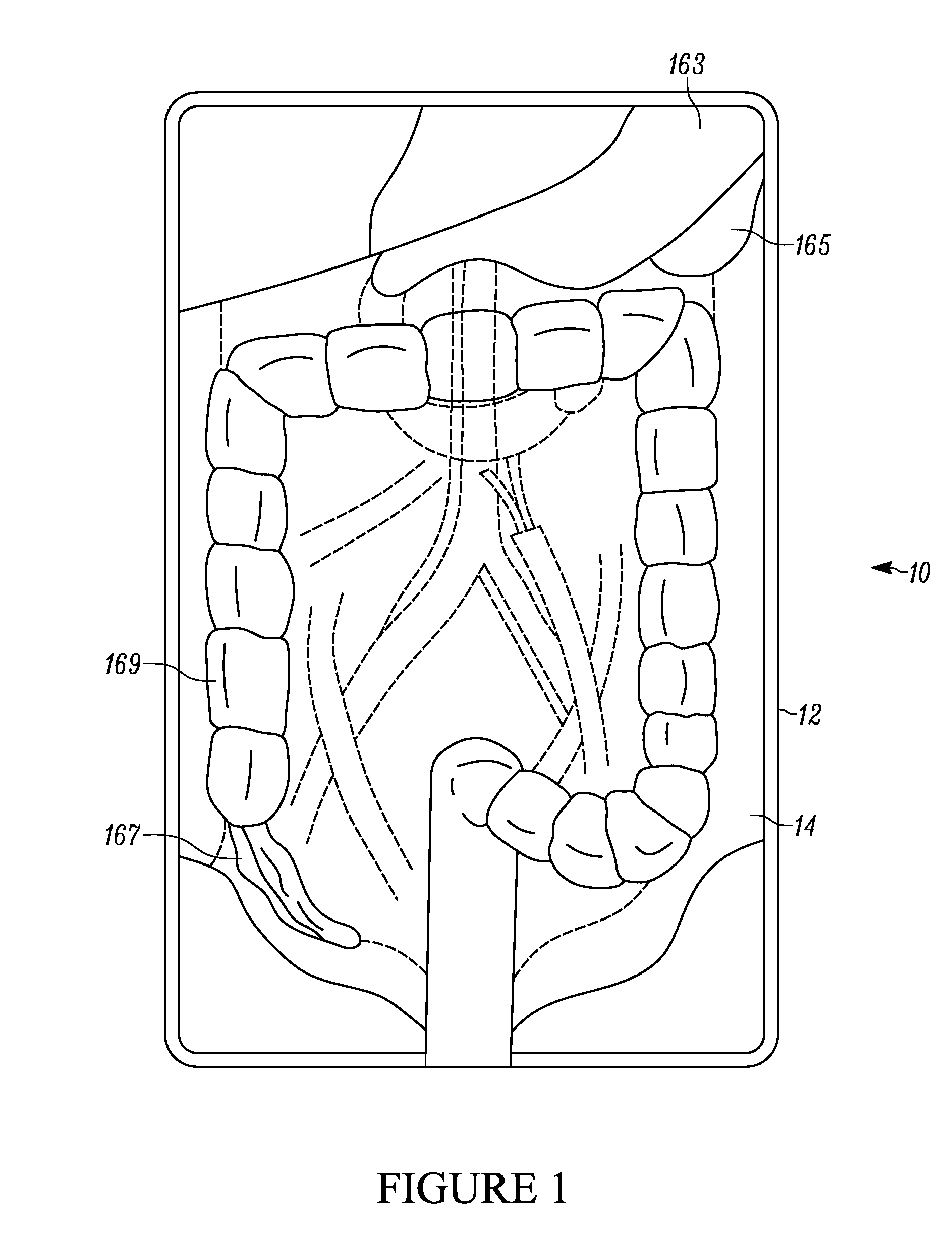
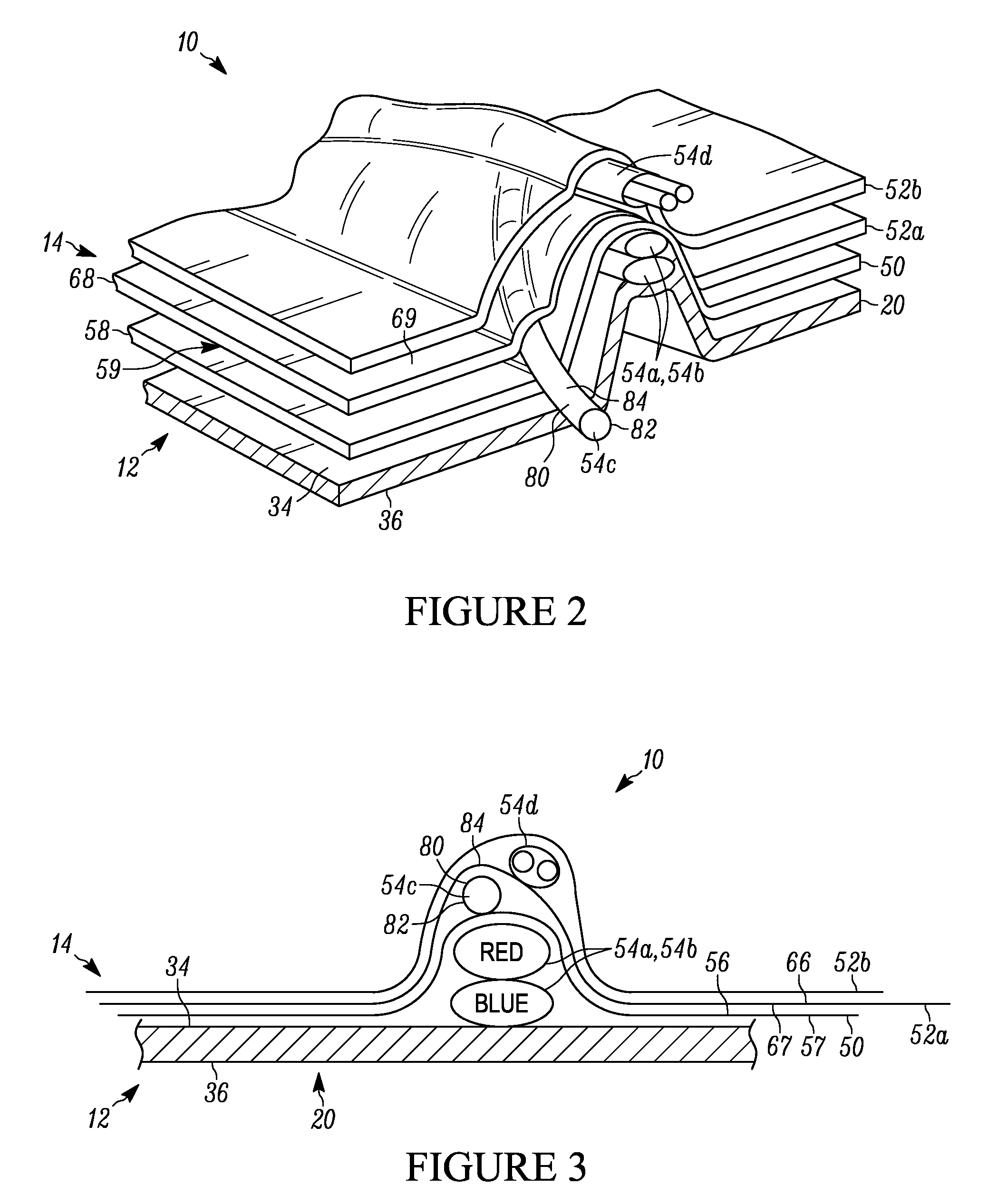
[0042]Referring now to the drawings and in particular to FIG. 1, a surgical training aid apparatus of the present invention is shown generally at 10. Apparatus 10 includes housing 12, and anatomical replicating assembly 14. Typically, the surgical t...


PUM


Abstract

A surgical training aid apparatus for facilitating the training of medical procedures comprising a housing and an anatomical replicating assembly. The housing includes a tray having a bottom surface and a top surface opposite the bottom surface. The anatomical replicating assembly is positioned upon the top surface of the tray of the housing, and includes a base layer and at least one body component. The base layer has a bottom surface and a top surface, and, overlays a portion of the top surface of the tray of the housing. The base layer further includes a tackiness. The body component is positioned between the top surface of the housing and the bottom surface of the base layer or on the base layer. The body component likewise includes a tackiness. The body component is releasably coupled to the base layer due to the tackiness of the two components, wherein the tackiness is overcome so as to separate the at least one body component relative to the base layer.

Description

BACKGROUND OF THE INVENTION[0001]1. Field of the Invention[0002]The invention relates in general to training aids, and more particularly, to a training aid for the medical field, wherein the training aid facilitates the teaching and practicing of various medical procedures.[0003]2. Background Art[0004]The use of practice aids or teaching aids in the medical field has been quite extensive. For example, rigid models of the body have been used as demonstration aids to teach both anatomy and procedures to students and practitioners. Such models may show anatomical relationships of organs but not the characteristics of normal and pathologic tissue planes that separate organs. These key fact limits the usefulness of current models to teach complex surgical skills.[0005]Typically, for practicing many medical procedures, and especially for practicing soft tissue procedures, it is quite common to use human cadavers. One problem with such an approach is that human cadavers vary relative to he...

Claims


Application Information

Patent Timeline
Patent Type & Authority Patents(United States)
IPC IPC(8): G09B23/28
CPCG09B23/285
Inventor SENAGORE, ANTHONYZIMMERMAN, WARREN M.
Owner SENAGORE ANTHONY
Learn More