Device and method for remote vessel ligation

a technology of remote vessel and ligation, applied in the field of cannula, can solve the problems of difficult procedure, difficult transecting of the vessel, and surgeons encountering several difficulties in ligation procedures, and achieve the effect of facilitating accurate positioning of the vessel and sutur

Inactive Publication Date: 2011-07-05
MAQUET CARDIOVASCULAR LLC
View PDF485 Cites 36 Cited by
  • Summary
  • Abstract
  • Description
  • Claims
  • Application Information

AI Technical Summary

Benefits of technology

[0009]In a further embodiment, a transecting device is positioned within the cannula. The distal end of the tension mount is positioned to allow the distal end to be proximal to the shoulder of the dissection cradle responsive to the shoulder being in an axial position relative to the tension mount. This results in the suture and vessel being reliably positioned within reach of the transecting device for transection of the vessel and cutting of the suture.
[0010]Finally, in a preferred embodiment, the retractor has a distal end having an axis skewed relative to the central axis of the cannula, thus facilitating accurate positioning of the vessel and suture for transection and cutting, and ensuring the proper displacement of the suture loop onto the vessel in response to the retraction of the retractor.

Problems solved by technology

However, surgeons encounter several difficulties in ligation procedures.
However, this procedure is difficult because the slipknot often catches on stumps of cut tributaries or other tissue, and then constricts around the vessel at the wrong position.
Also, there is no easy method for transecting the vessel after the suture loop is tied to the vessel without potentially prematurely severing the suture.

Method used


Image

  • Device and method for remote vessel ligation
  • Device and method for remote vessel ligation
  • Device and method for remote vessel ligation

Examples


Embodiment Construction

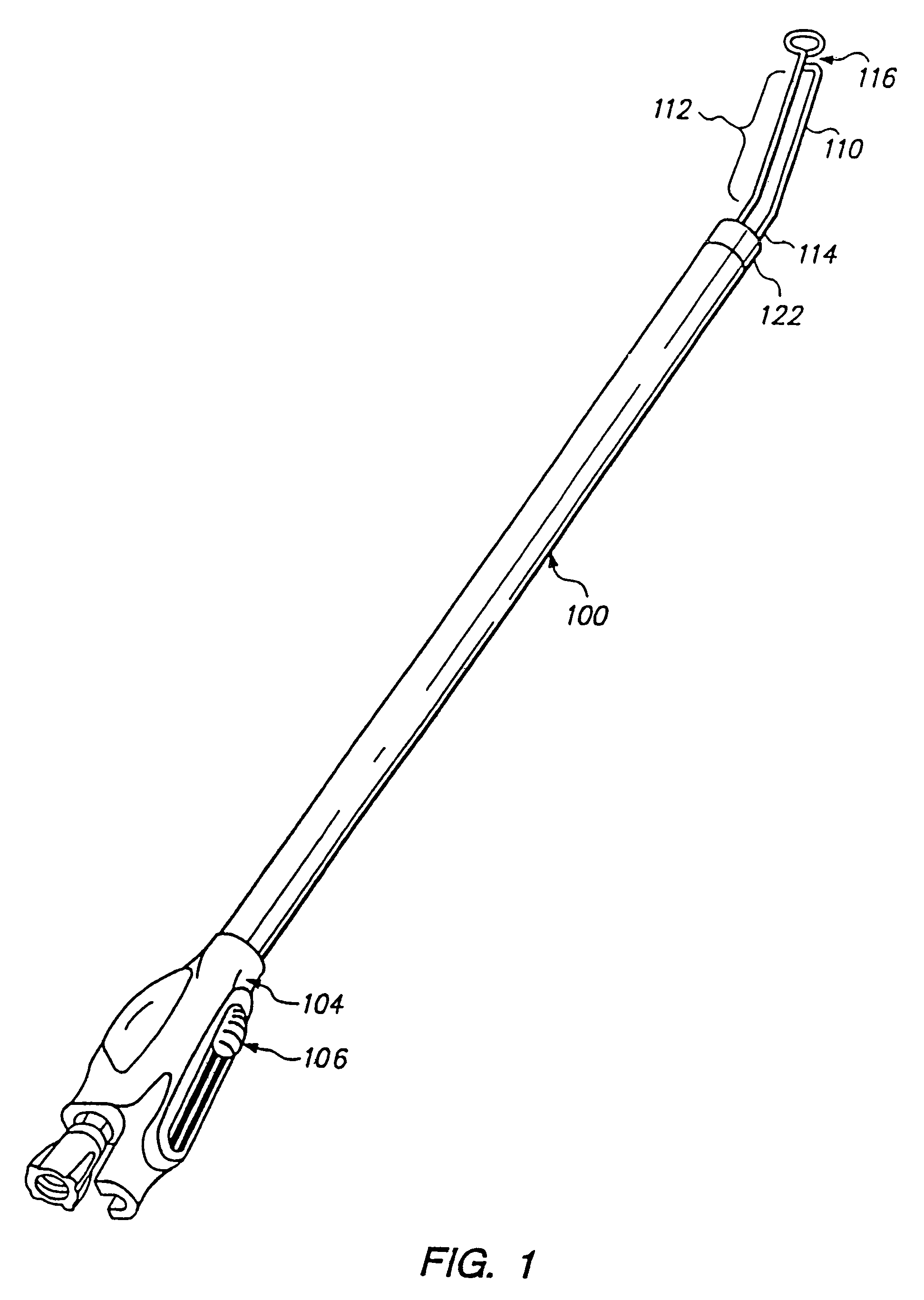
[0050]FIG. 1 illustrates a perspective view of a preferred embodiment of cannula 100 showing retractor 112 in an extended position. Cannula 100 includes an outer housing 102 of bioinert material such as polymed UD that may be approximately 12″ to 18″ in length. The proximal end of the cannula 100 is disposed in handle 104 that includes a button 106 which is coupled to retractor 112 for controlling the translational movement of retractor 112, as described in more detail below.

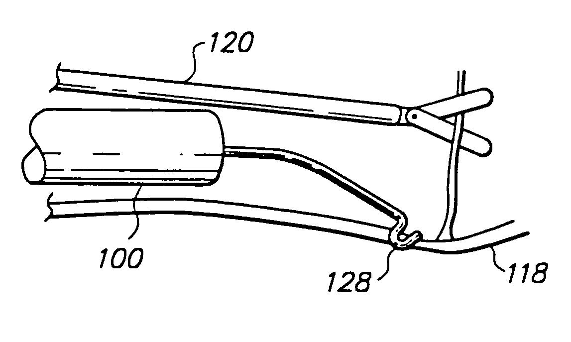
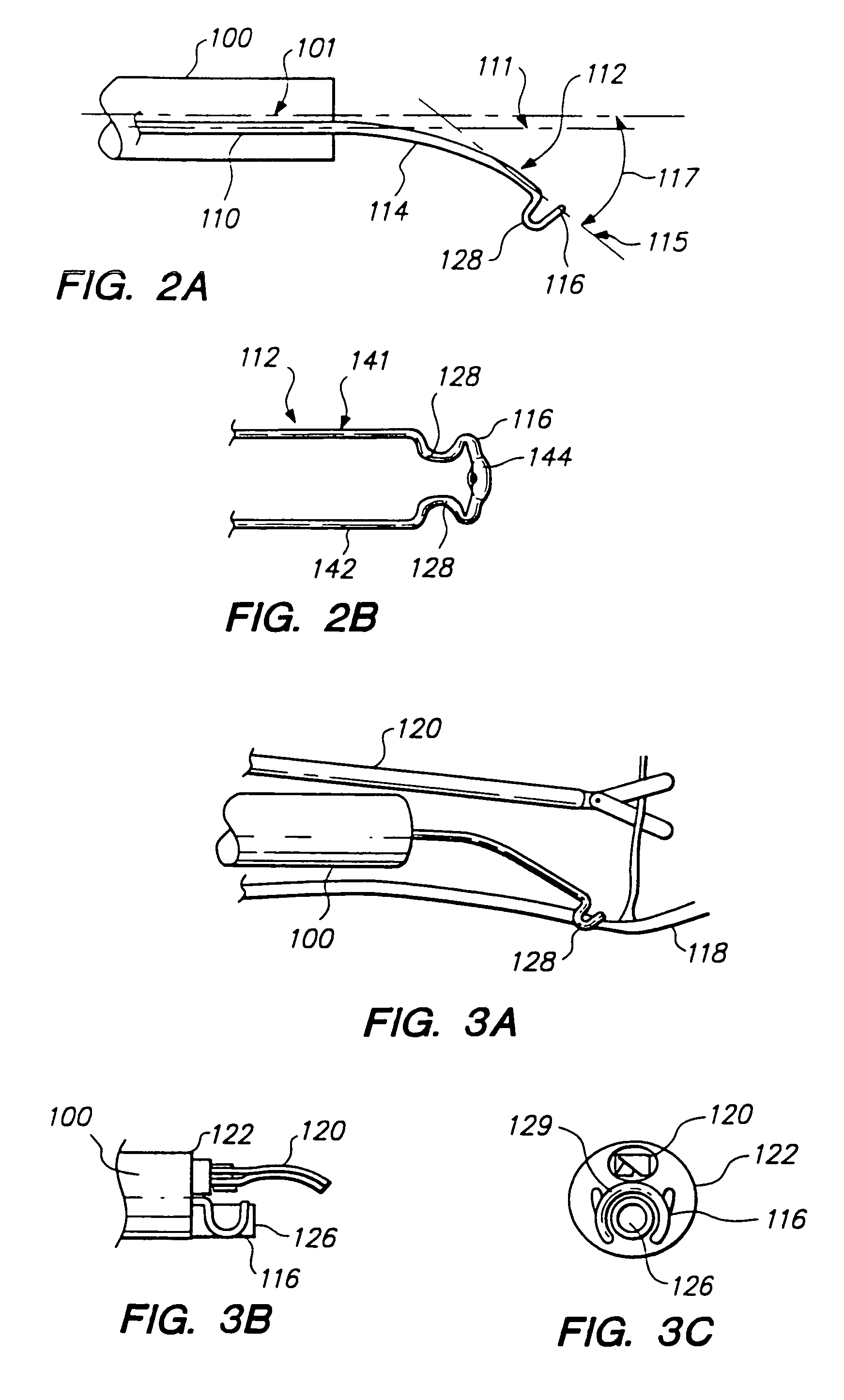
[0051]The distal end of the cannula houses a retractor 112, and optionally an endoscope 126 and a surgical tool 120, described below. FIG. 2a illustrates the retractor 112 in more detail. In one embodiment, retractor 112 is formed of resilient wire which has a smooth bend intermediate to a first portion 110 and a second portion 114 of the retractor. The retractor 112 is described as having two portions for ease of description, although the retractor 112 may be formed as an integrated structure. However, retracto...


PUM


Abstract

A retractor and a surgical tool are positioned at the distal end of the cannula. A dissection cradle is located at the distal end of a distal portion of the retractor that is resiliently skewed relative to the cannula axis, and includes two substantially parallel, spaced legs with the retractor shaped in a loop therebetween. The procedure includes locating a vessel and side branch of interest and extending the retractor to retain the vessel in the dissection cradle to urge the vessel away from the axis of the cannula in order to isolate a side branch for exposure to the surgical tool.

Description

RELATED APPLICATIONS[0001]This application is a continuation application of copending application Ser. No. 10 / 052,016 filed on Jan. 16, 2002 now U.S. Pat. No. 6,830,546, which is a continuation application of application Ser. No. 09 / 521,279, filed on Mar. 7, 2000, now U.S. Pat. No. 6,348,037, which is a continuation of application Ser. No. 09 / 200,218 filed on Nov. 25, 1998, now U.S. Pat. No. 6,162,173, which is a continuation-in-part application of application Ser. No. 09 / 102,723 filed on Jun. 22, 1998, now U.S. Pat. No. 5,895,353.FIELD OF THE INVENTION[0002]This invention relates to a cannula used for vessel retraction, and more particularly to a cannula and method that includes an endoscopic retractor for vessel ligation.BACKGROUND OF THE INVENTION[0003]Certain cannulas have surgical tools located within the cannula for performing surgical operations on a vessel of interest. The cannula is inserted into a surgical site with the distal end of the cannula positioned near the vessel ...

Claims


Application Information

Patent Timeline
Patent Type & Authority Patents(United States)
IPC IPC(8): A61B17/02A61B17/00
CPCA61B17/00008A61B17/0218
Inventor CHIN, ALBERT K.HLAVKA, EDWIN J.LUNSFORD, JOHN P.BAXTER, JEFFREY W.
Owner MAQUET CARDIOVASCULAR LLC
Why Eureka
Try Eureka