Mapping and diagnosis of macular edema by optical coherence tomography

a technology of optical coherence tomography and macular edema, applied in the field of ophthalmology, can solve the problems of visual loss, complicating the accurate and reproducible detection of macular edema areas, and affecting the accuracy of the diagnosis

Inactive Publication Date: 2011-08-16
UNIV OF SOUTHERN CALIFORNIA
View PDF1 Cites 30 Cited by
  • Summary
  • Abstract
  • Description
  • Claims
  • Application Information

AI Technical Summary

Benefits of technology

[0008]Accordingly, one object of the present invention is to provide better methods and tools for the detection and diagnosis of macular edema.

Problems solved by technology

Macular edema is an important cause of visual loss and legal blindness in patients with diabetic retinopathy.
Variations in the amount of stereopsis present in paired stereo photographs or in the threshold for thickening adopted by the observer may further complicate the accurate and reproducible detection of areas of edema.
Thus, there is potential for considerable variability and possible lack of sensitivity in prior art methods for identifying macular edema that were used in previous clinical studies.
Furthermore, the lack of sensitivity of the clinical examination for detection of mild edema has been demonstrated by a number of investigators, including Brown et al.
However, as these cases of subclinical edema were presumably not recognized in the ETDRS, the rationale for treating these lesions is presently uncertain, although it may change as data from ongoing clinical trials incorporating OCT imaging become available.
Although the system proposed by Brown et al. is useful for identification of foveal edema, it is not likely to identify cases of nonfoveal CSME.
Unfortunately, the commonly used macular scanning patterns on the Stratus OCT machine (Carl Zeiss Meditec, Inc., Dublin, Calif.) consist of radial lines that provide a high density of points near the fovea, but a relatively sparse pattern in more peripheral zones.

Method used


Image

  • Mapping and diagnosis of macular edema by optical coherence tomography
  • Mapping and diagnosis of macular edema by optical coherence tomography
  • Mapping and diagnosis of macular edema by optical coherence tomography

Examples


examples

Materials and Methods

1. Data Collection and Study Population

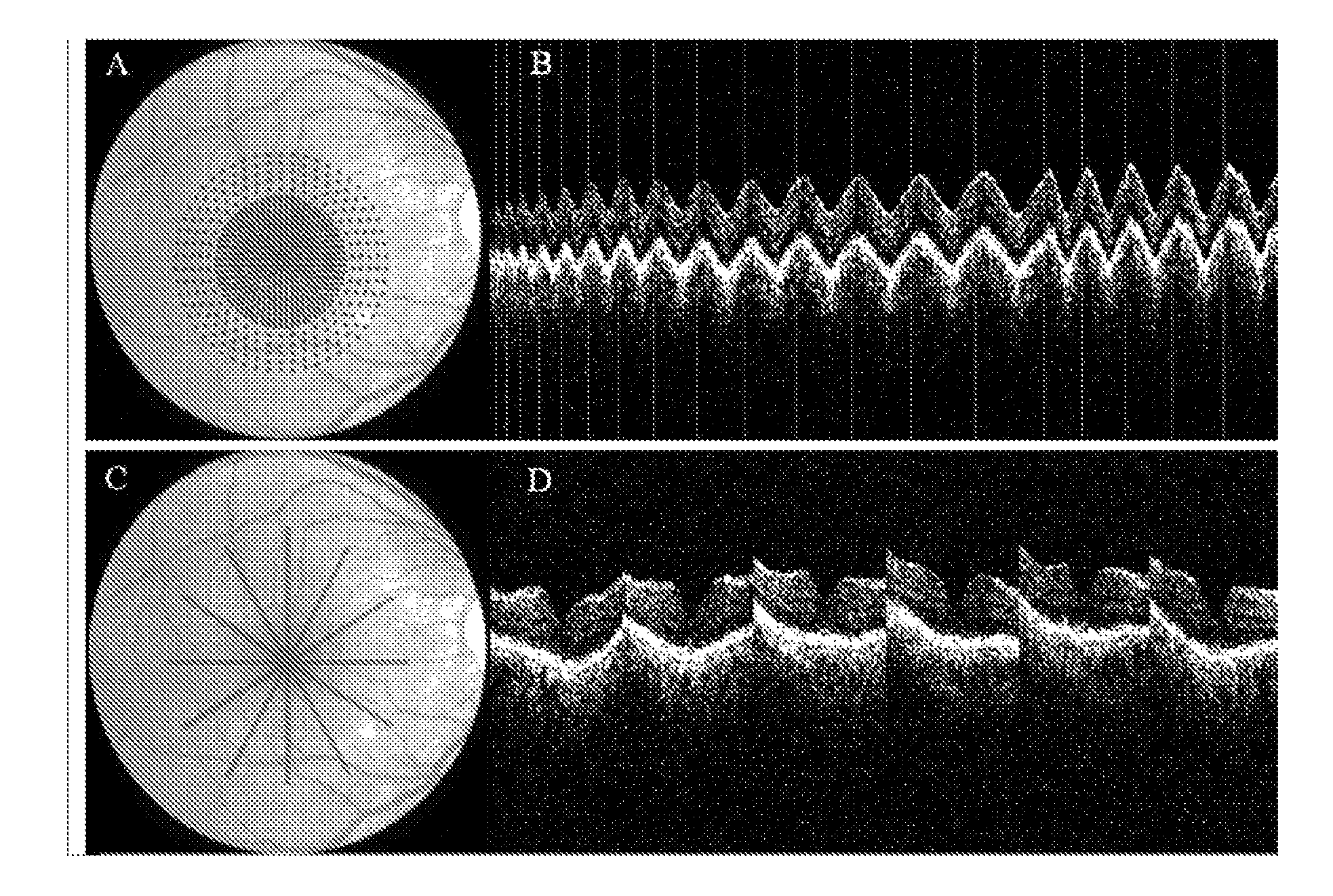
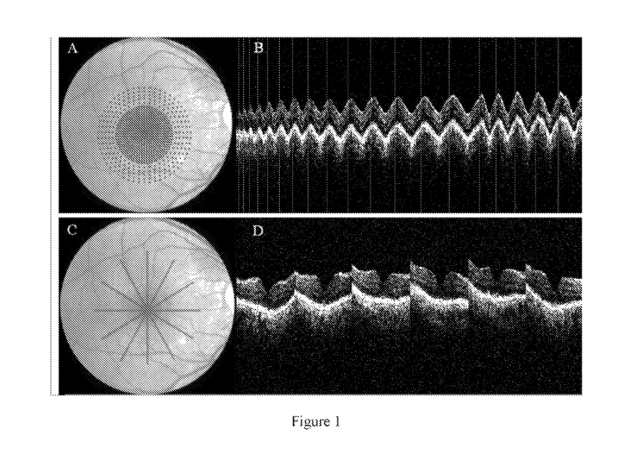
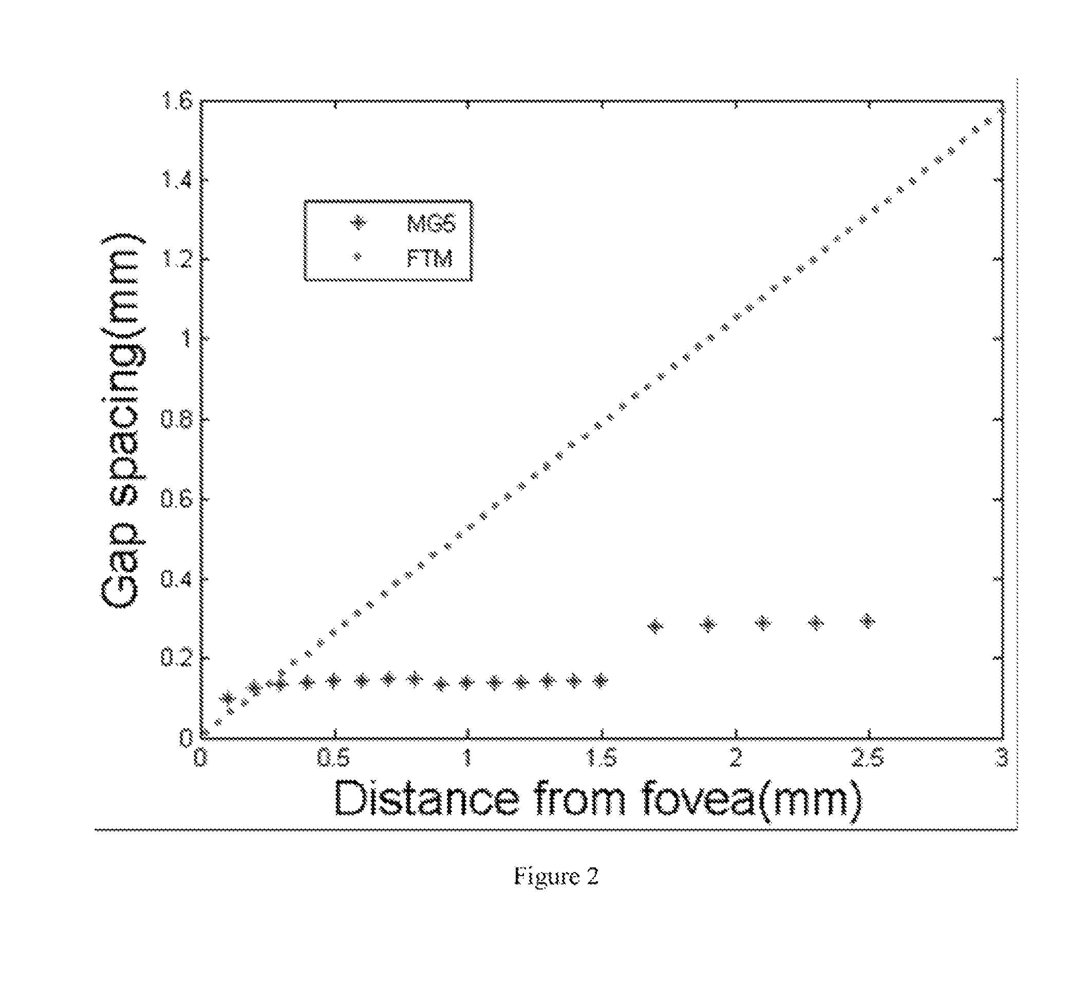
[0045]We retrospectively reviewed the clinical and imaging records of 71 eyes of 40 patients referred to the Doheny Ocular Imaging Unit with a diagnosis of diabetic macular edema (DME) who underwent OCT imaging using both the MG5 scan pattern (FIG. 1) and Fast Macular Thickness Map (FMTM). Approval for the analysis of these records was obtained from the institutional review board of the University of Southern California. For all OCT imaging studies, the Stratus OCT system with version 4.0 software was used to acquire the scan of the macula. Fast Macular Thickness Map and MG5 data from 65 normal subjects recruited from the Doheny Eye Institute and the University of Pittsburgh Medical Center were used as the reference baseline. The normal subjects were recruited as part of the prospective Advanced Imaging for Glaucoma Study. The study was approved by the institutional review boards of the University of Southern California and...


PUM


Abstract

The present invention discloses a method for comparing the detection of clinically significant diabetic macular edema by an optical coherence tomography (OCT) grid scanning protocol and biomicroscopic examination. Also provided are computer implemented, automated systems for performing the method thereof and computer readable media encoding the method thereof.

Description

CROSS-REFERENCE TO RELATED APPLICATIONS[0001]This application claims an invention which was disclosed in Provisional Application No. 60 / 796,811, filed on May 1, 2006. The benefit under 35 USC §119 (e) of the U.S. provisional application is hereby claimed. The above priority application is hereby incorporated herein by reference.STATEMENT REGARDING FEDERALLY SPONSORED RESEARCH AND DEVELOPMENT[0002]The present invention is made, at least in part, with the support of NIH grant R01 EY013516. The government has certain rights in the invention.FIELD OF THE INVENTION[0003]The invention pertains to the field of ophthalmology. More particularly, the invention pertains to mapping and diagnosing macular edema by optical coherence tomography.BACKGROUND OF THE INVENTION[0004]Macular edema is an important cause of visual loss and legal blindness in patients with diabetic retinopathy. In the Early Treatment Diabetic Retinopathy Study (ETDRS) [1], focal laser photocoagulation was demonstrated to re...

Claims


Application Information

Patent Timeline
Patent Type & Authority Patents(United States)
IPC IPC(8): A61B3/00A61B13/00
CPCA61B3/1005A61B3/102
Inventor HUANG, DAVIDSADDA, SRINIVAS R.TAN, OU
Owner UNIV OF SOUTHERN CALIFORNIA
Why Eureka
Try Eureka