Macular edema lesion area segmentation method based on deep neural network

A deep neural network and macular edema technology, which is applied in the field of medical image processing, can solve problems such as large time and labor costs, macular edema evaluation errors, and large image noise, so as to improve the effect, expand the diversity, and reduce the difference.

Pending Publication Date: 2019-12-10
成都智能迭迦科技合伙企业(有限合伙)
View PDF3 Cites 6 Cited by
  • Summary
  • Abstract
  • Description
  • Claims
  • Application Information

AI Technical Summary

Problems solved by technology

In order to improve the above "manually delineating the macular edema area will consume huge time and labor costs. In addition, due to the large noise of the OCT image and the

Method used


Image

  • Macular edema lesion area segmentation method based on deep neural network
  • Macular edema lesion area segmentation method based on deep neural network
  • Macular edema lesion area segmentation method based on deep neural network

Examples


Embodiment Construction

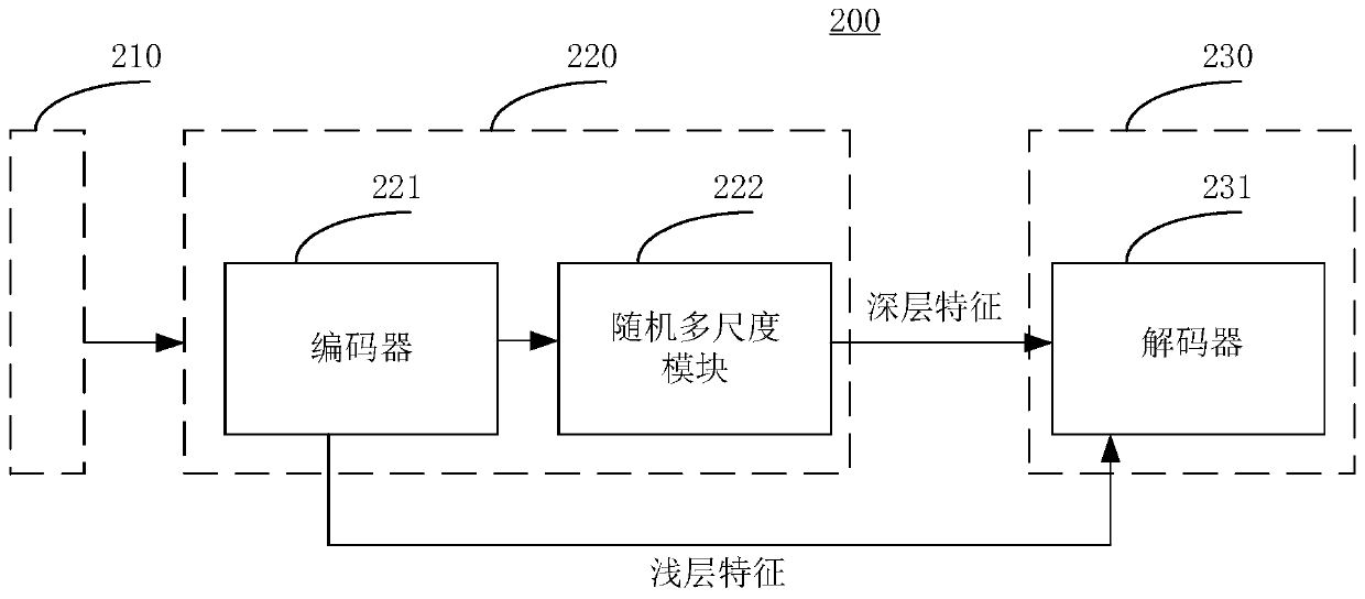
[0028] The technical solutions in the embodiments of the present application will be described below with reference to the drawings in the embodiments of the present application.

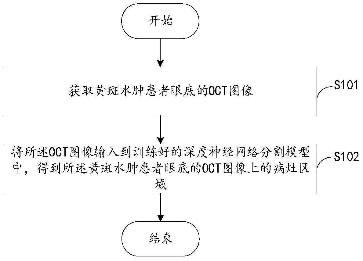
[0029] The macular area is the most sensitive part of the fundus to light. Macular edema refers to the inflammatory reaction and fluid infiltration in the macular area of ​​the fundus, resulting in edema. Diseases include: retinal pigment epithelial detachment (Pigment Epithelium Detachment, PED), subretinal edema (Subretinal Fluid, SRF), etc.

[0030] At present, patients are examined mainly by acquiring OCT (Optical coherence tomography, optical coherence tomography) images of the patients. OCT images use the interference principle of light to scan biological tissues to obtain micron-scale three-dimensional images. Compared with traditional fluorescein contrast examination methods, OCT images have the characteristics of non-contact, non-invasive, and high resolution.

[0031] In order to quantit...


PUM


Abstract

The invention provides a macular edema lesion area segmentation method based on a deep neural network. The macular edema lesion area segmentation method comprises the steps of acquiring an OCT image of an fundus of a macular edema patient; and inputting the OCT image into a trained deep neural network segmentation model to obtain a focus area on the OCT image of the fundus of the macular edema patient. The OCT image is input into the trained deep neural network segmentation model to obtain the focus area on the OCT image of the fundus of the macular edema patient. Compared with the prior art,a lot of manpower and material resources are saved. Meanwhile, a focus area on the OCT image of the fundus of the macular edema patient is acquired through the trained deep neural network segmentationmodel. The problem that the evaluation error of macular edema is large due to the fact that at present, due to the fact that OCT image noise is large and experience differences between doctors are large, sketching results of different doctors are often large in difference can be solved.

Description

technical field [0001] The present application relates to the technical field of medical image processing, in particular, to a method, device, electronic equipment and storage medium for macular edema lesion region segmentation based on a deep neural network. Background technique [0002] The macular area is the most sensitive part of the fundus to light. Macular edema refers to the inflammatory reaction and fluid infiltration in the macular area of ​​the fundus, resulting in edema. Diseases include: retinal pigment epithelial detachment (Pigment Epithelium Detachment, PED), subretinal edema (Subretinal Fluid, SRF), etc. At present, patients are examined mainly by acquiring OCT (Optical coherencetomography, optical coherence tomography) images of the patients. In order to quantitatively evaluate the patient's condition, the doctor will outline the area of ​​macular edema based on the OCT image, such as the specific area of ​​PED and SRF. Afterwards, the volume of the macul...

Claims


Application Information

Patent Timeline
IPC IPC(8): G06T7/11G06N3/04G06N3/08
CPCG06T7/11G06N3/08G06T2207/10101G06T2207/20081G06T2207/30041G06N3/044G06N3/045
Inventor 章毅陈媛媛郭际香胡俊杰张炜王璟玲
Owner 成都智能迭迦科技合伙企业(有限合伙)
Why Eureka
Try Eureka