System and method for computer aided detection of pulmonary embolism in tobogganing in CT angiography

a technology of pulmonary embolism and ct angiography, which is applied in the field of computer aided detection of pulmonary embolism in ct angiography, can solve the problems of difficult to separate critically ill patients suffering from pe, laborious manual reading of these slices, time-consuming and complicated,

Active Publication Date: 2011-10-11
SIEMENS MEDICAL SOLUTIONS USA INC
View PDF7 Cites 0 Cited by
  • Summary
  • Abstract
  • Description
  • Claims
  • Application Information

AI Technical Summary

Problems solved by technology

PE is a very serious condition that can cause sudden death in about one-third of the cases.
Therefore, a major clinical challenge, particularly in an emergency room scenario, is to quickly and correctly diagnose patients with PE and then send them on to treatment, as a prompt and accurate diagnosis is the key to survival.
However, PE is among the most challenging conditions to diagnosis because its primary symptoms are vague, non-specific, and may have a variety of other causes, making it hard to separate out the critically ill patients suffering from PE.
Clinically, manual reading of these slices is laborious, time consuming and complicated by various PE look-alikes (false positives) including respiratory motion artifacts, flow-related artifacts, streak artifacts, partial volume artifacts, stair step artifacts, lymph nodes, and vascular bifurcation, among many others.
The accuracy and efficiency of interpreting such a large image data set is also limited by human factors, such as attention span and eye fatigue.
However, in an ER (Emergency Room) scenario, radiologists only have a small time window (2-3 minutes) to read a case and make the diagnosis.
However, vessel segmentation is computationally time consuming and has been problematic for small vasculature where subsegmental PEs often occur.
This implies that one would not be able to obtain a watershed region till the whole image has been scanned and processed.
However, for large images, the underlying graph created in live-wire for search becomes large, the interativeness of livewire would be comprised due to fundamental limitations of Dijkstra's algorithm.
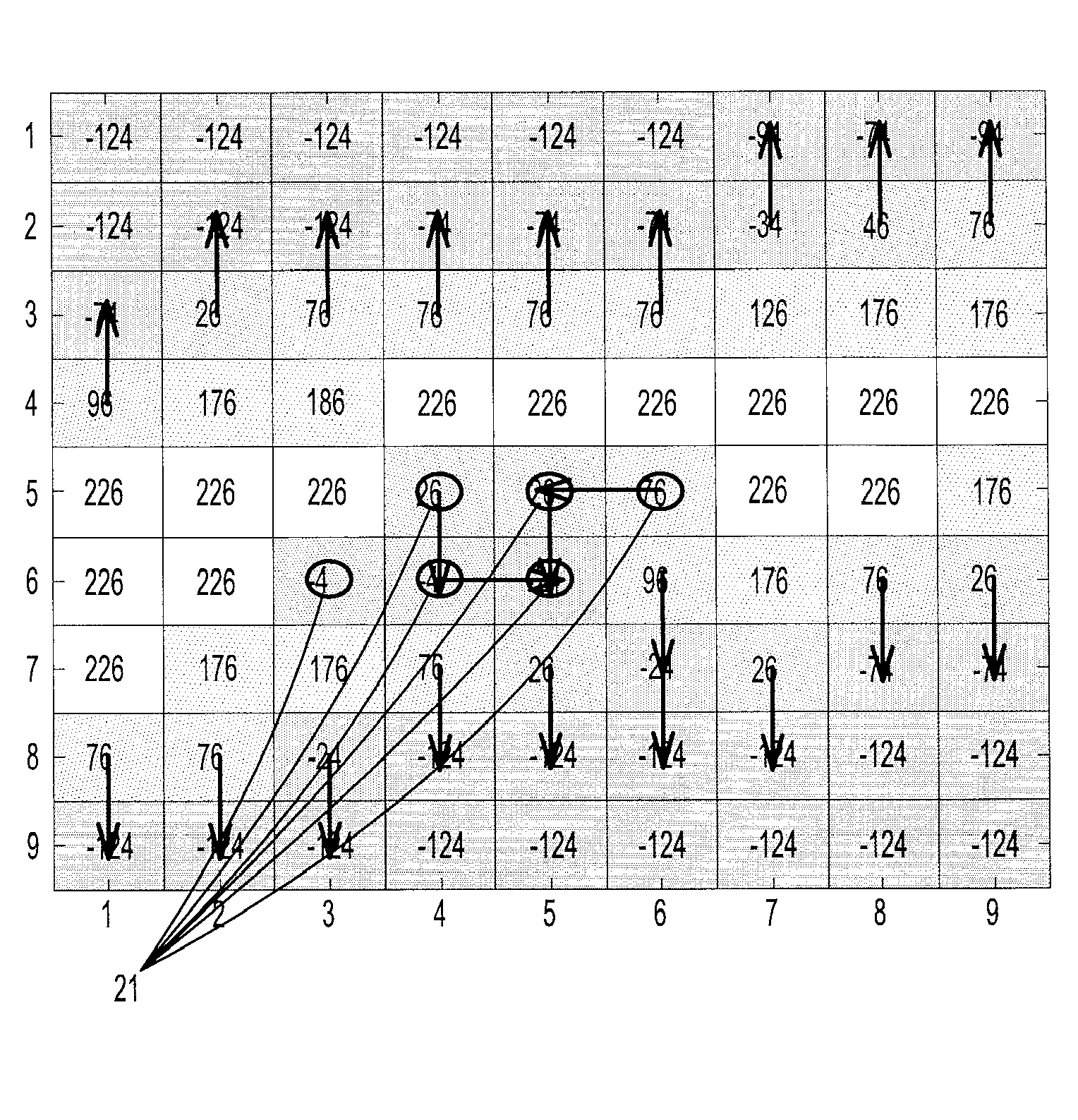
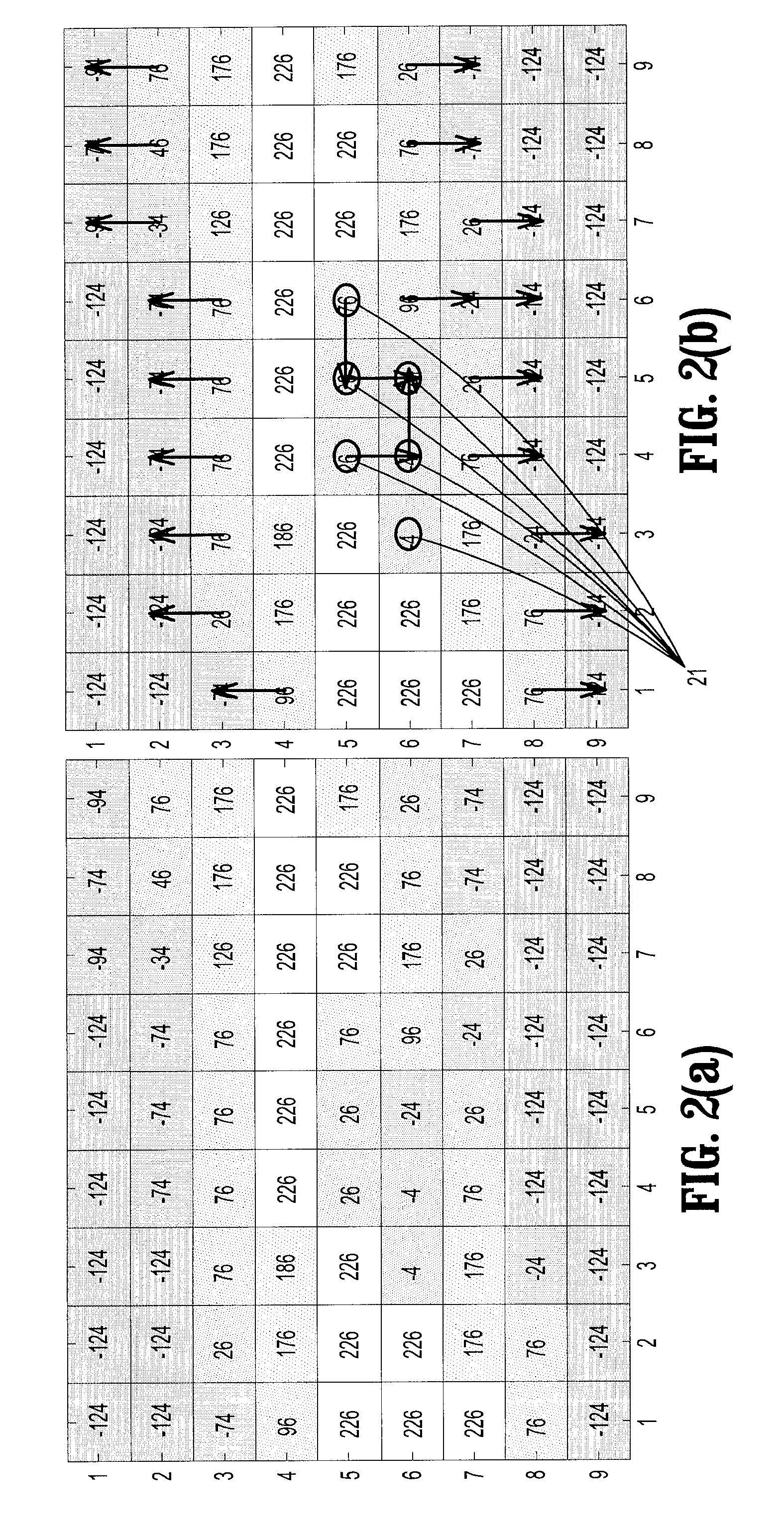
In short, none of the discussed algorithms can extract a toboggan cluster from an initial site without processing pixels beyond its external boundary.

Method used


Image

  • System and method for computer aided detection of pulmonary embolism in tobogganing in CT angiography
  • System and method for computer aided detection of pulmonary embolism in tobogganing in CT angiography
  • System and method for computer aided detection of pulmonary embolism in tobogganing in CT angiography

Examples


Embodiment Construction

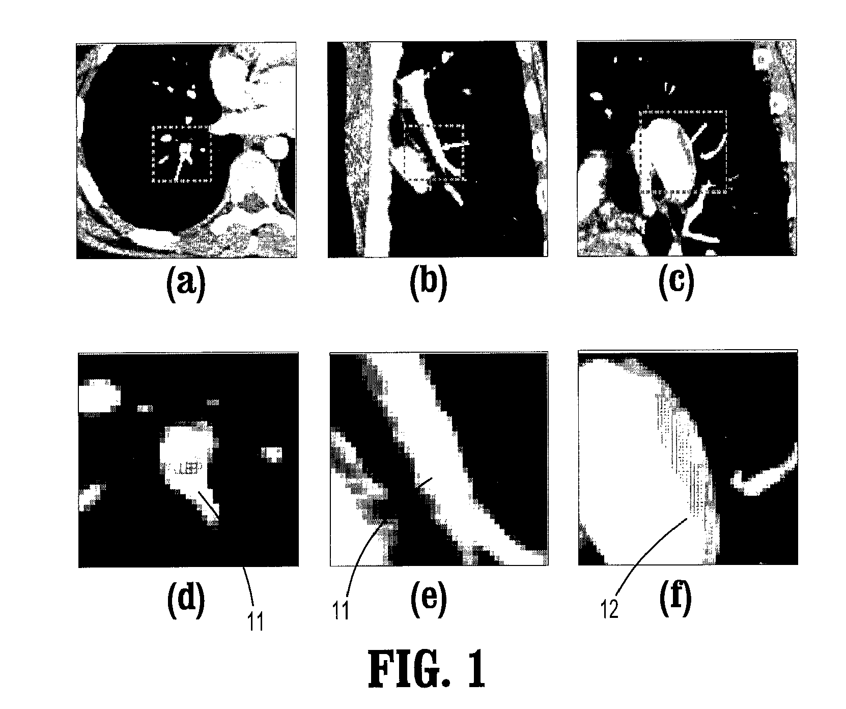
[0027]Exemplary embodiments of the invention as described herein generally include systems and methods for computer aided detection of pulmonary embolism with tobogganing in CT angiography. Accordingly, while the invention is susceptible to various modifications and alternative forms, specific embodiments thereof are shown by way of example in the drawings and will herein be described in detail. It should be understood, however, that there is no intent to limit the invention to the particular forms disclosed, but on the contrary, the invention is to cover all modifications, equivalents, and alternatives falling within the spirit and scope of the invention.

[0028]As used herein, the term “image” refers to multi-dimensional data composed of discrete image elements (e.g., pixels for 2-D images and voxels for 3-D images). The image may be, for example, a medical image of a subject collected by computer tomography, magnetic resonance imaging, ultrasound, or any other medical imaging syste...


PUM


Abstract

A method for detecting pulmonary embolisms in computed tomographic (CT) angiography includes providing a digitized (CT) image acquired from inside a pulmonary vessel, the image comprising a plurality of intensities corresponding to a 3-dimensional grid of voxels, for each voxel in the image, extracting a first pulmonary embolism (PE) candidate and PE boundary from the voxel, and for each voxel in the PE boundary, selecting a voxel from the PE boundary, extracting a subsequent PE candidate and PE boundary from the voxel, merging the subsequent PE candidate with the first PE candidate, and merging the subsequent PE boundary with the first PE boundary.

Description

CROSS REFERENCE TO RELATED UNITED STATES APPLICATIONS[0001]This application claims priority from “Computer Aided Detection of Pulmonary Embolism with Tobogganing in CT Angiography”, U.S. Provisional Application No. 60 / 888,165 of Jianming Liang, et al., filed Feb. 5, 2007, the contents of which are herein incorporated by reference.TECHNICAL FIELD[0002]This disclosure is directed to the detection of pulmonary embolisms in CT angiography.DISCUSSION OF THE RELATED ART[0003]Pulmonary embolism (PE) is the third most common cause of death in the US with at least 650,000 cases occurring annually. PE is a sudden blockage in a lung artery, and is caused by an embolus that is formed in one part of the body and travels in the bloodstream through the heart before reaching the lungs. PE is a very serious condition that can cause sudden death in about one-third of the cases. Most of those who die do so within 30 to 60 minutes after symptoms start. Treatment with anti-clotting medications is highly...

Claims


Application Information

Patent Timeline
Patent Type & Authority Patents(United States)
IPC IPC(8): G06K9/00G06K9/34
CPCG06K9/00201G06T7/0012G06T7/0081G06T2207/10081G06T2207/30061G06T2207/30101G06T7/11G06V20/64
Inventor LIANG, JIANMINGBI, JINBO
Owner SIEMENS MEDICAL SOLUTIONS USA INC
Why Eureka
Try Eureka