Self-expandable medical instrument for treating defects in a patient's heart

a medical instrument and self-expanding technology, applied in the field of self-expandable medical instruments for treating defects in patients' hearts, can solve the problems of high risk of surgical procedures, risk of heart valve implant malalignment, and no longer justifiable risks of such surgical procedures, and achieve the effect of high memory capacity

Active Publication Date: 2011-11-22
JENAVALVE TECH INC
View PDF426 Cites 488 Cited by
  • Summary
  • Abstract
  • Description
  • Claims
  • Application Information

AI Technical Summary

Benefits of technology

[0016]The advantages of the invention are in particular noted to be in the providing of a transvascularly introducible medical instrument, in particular for treating a patient's heart defects, whereby the medical instrument is suitable to be delivered by catheter to the is defect to be treated in the patient's heart. Because the medical instrument is configured as a self-expandable instrument and essentially consists of a stent made of a flexible mesh of thin wires or filaments, one particularly advantageous result achieved is that the medical instrument—regardless of the size of the heart valve to be treated and regardless of the diameter to the defective heart valve—can self-adapt to the defective heart valve, and in such a way that, on the one hand, the portions of the medical instrument protruding into the bloodstream flowing past the implanted medical instrument are as small as possible, while at the same time, an optimal positioning, secure anchorage and optimal lateral sealing of the implanted medical instrument is ensured.
[0030]It is of particular advantage for the stent made of flexible mesh, in particular wire mesh, with a self-expanding prosthetic heart valve arranged in its center area to be configured such that the stent with the prosthetic heart valve can be withdrawn back into the catheter system, and thus removable from the patient's body, at any time during the implantation of the medical instrument.

Problems solved by technology

This consequently makes the surgical procedure a risky one coupled with the associated risks for the patients and a lengthy post-operative treatment phase.
In particular, the risks of such a surgical procedure are often no longer justifiable in the case of multimorbid patients.
In the known solutions, however, there is a risk of heart valve implant malalignment.
Among other complications, an implantation malalignment of a less than optimally positioned prosthetic heart valve can lead to, for example, leakage or valvular regurgitation, which puts a substantial burden on the ventricle.
Should, for example, a prosthetic heart valve be implanted too far above the actual heart valve plane, this can lead to occlusion of the coronary artery origination (coronaries) and thus to a fatal coronary ischemia with myocardial infarction.
Although such a surgical introduction can be monitored and controlled, for example with fluoroscopy (Cardiac Catheterization Laboratory=CCL) or with ultrasound (Transesophageal Echocardiogram=TEE), oftentimes—due to the limited maneuverability of the prosthetic heart valve which is still in a collapsed state during the introduction procedure and despite being in the collapsed state is still of relatively large size—it is not possible to ensure the required positioning accuracy and especially the longitudinal placement to the prosthetic heart valve implant with the corresponding anchoring elements affixed thereto.
In particular—as a result of a possible coronary artery occlusion—an angle misalignment to the implanted prosthetic heart valve from the optimum site of deployment can pose a threat to the respective patient.
Moreover, explanting a previously implanted prosthetic heart valve in a minimally-invasive procedure or accordingly correcting an incorrectly positioned prosthetic heart valve has to date often only been possible with great effort, if at all.

Method used


Image

  • Self-expandable medical instrument for treating defects in a patient's heart
  • Self-expandable medical instrument for treating defects in a patient's heart
  • Self-expandable medical instrument for treating defects in a patient's heart

Examples


Embodiment Construction

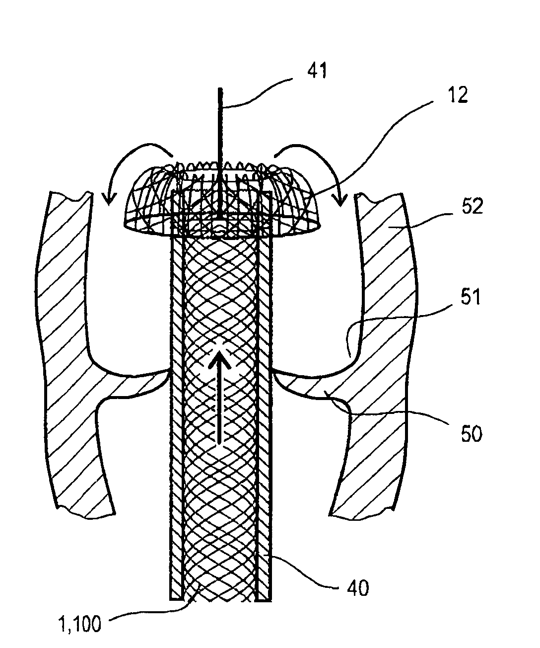
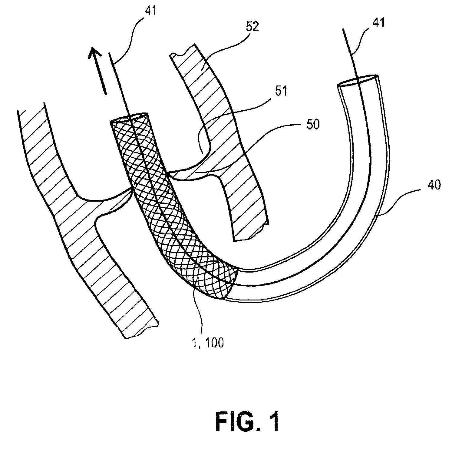
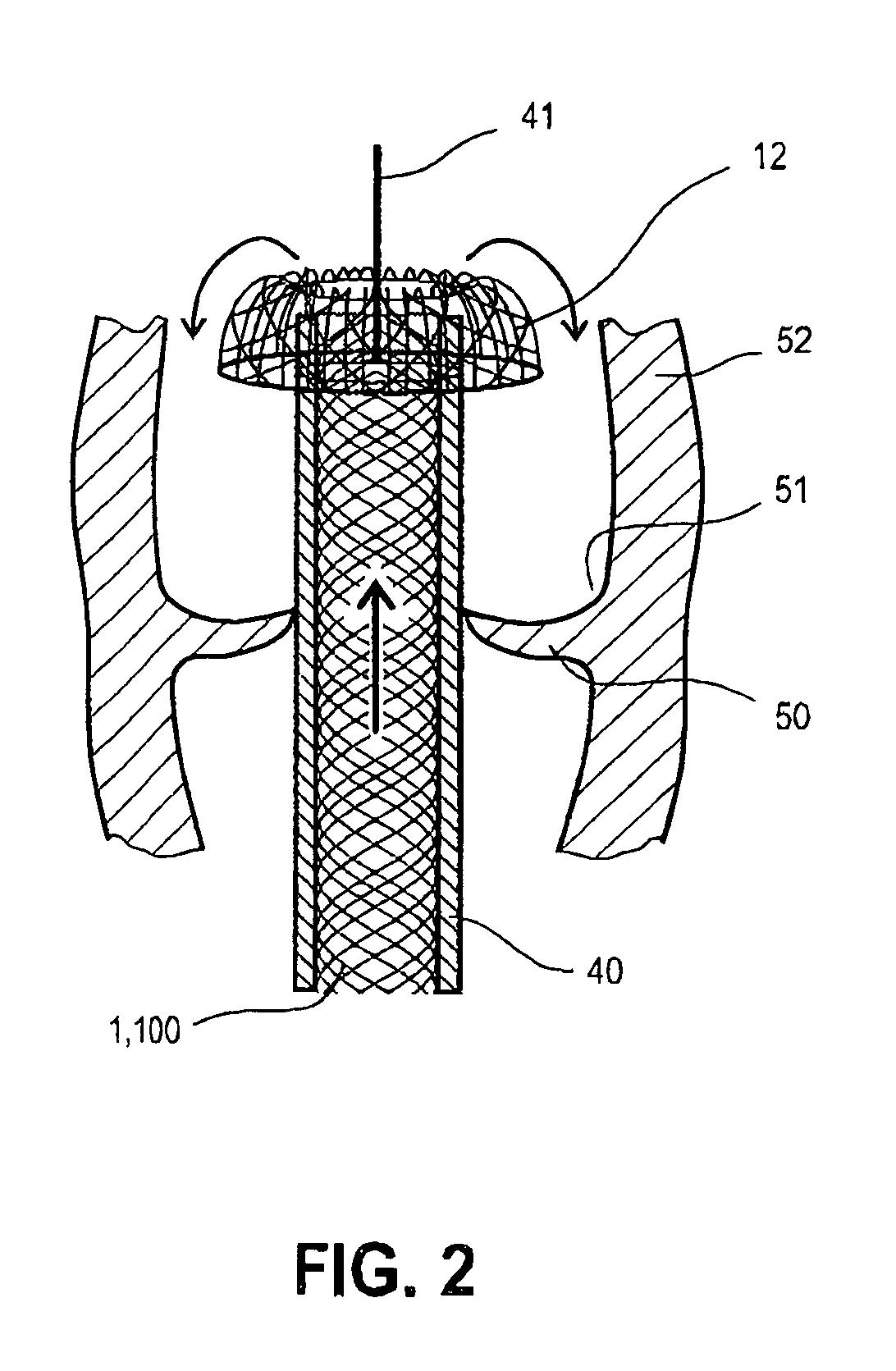
[0041]The embodiment depicted in the figures of the inventive self-expandable medical instrument 100 for treating defects of a patient's heart relates to a self-expandable medical instrument for the transvascular implantation of a prosthetic heart valve 30, wherein the medical instrument 100 can be introduced into a patient's body in minimally-invasive fashion by means of a catheter system 40 and consists of a stent 1 made from a flexible mesh (2) of thin wires or filaments 2′.

[0042]As FIG. 1 shows, the stent 1 configured from flexible mesh 2 is in a first predefined shape during the insertion of the medical instrument 100 into the patient's body. The stent 1 further exhibits a self-expandable prosthetic heart valve 30 at its center area 15, which is covered by mesh 2 in FIG. 1 and thus not explicitly shown. As will be described below, the self-expandable prosthetic heart valve 30 unfolds by itself upon the medical instrument 100, the stent 1 respectively, being released from the ca...


PUM


Abstract

The invention relates to a self-expandable medical instrument (100) for treating defects in a patient's heart, in particular for the transvascular implantation of a prosthetic heart valve (30), wherein the medical instrument (100) is introducible into the body of a patient in a minimally-invasive procedure using a catheter system (40) and comprises a stent (1) made of a flexible mesh (2) of thin wires or filaments (2′). In order to realize a positioning of medical instrument (100) in the patient's heart which is as precise as possible and to securely anchor same there, it is provided for the stent (1) composed of the flexible mesh (2) to exhibit in the expanded state of the medical instrument (100) a distal retention area (10) with a laterally-inverted beaded portion (12) which is engageable in at least one pocket (51) of the patient's defective heart valve (50), a proximal retention area (20), and a center area (15) positioned between the distal and the proximal retention area (10, 20).

Description

CROSS-REFERENCE TO RELATED APPLICATIONS[0001]The present application claims priority from German Patent Application No. DE 10 2005 052 628.4, which was filed on Nov. 4, 2005, by Hans-Reiner Figulla et al. for a SELF-EXPANDABLE MEDICAL INSTRUMENT FOR TREATING DEFECTS IN A PATIENT'S HEART and is hereby incorporated by reference.BACKGROUND OF THE INVENTION[0002]1. Field of the Invention[0003]The present invention relates to a self-expandable medical instrument for treating defects in a patient's heart, in particular for the transvascular implantation of a prosthetic heart valve, whereby the medical instrument can be introduced via a catheter system into the patient's body in a minimally-invasive procedure. In particular, the invention relates to a device for the transvascular replacement of diseased heart valves.[0004]2. Background Information[0005]A device of this type is known in principle to medical technology. At present, biological or mechanical valve models are available to subst...

Claims


Application Information

Patent Timeline
Patent Type & Authority Patents(United States)
IPC IPC(8): A61F2/06A61F2/95
CPCA61F2/2418A61F2/2436A61F2230/008A61F2230/0078A61F2002/9528
Inventor FIGULLA, HANS-REINERFERRARI, MARKUS
Owner JENAVALVE TECH INC
Why Eureka
Try Eureka