Endoscope system having retaining instrument

a technology of endoscope and retaining instrument, which is applied in the field of endoscope system, can solve the problems of difficult accurate grasping, for example, the positional relation between the treatment instrument and the organ

Active Publication Date: 2012-05-22
OLYMPUS CORP
View PDF28 Cites 67 Cited by
  • Summary
  • Abstract
  • Description
  • Claims
  • Application Information

AI Technical Summary

Benefits of technology

[0005]In recent years, a surgical operation for, without dissecting the abdomen of a patient in order to reduce invasion to a patient, penetrating, into the abdomen, a trocar that leads an endoscope for observation into a body cavity and a trocar that leads a treatment instrument to a treatment region and performing treatment while observing the treatment instrument and the treatment region with the endoscope, a so-called laparoscopic surgical operation, has been performed. With this method, a range of a visual field that can be actually observed by the endoscope is relatively narrow. Therefore, since it is difficult to extensively observe the entire treatment region in the abdominal cavity, it is difficult to accurately grasp, for example, a positional relation between the treatment instrument and organs.

Problems solved by technology

Therefore, since it is difficult to extensively observe the entire treatment region in the abdominal cavity, it is difficult to accurately grasp, for example, a positional relation between the treatment instrument and organs.

Method used


Image

  • Endoscope system having retaining instrument
  • Endoscope system having retaining instrument
  • Endoscope system having retaining instrument

Examples


first embodiment

(First Embodiment)

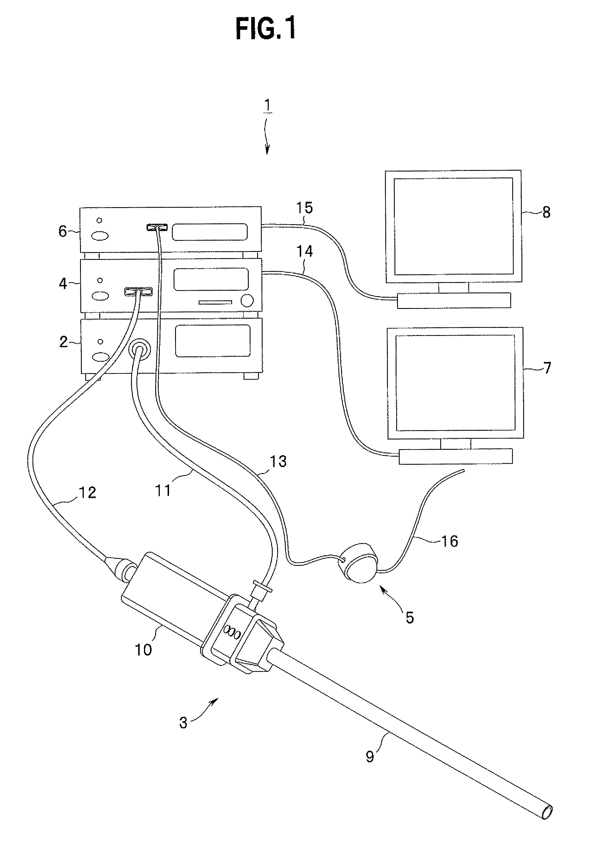
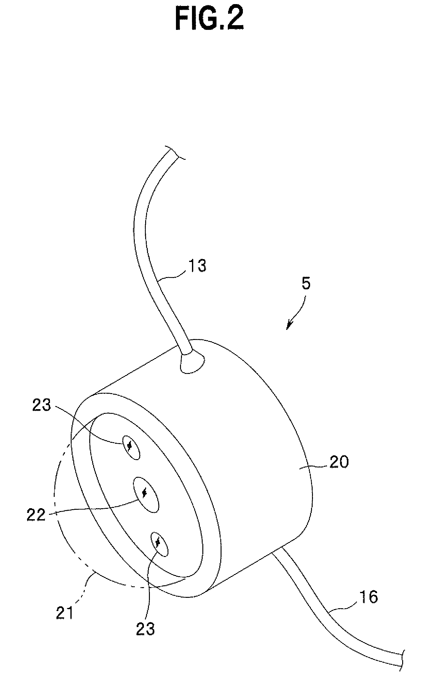
[0023]First, an endoscope system according to a first embodiment of the present invention is explained below. FIGS. 1 to 8 relate to the first embodiment of the present invention. FIG. 1 is a diagram showing a configuration of the endoscope system. FIG. 2 is a perspective view showing a configuration of an intra-body cavity set camera. FIG. 3 is a diagram showing a state in which the endoscope system is set on a patient. FIGS. 4 to 7 are diagrams for explaining a procedure for leading the intra-body cavity set camera into a body cavity. FIG. 8 is a diagram showing view angles of a rigid endoscope and the intra-body cavity set camera set in the body cavity.

[0024]As shown in FIG. 1, an endoscope system I according to the present embodiment for performing a laparoscopic surgical operation mainly includes a light source device 2, a rigid endoscope 3 as a first photographing device, a first camera control unit (hereinafter abbreviated as CCU) 4 as a first signal process...

second embodiment

(Second Embodiment)

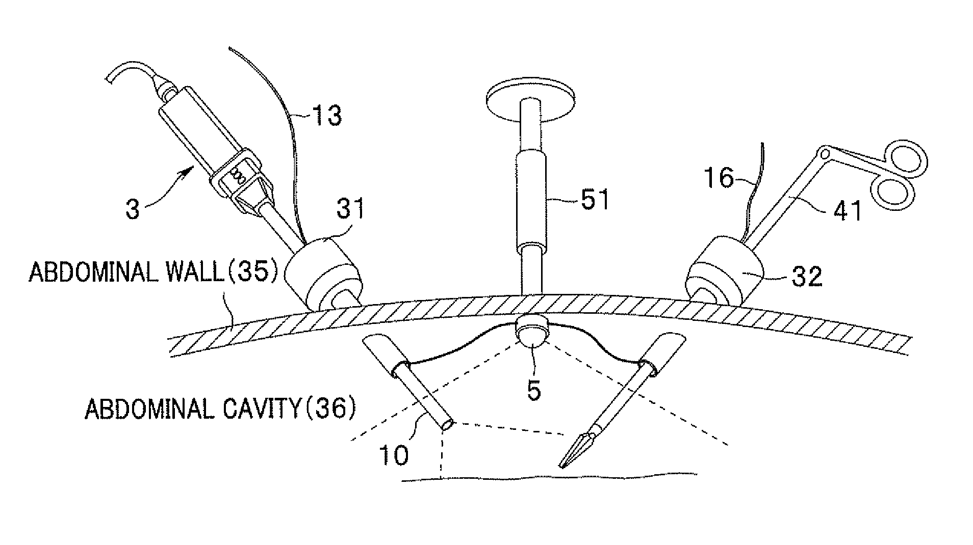
[0050]An endoscope system according to a second embodiment of the present invention is explained below with reference to FIGS. 9 and 10. FIGS. 9 and 10 relate to the second embodiment of the present invention. FIG. 9 is a diagram showing a state in which the endoscope system is set on a patient. FIG. 10 is a sectional view showing a state in which an intra-body cavity set camera is fixed by an abdominal cavity needle through an abdominal wall. In the following explanation, components identical with those of the endoscope system 1 according to the first embodiment are denoted by the same reference numerals. Detailed explanation of the components is omitted.

[0051]As shown in FIG. 9, the endoscope system 1 according to the present embodiment includes an abdominal cavity needle 51 that is a retaining instrument for fixing and retaining the camera 5 on an inner surface of the abdominal wall 35. As shown in FIG. 10, the abdominal cavity needle 51 includes a needle secti...

third embodiment

(Third Embodiment)

[0054]An endoscope system according to a third embodiment of the present invention is explained below with reference to FIGS. 11 and 12. FIGS. 11 and 12 relate to the third embodiment of the present invention. FIG. 11 is a diagram showing a state in which the endoscope system is set on a patient. FIG. 12 is a block diagram showing a state in which an intra-body cavity set camera and a receiving device are fixed by a magnetic force through an abdominal wall. In the following explanation, as in the above explanation, components identical with those of the endoscope system 1 according to the first embodiment are denoted by the same reference numerals. Detailed explanation of the components is omitted.

[0055]The endoscope system 1 according to the present embodiment transits an image of the inside of the abdominal cavity 36 photographed by the camera 5 to the outside of the abdominal cavity 36 by radio. Specifically, as shown in FIG. 11, the camera 5 according to the pr...


PUM


Abstract

An endoscope system having a first image pickup device including at least one image pickup unit that is capable of picking up an image of an object, a second image pickup device including at least one image pickup unit, the second image pickup device being different from the first image pickup device, a retaining member being provided for retaining the second image pickup device in a body cavity, and including a camera cable that is connected to the second image pickup device and transmits an image pickup signal obtained by the second image pickup device, and a wire connected to the second image pickup device, a signal processing device that is provided outside the body cavity and processes image pickup signals obtained by the first image pickup device and the second image pickup device, and a display device that displays an image signal outputted from the signal processing device.

Description

CROSS REFERENCE TO RELATED APPLICATION[0001]This application claims benefit of Japanese Patent Application No. 2007-157948 filed on Jun. 14, 2007, the contents of which are incorporated by this reference.BACKGROUND OF THE INVENTION[0002]1. Field of the Invention[0003]The present invention relates to an endoscope system that can extensively view the inside of an abdominal cavity and includes an image pickup device fixed on an inner side of the abdominal cavity.[0004]2. Description of the Related Art[0005]In recent years, a surgical operation for, without dissecting the abdomen of a patient in order to reduce invasion to a patient, penetrating, into the abdomen, a trocar that leads an endoscope for observation into a body cavity and a trocar that leads a treatment instrument to a treatment region and performing treatment while observing the treatment instrument and the treatment region with the endoscope, a so-called laparoscopic surgical operation, has been performed. With this metho...

Claims


Application Information

Patent Timeline
Patent Type & Authority Patents(United States)
IPC IPC(8): A61B1/04
CPCA61B1/0005A61B1/042A61B1/05A61B1/051A61B1/3132A61B1/0661A61B19/5202A61B2017/00283A61B2017/00221A61B2017/00734A61B2017/00876A61B2017/308A61B19/5212A61B90/30A61B90/361
Inventor HANDA, KEIJIKARASAWA, HITOSHIASADA, DAISUKE
Owner OLYMPUS CORP
Why Eureka
Try Eureka