Dual Action Aspiration Biopsy Needle

a biopsy needle and needle body technology, applied in the field of aspiration biopsy needles, can solve the problems of inability to immediately examine under microscope, high cost, and inability to perform a biopsy, and achieve the effect of improving the ability to collect cellular materials

Inactive Publication Date: 2005-04-28
MEDICAL DEVICE TECH
View PDF5 Cites 54 Cited by
  • Summary
  • Abstract
  • Description
  • Claims
  • Application Information

AI Technical Summary

Benefits of technology

[0005] There are three main types of biopsy procedures. In a first type, a conventional surgical incision is made and the patient's body is opened so that a surgeon may retrieve one or more large pieces of the tumor or lesion to be tested for malignancy. This type of biopsy is very invasive, expensive to perform, and requires a considerable recovery time. Inventive endeavors in the field have resulted in two improved procedures that substantially reduce the invasiveness of the biopsy procedure, as well as the expense of the procedure and the length of the recovery time.
[0009] More particularly, in the Wang device, a hook engages the pleural tissue and an outer cannula is advanced to cut off the hooked piece of pleural tissue. This two needle system is designed to reduce the chances of a pneumothorax and thus represents a significant improvement over earlier biopsy techniques. However, the diameter of the Wang needle as described is about 4.5 mm, which is considerably larger than an FNA biopsy needle of the novel type disclosed herein which is typically no larger than 20-22 gauge. The Wang needle is not designed as a fine needle aspiration device but is designed to cut off pieces of lung pleura. A large diameter needle like the Wang device measuring about 4.5 mm in diameter is inappropriate for fine needle biopsy procedures due to the high risk of complications from a large tissue puncture including bleeding, pneumothorax and bile leakage.
[0014] The third technique is called fine needle aspiration (FNA) biopsy and is performed with an FNA biopsy needle. An FNA biopsy needle scrapes cells from the tumor or lesion that are so small as to be invisible to the unaided eye. Advantageously, the cellular material is already very thin when it is collected so there is no need to slice it to a thinner size prior to viewing it on a microscope slide because light can already pass through the thin cell layer. The cellular material is deposited onto a microscope slide directly from the FNA biopsy needle, stained, and viewed so that there is less time to the final biopsy report because the slicing machine and all of the handling steps necessitated thereby are eliminated. Trauma to the patient is greatly reduced because of smaller needle diameter and because cells are scraped instead of tissue being cut in thick pieces. Complication risk is also minimized or eliminated due to smaller nozzle diameters.
[0019] Although FNA biopsy needles represent a significant improvement over more invasive procedures for performing biopsies on suspect lesions or tumors, there remains a need for an improved FNA biopsy needle that increases the amount of sample that may be recovered per entrance into the lesion with one needle insertion so that multiple insertions need not be performed to complete a biopsy procedure.
[0021] The longstanding but heretofore unfulfilled need for a biopsy needle having enhanced cellular material collection capability is now met by a new, useful, and nonobvious invention. The novel aspiration biopsy needle includes a single needle of elongate, hollow construction having a proximal end and a beveled distal end. The beveled distal end of the needle is open and therefore forms a first sharp edge adapted to scrape cellular material when the needle is inserted into tissue. The needle is displaced from a proximal position to a distal position during insertion. The needle has a uniform diameter along a substantial part of its extent. A slot is formed in the needle near the beveled distal end; the slot is transversely disposed relative to a longitudinal axis of the needle. The slot is also angled relative to a transverse axis of the needle such that a bottom of the slot is positioned distal to an opening of the slot. The opening of the slot includes a second sharp edge adapted to scrape tissue of cellular size when the needle is displaced from a distal position to a proximal position. The novel structure further includes conventional means for applying a vacuum to a proximal end of the needle so that cellular material removed by the first sharp edge during proximal-to-distal travel of the needle is pulled into a lumen of the needle and so that cellular material removed by the second sharp edge during distal-to-proximal travel of the needle is also pulled into the lumen. However, the novel structure also has utility when no vacuum means is employed, i.e., even if it is not affixed to a syringe barrel.
[0031] In additional embodiments, the second sharp edge is mounted for pivotal movement about a transversely disposed hinge. The hinge enables the second sharp edge to open wider relative to its non-hinged position so that it may scrape off larger numbers of cellular material as the needle is displaced in a distal-to-proximal direction.

Problems solved by technology

This type of biopsy is very invasive, expensive to perform, and requires a considerable recovery time.
They cannot be immediately examined under a microscope because they are too thick for light to pass therethrough.
Accordingly, histopathological examination and diagnosis of breast lesions may be much more time-consuming than the histopathological examination and diagnosis of other types of lesions.
However, the need to slice the specimens causes a delay in providing the diagnosis, due to processing.
Additionally, the diameter of core biopsy needles are typically larger than the diameter of fine needle aspiration (FNA) needles, thereby increasing the risk of procedure-related complications including bleeding, pneumothorax, and bile leakage.
A large diameter needle like the Wang device measuring about 4.5 mm in diameter is inappropriate for fine needle biopsy procedures due to the high risk of complications from a large tissue puncture including bleeding, pneumothorax and bile leakage.
Thus, the Wang needle collects relatively large samples in a knifing action, and performs no scraping action capable of collecting samples at the cellular level.
Neither Wang nor any other known two needle systems include both an open distal end and a port for cutting tissue nor is any core biopsy system capable of collecting samples of cellular thickness.
Sometimes, however, the FNA biopsy procedure fails to collect a sample of sufficient size to enable definitive pathological analysis.
When that happens, the physician must repeat the procedure, causing additional trauma to the body part undergoing biopsy and creating an additional risk of an adverse event.

Method used


Image

  • Dual Action Aspiration Biopsy Needle
  • Dual Action Aspiration Biopsy Needle
  • Dual Action Aspiration Biopsy Needle

Examples


first embodiment

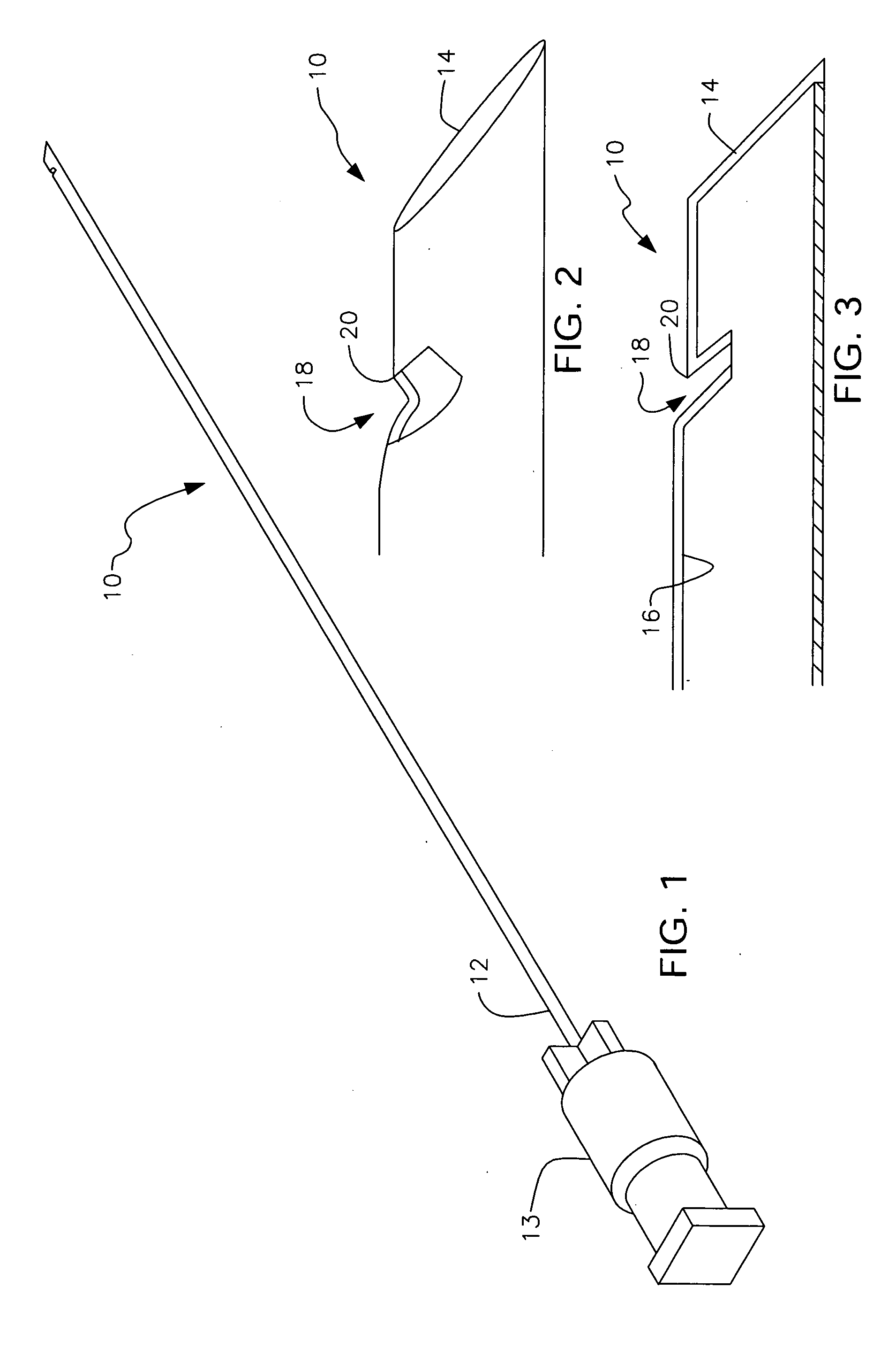
[0062] As perhaps best depicted in FIG. 3, second sharp edge 20 is coincident or flush with the exterior surface of needle 10 in this

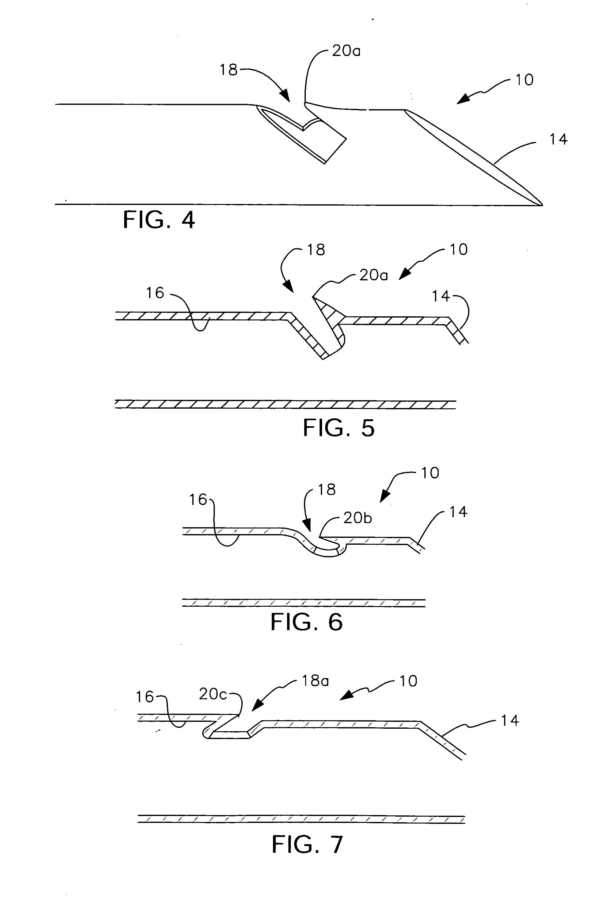
[0063]FIG. 4 provides a perspective view of a second embodiment of needle 10 where second sharp edge 20a is elevated with respect to the exterior surface of needle 10 and FIG. 5 provides a longitudinal sectional view of said second embodiment. The protrusion of second sharp edge 20a above the exterior surface of the needle ensures that the cellular material collected when using this second embodiment should be greater than the amount of cellular material collected when using the first embodiment.

third embodiment

[0064]FIG. 6 provides a longitudinal sectional view of a third embodiment where second sharp edge 20b is recessed with respect to said exterior surface. The lesion, not shown, is under compression as needle 10 penetrates it. Accordingly, an amount of tissue will enter into slot 18 and be scraped off during distal-to-proximal travel of needle 10, even though sharp edge 20b is recessed with respect to the exterior surface of the needle.

[0065]FIG. 7 depicts a fourth embodiment where a slot 18a is formed transversely to the longitudinal axis of needle 10 as in the first embodiment, but the axis of symmetry of slot 18a is normal to the axis of symmetry of slot 18. In other words, the bottom of slot 18a is proximal to the open upper end of slot 18a. Accordingly, a third sharp edge, denoted 20c, is formed. Third sharp edge 20c is auxiliary to first sharp edge 14 in that said third sharp edge scrapes cellular material from a lesion during proximal-to-distal travel of needle 10.

[0066]FIGS. ...


PUM


Abstract

A dual action biopsy needle scrapes tissue of cellular thickness from a lesion during proximal-to-distal travel of the needle along its longitudinal axis of symmetry. A first sharp edge, formed by a beveled distal end of the needle, scrapes tissue during proximal-to-distal travel of the needle. A transversely disposed slot formed in the needle near the first sharp edge provides a second sharp edge. A bottom of the slot is proximal to the opening of the slot. The second sharp edge also scrapes tissue during proximal-to-distal travel of the needle. The second sharp edge is coincident with an exterior surface of the needle. A vacuum pulls the scraped cellular material into the lumen of the needle.

Description

CROSS-REFERENCE TO RELATED DISCLOSURES [0001] This disclosure is a divisional application claiming the benefit of the filing date of pending U.S. patent application Ser. No. 10 / 708,710, filed Mar. 19, 2004 by the same inventor and having the same title, which is a pending divisional U.S. patent application claiming the benefit of the filing date of U.S. patent application Ser. No. 10 / 065,155, now U.S. Pat. No. 6,709,408, filed Sep. 23, 2002 by the same inventor and having the same title, which is a continuation-in-part of U.S. patent application Ser. No. 09 / 682,252, now abandoned, filed Aug. 9, 2001 by the same inventor and having the same title.BACKGROUND OF THE INVENTION [0002] 1. Field of the Invention [0003] This invention relates, generally, to aspiration biopsy needles. More particularly, it relates to an aspiration biopsy needle having an enhanced cellular material collection capability. [0004] 2. Description of the Prior Art [0005] There are three main types of biopsy proced...

Claims


Application Information

Patent Timeline
Patent Type & Authority Applications(United States)
IPC IPC(8): A61B10/00A61B10/02A61B10/04
CPCA61B10/0266A61B10/04A61B10/0283A61B10/0275
Inventor FISHER, JOHN S.
Owner MEDICAL DEVICE TECH
Why Eureka
Try Eureka