Peptide homing to brain tumors
a brain tumor and peptide technology, applied in the field of peptides, can solve the problems of poor prognosis, poor treatment effect, and difficulty in detecting low-grade astrocytomas
- Summary
- Abstract
- Description
- Claims
- Application Information
AI Technical Summary
Benefits of technology
Problems solved by technology
Method used
Image
Examples
example 1
Homing of the Phage Displaying Peptide Sequence CGLSGLGVA to the Intracranial Islet Tumors
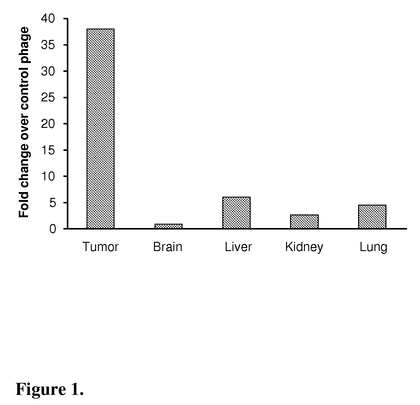
[0033]CGLSGLGVA or a control phage (109 pfu=plaque forming unit) were injected into the tail vein of intracranial HIF-1alfa KO (HIFko) tumor-bearing mice. After 15 min of circulation the mice were perfused through the heart with saline to remove unbound phage followed by excision of tumor containing brain, normal brain, liver, kidney and lungs. The number of phage homed to those organs were determined by titration. FIG. 1 shows as a bar graph that the peptide very specifically homes to the intracranial, early stage astrocytoma model (HIFko) that grows as islets and harbors co-opted tumor vessels in the brain.
example 2
Homing of the Fluorescein-Conjugated CGLSGLGVA Peptide to the Intracranial Islet Tumors
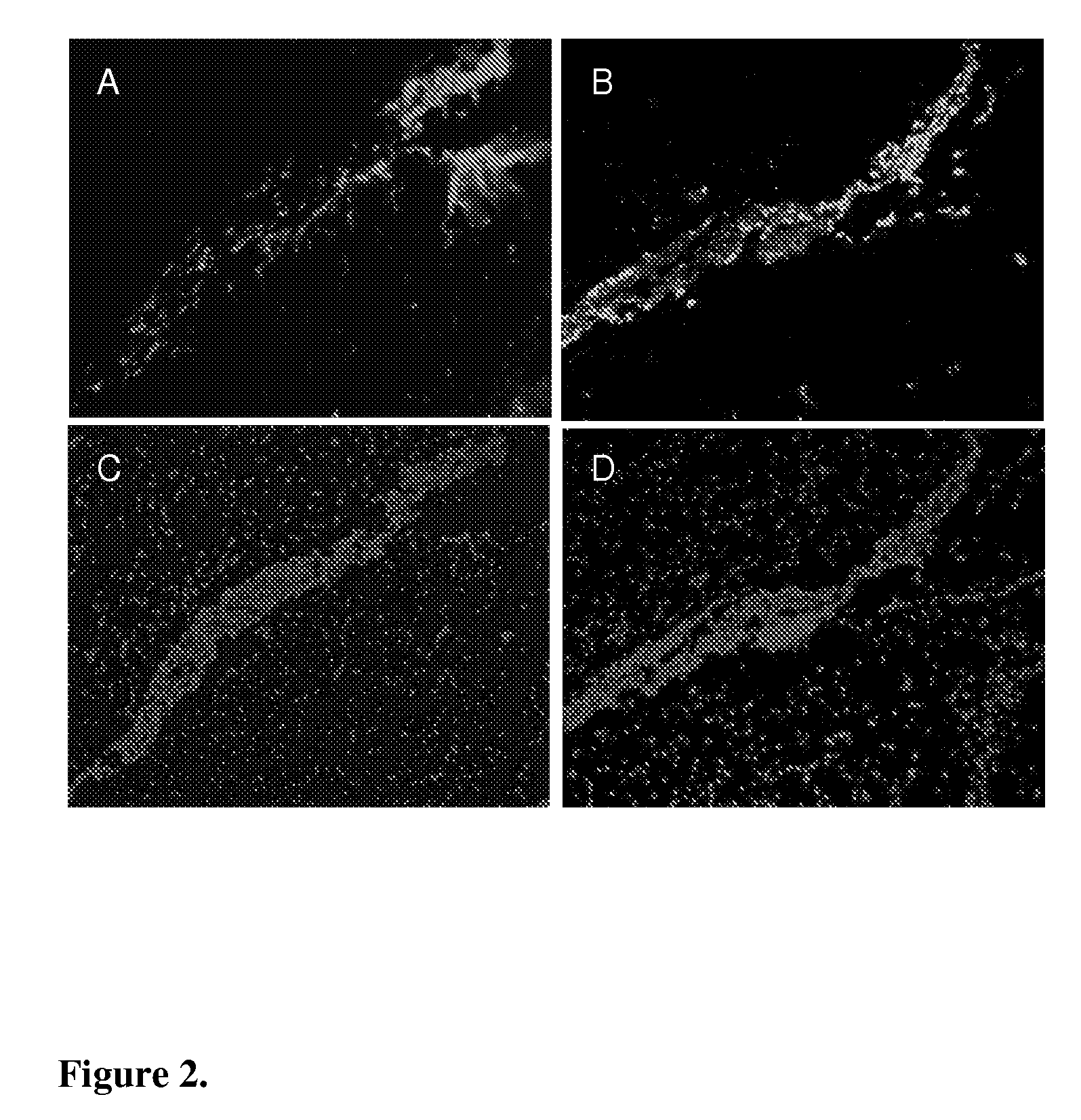
[0034]Fluorescein-conjugated CGLSGLGVA peptide (10 μg) was injected into tail vein of HIFko tumor-bearing mice. After 60 min of circulation tumor-containing brain was excised and prepared for histological analysis. Peptide was visualized by using an antibody against fluorescein (rabbit anti-FITC, Zymed Laboratories). FIG. 2 shows that the peptide (red in FIG. 2A) was detected only in the tumor islets and not in the surrounding normal brain tissue. Presence of the tumor cells was confirmed by staining for the SV40 T-antigen specific for tumor cells from a subsequent section (green in FIG. 2B). FIGS. 2C and 2D show the same microscopic field as 2A and 2B. Nuclei were visualized by DAPI staining (blue).
example 3
Fluorescein-Conjugated CGLSGLGVA Peptide Does Not Home to the Intracranial VEGF-Overexpressing Tumors
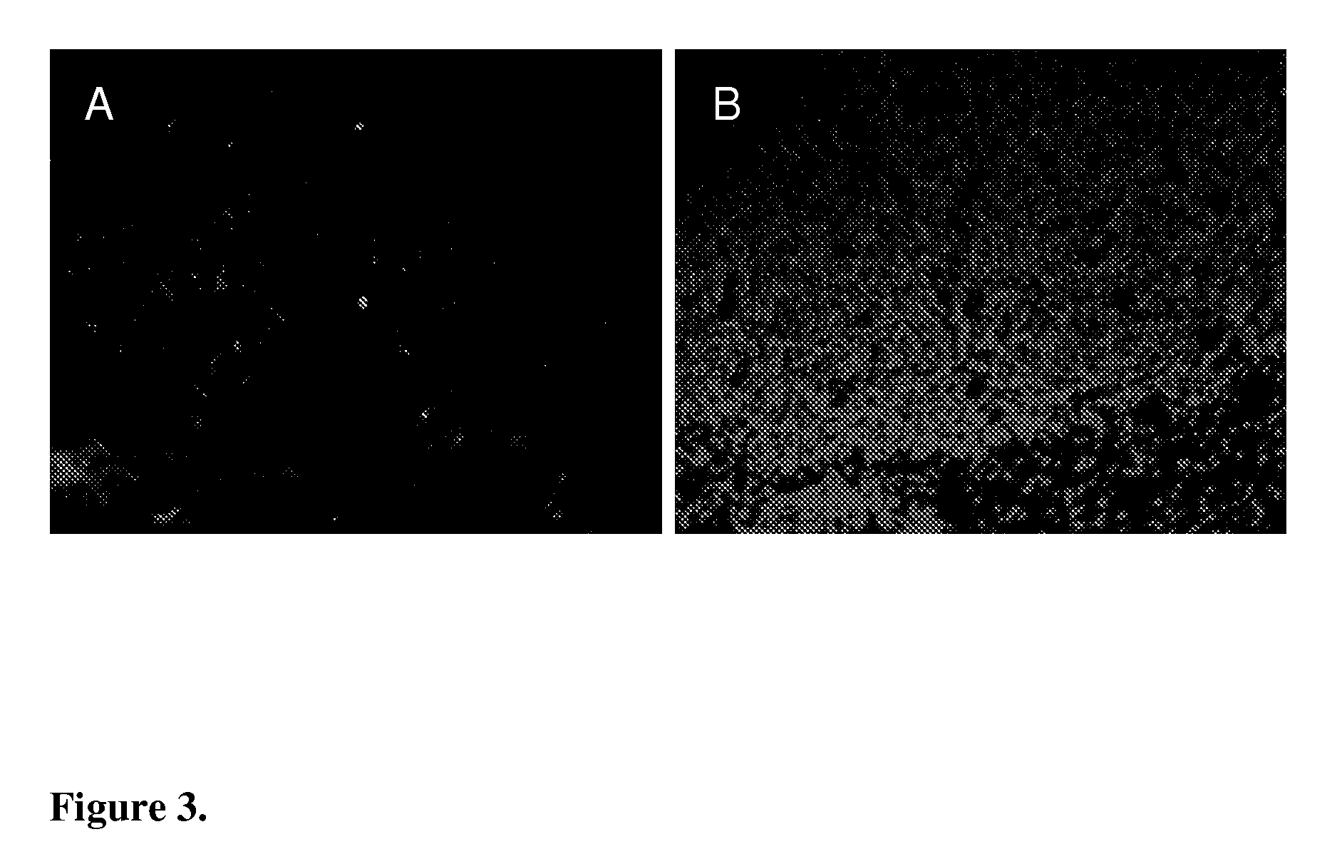
[0035]Fluorescein-conjugated CGLSGLGVA peptide (10 μg) was injected into the tail vein of VEGF overexpressing (VEGF+) tumor-bearing mice. After 60 min of circulation tumor was excised and prepared for histological analysis. Peptide was visualized by using an antibody against fluorescein (rabbit anti-FITC, Zymed Laboratories). FIG. 3A. Peptide was not detected in the tumor tissue. 3B: Same microscopic field as in 3A. Nuclei were visualized by DAPI staining (blue).
PUM
| Property | Measurement | Unit |
|---|---|---|
| imaging | aaaaa | aaaaa |
| distances | aaaaa | aaaaa |
| size | aaaaa | aaaaa |
Abstract
Description
Claims
Application Information
 - R&D
- Intellectual Property
- Life Sciences
- Materials
- Tech Scout
- Unparalleled Data Quality
- Higher Quality Content
- 60% Fewer Hallucinations
Browse by: Latest US Patents, China's latest patents, Technical Efficacy Thesaurus, Application Domain, Technology Topic, Popular Technical Reports.
© 2025 PatSnap. All rights reserved.Legal|Privacy policy|Modern Slavery Act Transparency Statement|Sitemap|About US| Contact US: help@patsnap.com


