Breast tissue density measure

a breast tissue density and measurement technology, applied in the field of breast tissue density measurement, can solve the problems of difficult breast cancer detection, room for error or misjudgment, etc., and achieve the effect of more accurate and sensitive measurement of breast density changes

Active Publication Date: 2012-07-10
BIOCLINICA
View PDF8 Cites 9 Cited by
  • Summary
  • Abstract
  • Description
  • Claims
  • Application Information

AI Technical Summary

Problems solved by technology

Detection of breast cancer can be very difficult, since the first signs of breast cancer are often asymptomatic.
Naturally, where human intervention is required, there is room for error or misjudgment.
A currently frequently discussed issue related to breast cancer risk is the potential influence of hormone replacement therapy taken after the menopause.

Method used


Image

  • Breast tissue density measure
  • Breast tissue density measure
  • Breast tissue density measure

Examples


Embodiment Construction

[0099]We shall describe first an embodiment of the invention based on classifying pixels of an image using a trained classifier which has been trained by unsupervised learning.

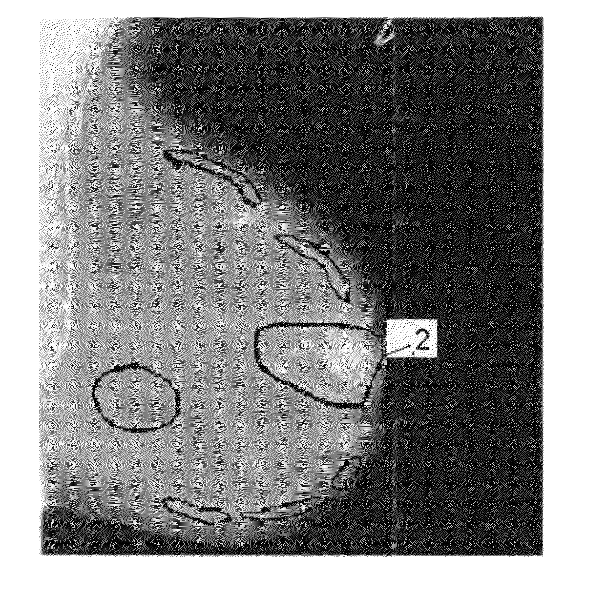
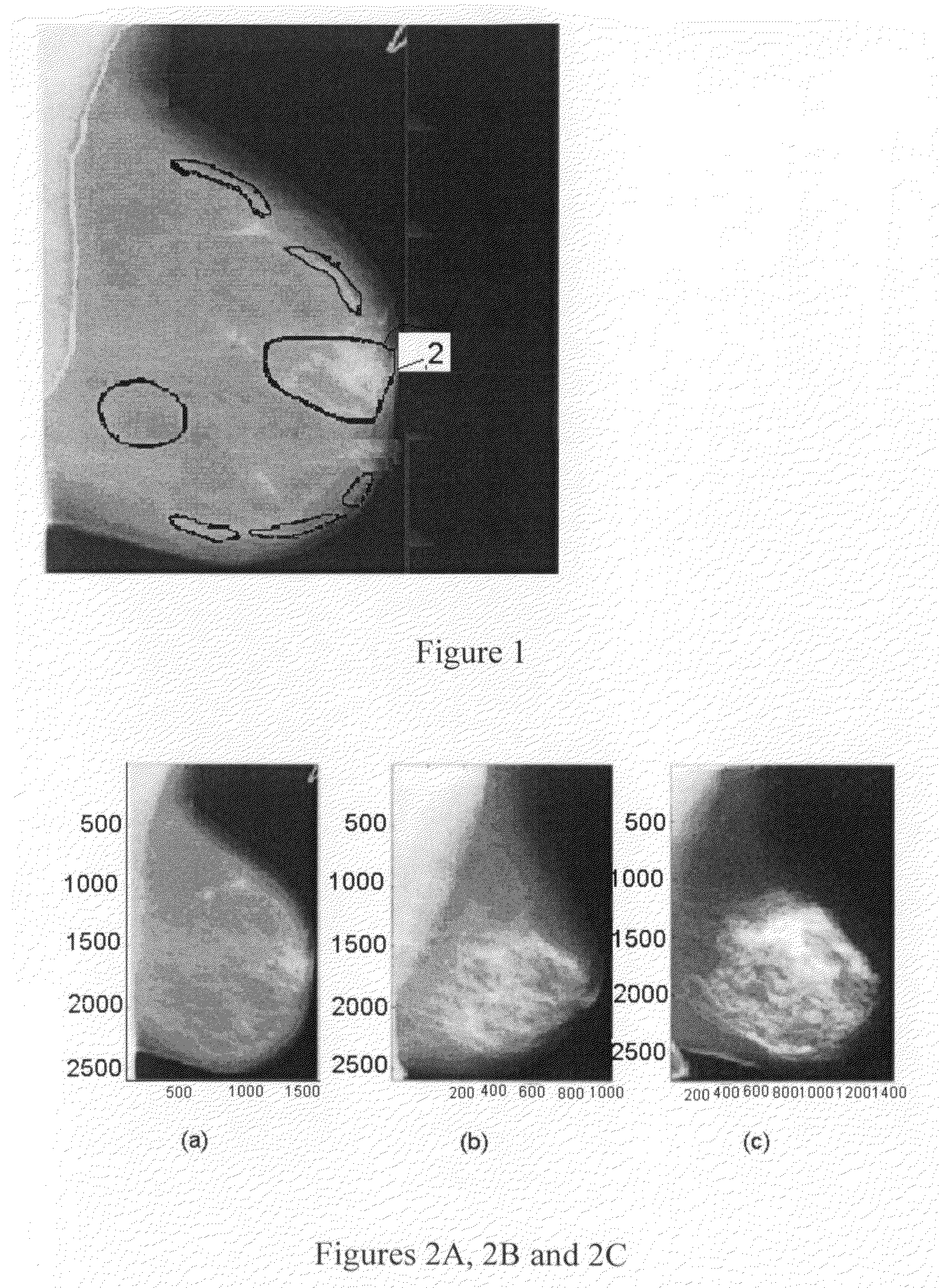
[0100]Numerous studies have investigated the relation between mammographic density and breast cancer risk, and women with high breast density appear to have a four to six fold increase in breast cancer risk. Therefore the density is an important feature embedded in a mammogram. In this context and as shown in FIG. 1, the density refers to a specialist's assessment (typically a radiologist) of the projected area 2 of fibro glandular tissue—sometimes called dense tissue.

[0101]As an example, FIGS. 2A to 2C respectively show three example mammograms depicting low, medium and high mammographic densities.

[0102]Typically a mammogram is classified into one of four or five density categories, e.g. Wolfe patterns and BI-RADS. These classifications are subjective and sometimes crude. They may be sufficient in some cases ...


PUM


Abstract

A method of processing a mammogram image to derive a value for a parameter useful in detecting differences in breast tissue in subsequent images of the same breast or relative to a control group of such images, said derived parameter being an aggregate probability score reflecting the probability of the image being a member of a predefined class of mammogram images, comprises computing for each of a multitude of pixels within a large region of interest within the image a pixel probability score assigned by a trained statistical classifier according to the probability of said pixel belonging to an image belonging to said class, said pixel probability being calculated on the basis of a selected plurality of features of said pixels, and computing said parameter by aggregating the pixel probability scores over said region of interest. Said features may include the 3-jet of said pixels.

Description

CROSS-REFERENCE TO RELATED APPLICATIONS[0001]This is a continuation in part of application Ser. No. 12 / 223,550 filed 9 Feb. 2007.BACKGROUND OF THE INVENTION[0002]The present invention relates to a method of detecting differences in breast tissue in subsequent images of the same breast.[0003]Breast cancer is one of the largest serious diseases among women in the western world. It is the most common cancer in women accounting for nearly one out of every three cancers diagnosed in the United States. It is also the most common and deadly cancer for women on a global scale, where breast cancer accounts for 21% of all cancer cases and 14% of all cancer deaths.[0004]However, if detected sufficiently early, there is a high probability of survival. Detection of breast cancer can be very difficult, since the first signs of breast cancer are often asymptomatic.[0005]Mammograms have thus far been found to be the most effective way to detect breast cancer early, sometimes up to two years before ...

Claims


Application Information

Patent Timeline
Patent Type & Authority Patents(United States)
IPC IPC(8): G06T7/00G06V10/52
CPCG06K9/527G06K9/6223G06V10/52G06V10/763G06F18/23213
Inventor RAUNDAHL, JAKOBLOOG, MARCONIELSEN, MADS
Owner BIOCLINICA
Why Eureka
Try Eureka