Methods, systems, and computer-program products for estimating scattered radiation in radiographic projections

a radiographic projection and radiographic technology, applied in material analysis using wave/particle radiation, instruments, nuclear engineering, etc., can solve the problems of numerical errors in image reconstruction algorithms, phantom images, loss of resolution and contrast in ct image slices produced,

Active Publication Date: 2012-12-04
VARIAN MEDICAL SYSTEMS
View PDF6 Cites 32 Cited by
  • Summary
  • Abstract
  • Description
  • Claims
  • Application Information

AI Technical Summary

Problems solved by technology

The scattered radiation causes artifacts (e.g., phantom images) and loss of resolution and contrast in the CT image slices produced by the radiation imaging system.
The scattered radiation can also cause numerical errors in the image reconstruction algorithms (generally referred to as “CT number problems” in the art).
All of the foregoing lead to image degradation.

Method used


Image

  • Methods, systems, and computer-program products for estimating scattered radiation in radiographic projections
  • Methods, systems, and computer-program products for estimating scattered radiation in radiographic projections
  • Methods, systems, and computer-program products for estimating scattered radiation in radiographic projections

Examples


Embodiment Construction

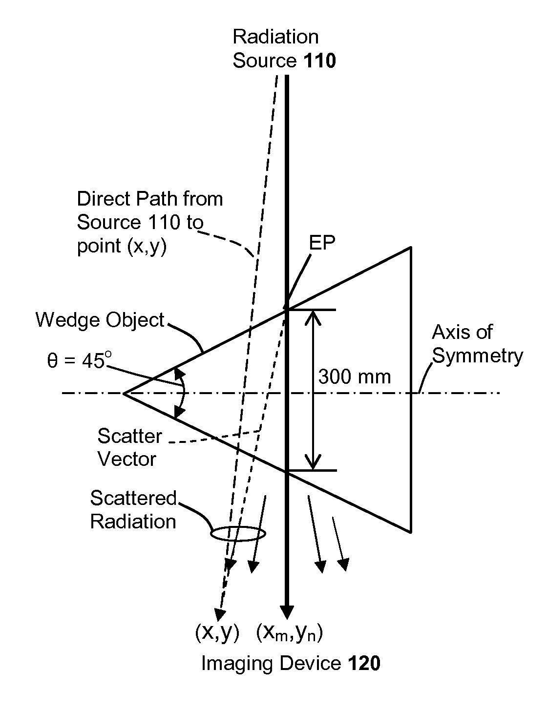
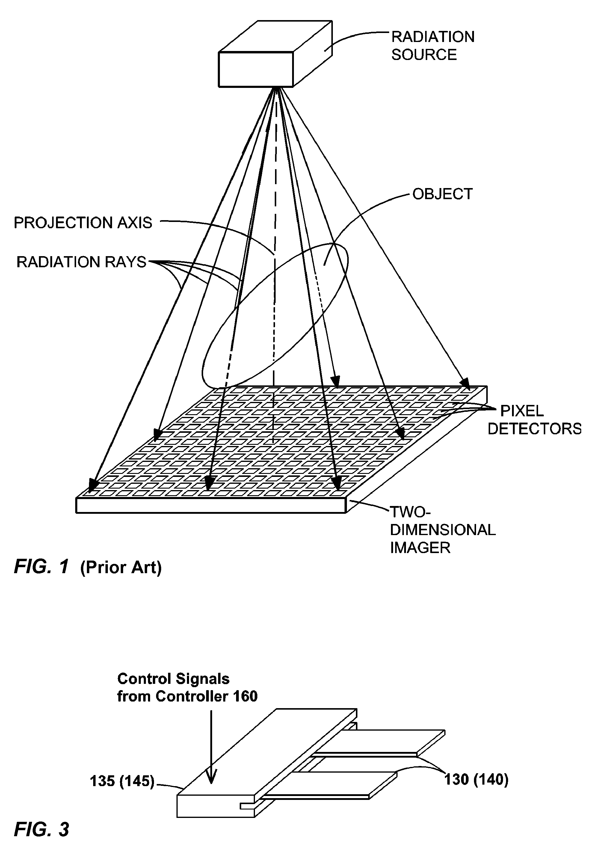
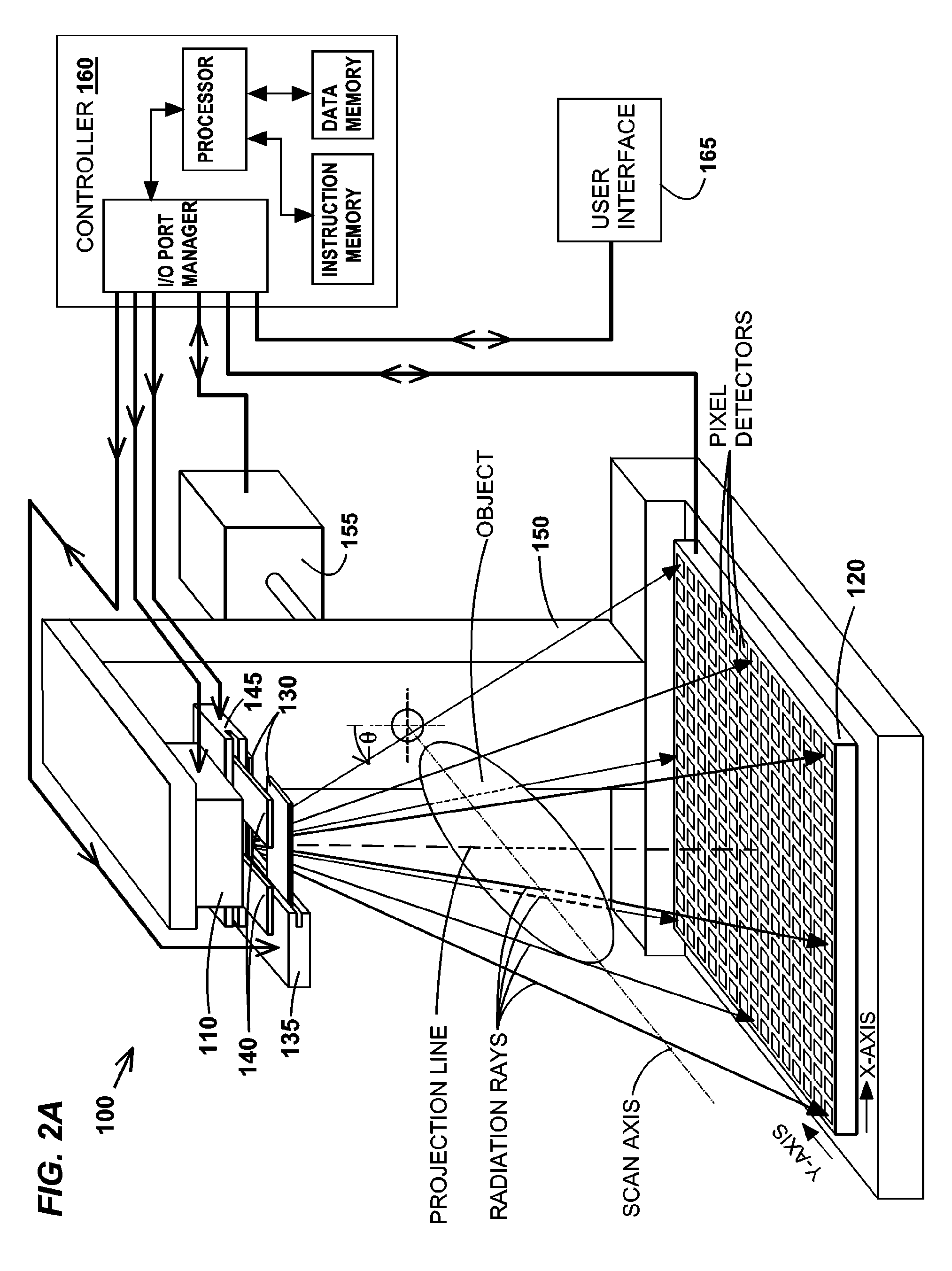
[0045]System Overview. FIG. 2A is a schematic diagram of a first exemplary imaging system 100 according to the system inventions of the present application. System 100 comprises a radiation source 110, a two-dimensional imaging device 120 disposed opposite to radiation source 110 along a projection line, a first set of fan blades 130 disposed between the radiation source and the two-dimensional imaging device, a first fan-blade drive 135 that holds fan blades 130 and sets their positions. The edges of fan blades 130 are oriented substantially perpendicular to the scan axis (defined below), and are substantially parallel with the trans-axial dimension (defined below) of imaging device 120. As an option, system 100 may further comprise a second set of fan blades 140 disposed between the radiation source and the two-dimensional imaging device, and a second fan-blade drive 145 that holds fan blades 140 and sets their positions. The edges of fan blades 140 are oriented substantially para...


PUM


Abstract

Several related inventions for estimating scattered radiation in radiographic projections are disclosed. Several of the inventions use scatter kernels of various forms, including symmetric and asymmetric forms. The inventions may be used alone or in various combinations with one another. The resulting estimates of scattered radiation may be used to correct the projections, which can improve the results of tomographic reconstructions. Still other inventions of the present application generate estimates of scattered radiation from shaded or partially shaded regions of a radiographic projection, which may be used to correct the projections or used to adjust the estimates of scattered radiation generated according to inventions of the present application that employ kernels.

Description

CROSS-REFERENCES TO RELATED APPLICATIONS[0001]Not ApplicableBACKGROUND OF THE INVENTION[0002]Computerized tomography (CT) involves the imaging of the internal structure of an object by collecting several projection images (“radiographic projections”) in a single scan operation (“scan”), and is widely used in the medical field to view the internal structure of selected portions of the human body. Typically, several two-dimensional projections are made of the object, and a three-dimensional representation of the object is constructed from the projections using various tomographic reconstruction methods. From the three-dimensional image, conventional CT slices through the object can be generated. The two-dimensional projections are typically created by transmitting radiation from a “point source” through the object, which will absorb some of the radiation based on its size, density, and atomic composition, and collecting the non-absorbed radiation onto a two-dimensional imaging device,...

Claims


Application Information

Patent Timeline
Patent Type & Authority Patents(United States)
IPC IPC(8): G06K9/00G01B15/02G01N23/00G01N23/201
CPCG06T11/005G06T7/0012
Inventor STAR-LACK, JOSHSUN, MINGSHANPAVKOVICH, JOHN MILAN
Owner VARIAN MEDICAL SYSTEMS
Why Eureka
Try Eureka