Method for optically imaging an interior of a turbid medium, method for reconstructing an image of an interior of a turbid medium, device for imaging an interior of a turbid medium, medical image acquisition device and computer program

a technology of optical imaging and interior, which is applied in the field of optical imaging an interior of a turbid medium, can solve the problems of not all of the excitation light excites the fluorescent agent, complex and time-consuming, and difficult to achieve the effect of enabling examination, improving the quality and enhancing the contrast of the image reconstructed according to the method according to the invention

Inactive Publication Date: 2013-02-12
KONINKLIJKE PHILIPS ELECTRONICS NV
View PDF16 Cites 17 Cited by
  • Summary
  • Abstract
  • Description
  • Claims
  • Application Information

AI Technical Summary

Benefits of technology

[0018]An embodiment of the method according to the invention is characterized in that the dataset, as a function of a chosen variable, has a measured trend, in that the input assumption requires a dataset to have a required trend as a function of the chosen variable, and in that the dataset is changed into the further dataset by mapping the dataset onto the required trend. This embodiment has the advantage that it enables a dataset to be changed into a further dataset that satisfies an input assumption underlying the image reconstruction algorithm in situations in which the input assumption implies that there is a specific dependence, for instance, a linear dependence of the dataset on the chosen variable.
[0040]a data processing unit for carrying out the method according to any one of the previous embodiments. This embodiment has the advantage that a medical image acquisition device would benefit from any one of the previous embodiments of the method according to the invention.

Problems solved by technology

However, this is often very complex and time-consuming.
However, not all of the excitation light will actually excite the fluorescent agent.

Method used


Image

  • Method for optically imaging an interior of a turbid medium, method for reconstructing an image of an interior of a turbid medium, device for imaging an interior of a turbid medium, medical image acquisition device and computer program
  • Method for optically imaging an interior of a turbid medium, method for reconstructing an image of an interior of a turbid medium, device for imaging an interior of a turbid medium, medical image acquisition device and computer program
  • Method for optically imaging an interior of a turbid medium, method for reconstructing an image of an interior of a turbid medium, device for imaging an interior of a turbid medium, medical image acquisition device and computer program

Examples


Embodiment Construction

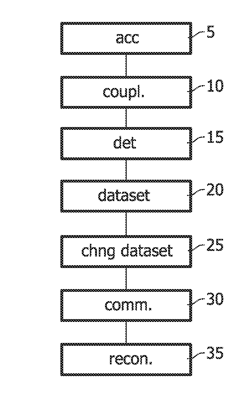
[0051]FIG. 1 illustrates an embodiment of the method of imaging an interior of a turbid medium according to the invention. In step 5 the turbid medium is accommodated inside a receiving volume. Next, in step 10, light from a light source is coupled into the receiving volume. The light from the light source is chosen to excite a fluorescent agent comprised in the turbid medium. Alternatively, the light from the light source may be chosen such that the light is able to propagate through the turbid medium without giving rise to fluorescence light. Light emanating from the receiving volume as a result of coupling light from the light source into the receiving volume is detected through use of a photodetector unit. This is done in step 15. On the basis of the detected light a dataset is obtained in step 20. The dataset may, for instance, comprised data relating to the intensity of fluorescence light emanating from the receiving volume as a function of the distance between the position at...


PUM


Abstract

The invention relates to a method of imaging an interior of a turbid medium, a device for imaging an interior of a turbid medium, and a medical image acquisition device. A turbid medium is accommodated in a receiving volume (5), light from a light source is coupled into (10) and out of the receiving volume and detected (15) after which a dataset is obtained from the detected light (20). The dataset is then communicated to an image reconstruction algorithm (30) and an image of an interior of the turbid medium is reconstructed based on the detected light (35). According to the invention the dataset is changed prior to communicating the dataset to the image reconstruction algorithm into a further dataset (25), with a further dataset satisfying an input assumption underlying the image reconstruction algorithm.

Description

FIELD OF INVENTION[0001]The invention relates to a method for optically imaging an interior of a turbid medium employing an image reconstruction algorithm[0002]The invention also relates to a method for reconstructing an image of an interior of a turbid medium.[0003]The invention also relates to a device for imaging an interior of a turbid medium.[0004]The invention also relates to a medical image acquisition device.[0005]The invention also relates to computer program products for use in the abovementioned methods and devices.BACKGROUND OF THE INVENTION[0006]An embodiment of methods and devices of this kind is known from European patent application 05111164.9. The described method and device can be used for imaging an interior of a turbid medium, such as biological tissue using diffuse optical tomography. In medical diagnostics, the method and device may be used for imaging, for instance, an interior of a female breast. The receiving volume receives a turbid medium, such as a breast...

Claims


Application Information

Patent Timeline
Patent Type & Authority Patents(United States)
IPC IPC(8): G06K9/00
CPCA61B5/0091A61B5/4312
Inventor JOCHEMSEN, ROBERTNIELSEN, TIM
Owner KONINKLIJKE PHILIPS ELECTRONICS NV
Why Eureka
Try Eureka