System and method for segmentation of anatomical structures in MRI volumes using graph cuts

a technology of graph cuts and anatomical structures, applied in the field of 3d segmentation of anatomical structures of the brain in mri volumes, can solve the problems of poor contrast, inability to reliably segment image segmentation methods such as active contours or region growth, and difficulty in segmentation of anatomical structures of the brain

Inactive Publication Date: 2013-02-19
SIEMENS MEDICAL SOLUTIONS USA INC
View PDF22 Cites 10 Cited by
  • Summary
  • Abstract
  • Description
  • Claims
  • Application Information

AI Technical Summary

Problems solved by technology

The segmentation of the anatomical structures of the brain can be difficult due to problems such as lack of boundaries between the anatomical structures, poor contrast in medical images of the brain, and noise in the images, which is mainly attributed to the image acquisition systems (e.g., MRI) and partial volume effects.
Accordingly, because of such problems, image segmentation methods such as active contours or region growing are subject to leakage issues and are not reliable.
For an atlas to be accurate, the atlas typically must be very complex.
Although these techniques can be successful, there typically is a high computational cost and it is difficult to construct an accurate atlas.
Thus, these techniques can be time consuming and expensive.
In this technique, it can be difficult for a user to accurately mark the object and background.
Accordingly, graph cuts segmentation techniques can lead to erroneous segmentations.

Method used


Image

  • System and method for segmentation of anatomical structures in MRI volumes using graph cuts
  • System and method for segmentation of anatomical structures in MRI volumes using graph cuts
  • System and method for segmentation of anatomical structures in MRI volumes using graph cuts

Examples


Embodiment Construction

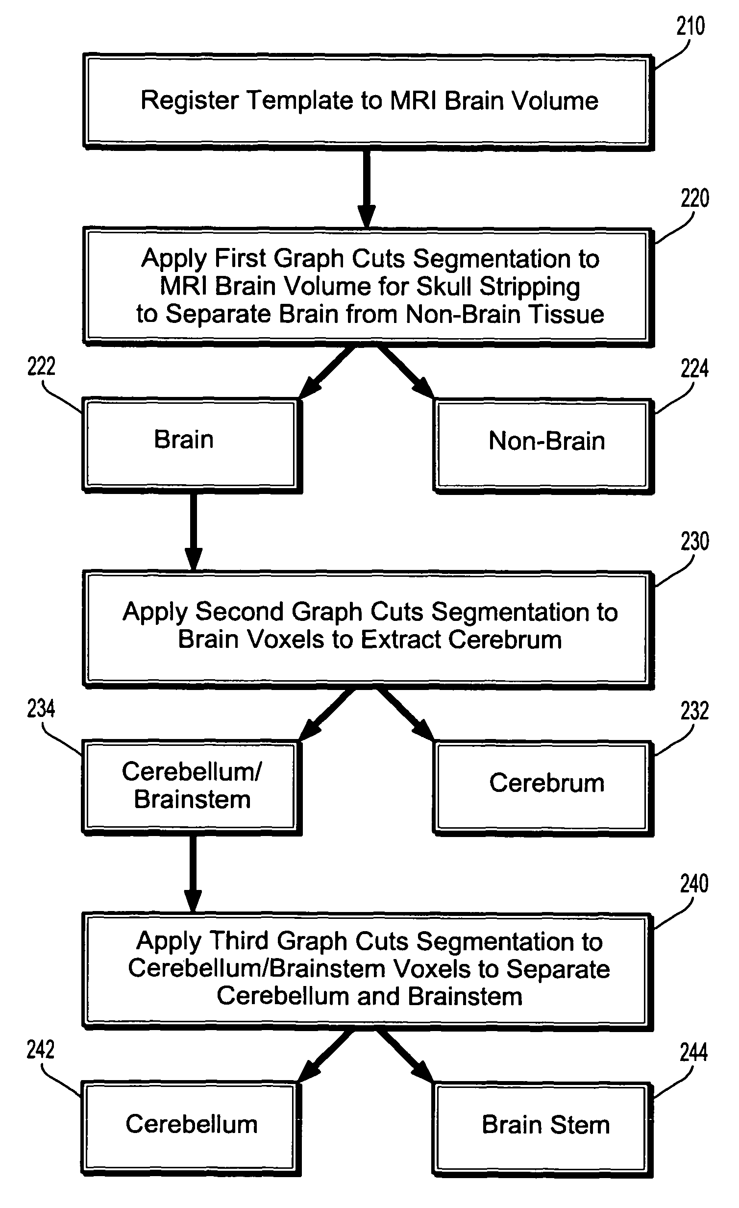
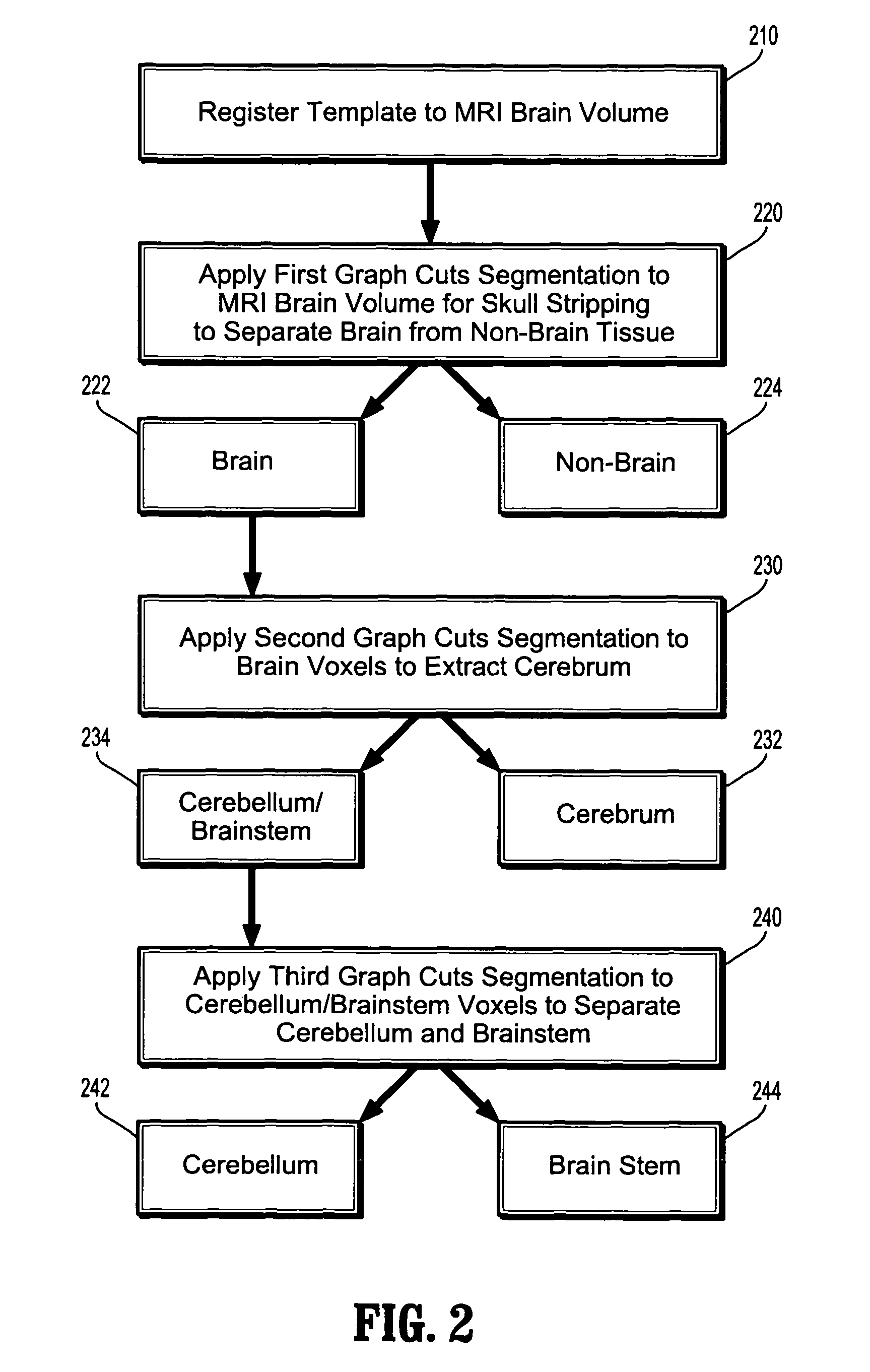
[0017]According to an embodiment of the present invention, anatomical structures are segmented in MRI volume data using graph cuts based on an anatomical template. As described herein, the method is implemented to segment anatomical brain structures in MRI brain volumes, however, the present invention is not limited thereto, and may be applied to other types of anatomical structures in various regions of the body as well.

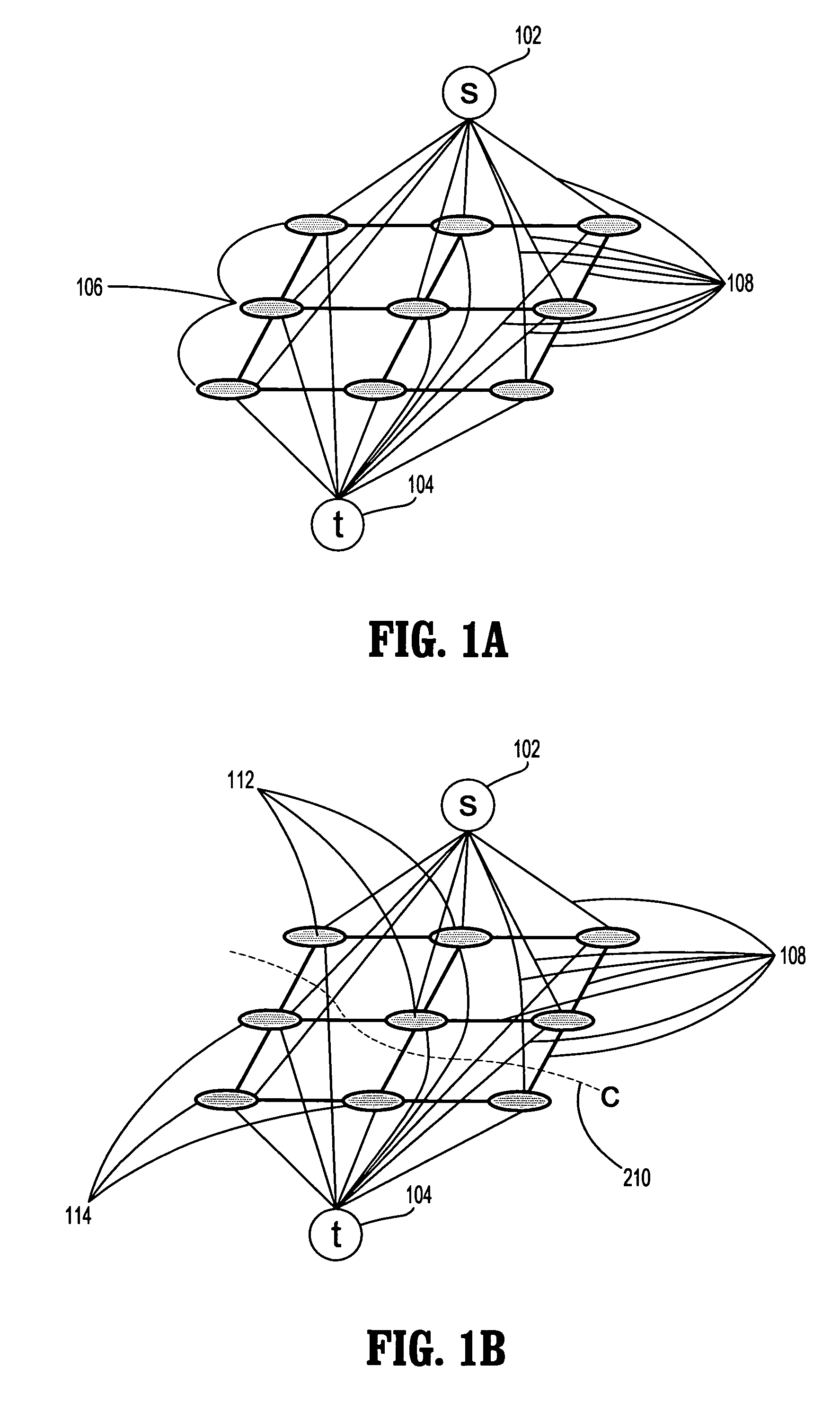
[0018]Before discussing specific aspects of the graph cut segmentation algorithm using an anatomical template, graph cut theory will be discussed. In particular, an undirected graph G=V,E consists of vertices V and undirected edges E that connect the vertices. Each edge eεE is assigned a non-negative cost c. There are two special vertices (referred to herein as “terminals”) in the graph that are identified as the source s and the sink t. With the exception of the terminals s and t, the vertices are comprised of pixels P of an image to be segmented. The image to be s...


PUM


Abstract

A system and method for segmentation of anatomical structures in MRI volumes using graph cuts is disclosed. In this method, a template is registered to an MRI brain volume. The template identifies seed points of anatomical brain structures, such as the cerebrum, the cerebellum, and the brain stem, in the MRI brain volume. Any or all of the anatomical brain structures can be segmented using graph cuts segmentation initialized based on the seed points identified by the template. It is possible to segment each of the anatomical brain structures by performing a hierarchical three-phase segmentation process including brain / non-brain segmentation, cerebrum / cerebellum and brain stem segmentation, and cerebellum / brain stem segmentation.

Description

[0001]This application claims the benefit of U.S. Provisional Application No. 60 / 758,407, filed Jan. 12, 2006, the disclosure of which is herein incorporated by reference.BACKGROUND OF THE INVENTION[0002]The present invention relates to segmentation of anatomical structures in magnetic resonance imaging (MRI) volumes, and more particularly, to 3D segmentation of anatomical structures of the brain in MRI volumes using graph cuts.[0003]The quantitative analysis of anatomical structures, such as the cerebrum, cerebellum, and the brain stem in MRI brain volumes is important in the study and detection of cerebral disease. In particular, volumetric quantification of cerebral and cerebellar tissues is important in image-based assessment of neuroanatomical disorders such as autism and Asperger's syndrome. The segmentation of the anatomical structures of the brain can be difficult due to problems such as lack of boundaries between the anatomical structures, poor contrast in medical images of...

Claims


Application Information

Patent Timeline
Patent Type & Authority Patents(United States)
IPC IPC(8): G06K9/00G06K9/34
CPCG06K9/342G06K9/6224G06K2209/05G06V10/267G06V2201/03G06V10/7635G06F18/2323
Inventor SLABAUGH, GREGORY G.UNAL, GOZDEFANG, TONGDOAN, HUY-NAM
Owner SIEMENS MEDICAL SOLUTIONS USA INC
Why Eureka
Try Eureka