Systems and methods for tomographic imaging in diffuse media using a hybrid inversion technique

a hybrid inversion and imaging technology, applied in the field of in vivo imaging systems and methods, can solve the problems of inability to use matrix-related inversion methods such as singular value decomposition, inability to achieve long computational times, and inability to achieve fast reconstruction of tomographic images. , to achieve the effect of fast reconstruction time, good tomographic reconstruction performance, and fast computational tim

Active Publication Date: 2013-03-19
VISEN MEDICAL INC
View PDF46 Cites 8 Cited by
  • Summary
  • Abstract
  • Description
  • Claims
  • Application Information

AI Technical Summary

Benefits of technology

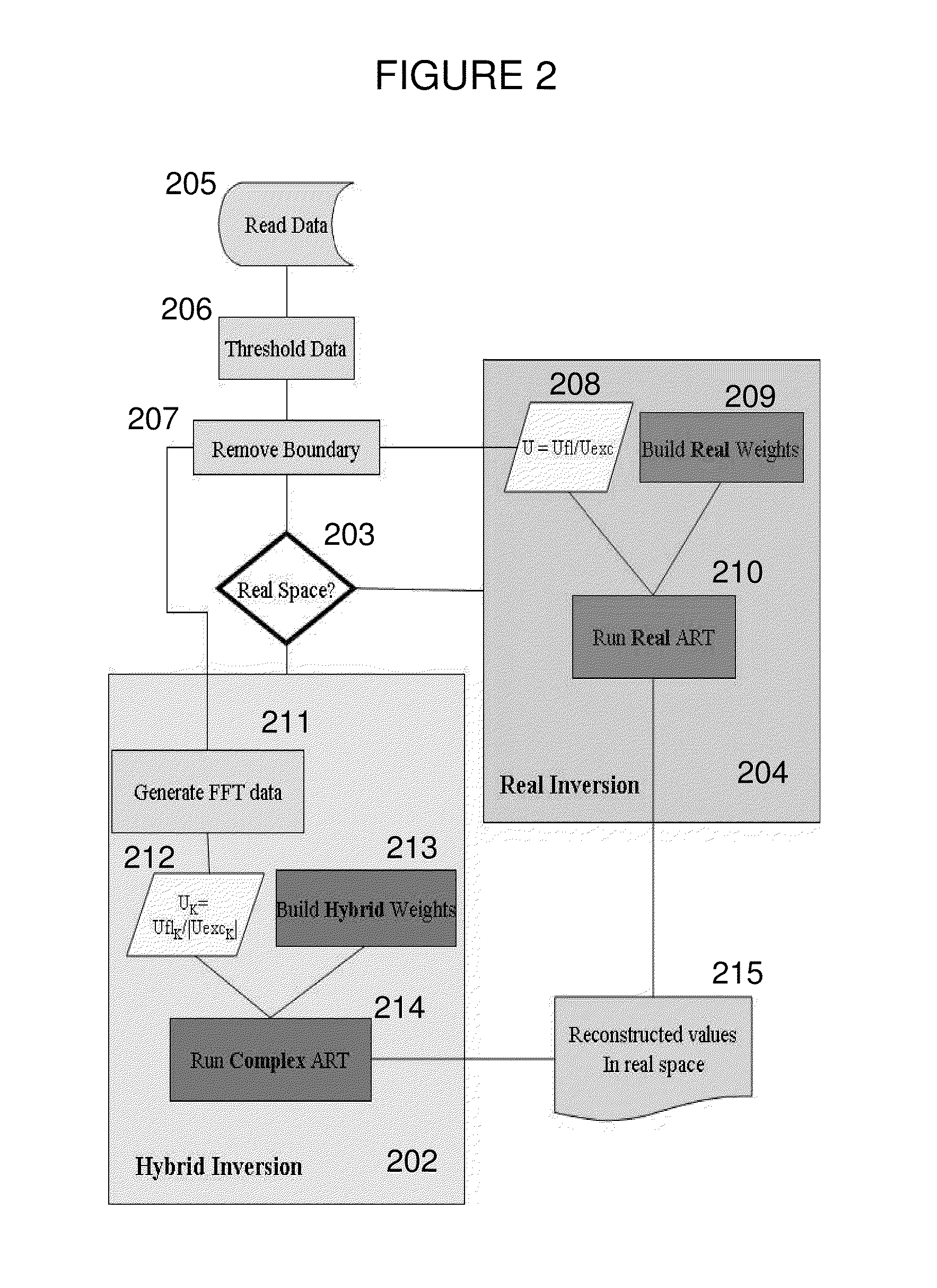
[0008]The invention presents a hybrid approach for fast reconstruction of tomographic images that offers advantages over backprojection and direct inversion techniques. In this hybrid approach, one or more subsets of large tomographic datasets are selected in frequency space (e.g., Fourier space) while one or more subsets are maintained in real space, then the weight matrix is inverted to obtain the tomographic representation of a region of interest within the subject in real space. This achieves fast computational times while maintaining good tomographic reconstruction performance.
[0009]For example, in preferred embodiments, the detector data is Fourier-transformed, while the sources and reconstructions are maintained in real-space. This enables the use of very large detector sets while still using lower numbers of sources (e.g., less than 102) than the complete Fourier (direct inversion) approaches, and does not present the typical Fourier artifacts in the reconstruction because data is reconstructed in real space. This additionally enables full body imaging and the imaging of larger anatomies, since the total number of measurements in Fourier Space is very low but still represents the full body being imaged. Fast computation in larger scan fields is made possible, with both satisfactory spatial resolution and computation speed, allowing fluorescence molecular tomographic imaging of not only mice and rats, but also larger animals such as guinea pigs, rabbits, non-human primates, other mammals, and humans.
[0014]In certain embodiments, in the forward model, a surface of the subject is identified and boundary conditions are established for the surface. In addition, in the forward model, boundary removal equations can be used to convert data corresponding to the surface into a simulated infinite homogeneous medium, thereby simplifying the forward problem (see for example, Ripoll and Ntziachristos, “From Finite to Infinite Volumes: Removal of Boundaries in Diffuse Wave Imaging”, Physical Review Letters 96, 173903, 2006). In certain embodiments, the data corresponding to the surface of the subject comprises an experimental measurement of surface flux distribution.
[0024]In another aspect, the present invention provides a method for imaging using a hybrid inversion technique to image the distribution of a fluorescence within a region of a subject, including but not limited to endogenous fluorescence, bioluminescence or fluorescent proteins, the method comprising: (a) directing excitation light into the subject at multiple locations to transilluminate through or reflect from at least a portion of the region of the subject containing the fluorescence; (b) optionally detecting excitation light transmitted through or reflected from the region of the subject; (c) detecting fluorescent light emitted from within the subject; and (d) processing data corresponding to the detected fluorescent light and the optionally detected excitation light to provide a tomographic representation of the region of the subject, wherein the processing step comprises (i) establishing a forward model of excitation light propagation from an excitation light source to the light source within the subject and of emission light propagation from the light source of the subject to a detector using the data corresponding to the optionally detected excitation light and the detected fluorescent light, wherein: (A) a surface of the subject is identified and boundary conditions are established for the surface, or, alternatively, boundary removal equations are used to convert data corresponding to the surface of the subject into a simulated infinite homogeneous medium, thereby simplifying the forward problem; (B) the excitation light source is represented in real space; (C) the detected fluorescent light and the optionally detected excitation light are represented in frequency space; and (D) the forward model is established as a discretized weight matrix of normalized elements; and (ii) inverting the weight matrix to obtain the tomographic representation of the region of the subject in real space.

Problems solved by technology

These large data sets reduce the ill-posed nature of the inversion, but also present an inherently large computational burden for reconstruction of tomographic images.
Using traditional real-space weight matrix and Algebraic Reconstruction Techniques (ART) for the inversion yields impractically long computational times, in some instances longer that 24 hours.
Similarly, using matrix-related inversion methods such as Singular Value Decomposition is not viable due to the amount of memory required.
Backprojection suffers from being non-quantitative, low in resolution and incapable of good depth-discrimination [Matson, C. L., N. Clark, et al.
Complete Fourier approaches, also termed Direct Inversion, present severe reconstruction artifacts and generally are not applicable to datasets with fewer than O(103) source positions [(Schotland, J. C. and V. A. Markel (2001).

Method used


Image

  • Systems and methods for tomographic imaging in diffuse media using a hybrid inversion technique
  • Systems and methods for tomographic imaging in diffuse media using a hybrid inversion technique
  • Systems and methods for tomographic imaging in diffuse media using a hybrid inversion technique

Examples


Embodiment Construction

[0051]It is contemplated that methods, systems, and processes described herein encompass variations and adaptations developed using information from the embodiments described herein.

[0052]Throughout the description, where systems and compositions are described as having, including, or comprising specific components, or where processes and methods are described as having, including, or comprising specific steps, it is contemplated that, additionally, there are systems and compositions of the present invention that consist essentially of, or consist of, the recited components, and that there are processes and methods of the present invention that consist essentially of, or consist of, the recited processing steps.

[0053]The mention herein of any publication, for example, in the Background section, is not an admission that the publication serves as prior art with respect to any of the claims presented herein. The Background section is presented for purposes of clarity and is not meant a...


PUM


Abstract

The invention relates to systems and methods for tomographic imaging in diffuse media employing a fast reconstruction technique. A hybrid Fourier approach is presented that enables the fast tomographic reconstruction of large datasets. In certain embodiments, the invention features methods of in vivo fluorescence molecular tomographic (FMT) reconstruction of signals, reporters and / or agents (i.e., contrast agents or probes) in a diffusive medium (e.g., a mammalian subject). The method preserves the three-dimensional fluorophore distribution and quantitative nature of the FMT approach while substantially accelerating its computation speed, allowing FMT imaging of larger anatomies.

Description

RELATED APPLICATION[0001]This application claims priority to and the benefit of, and incorporates herein by reference in its entirety, U.S. Provisional Patent Application No. 61 / 238,075, which was filed on Aug. 28, 2009.FIELD OF THE INVENTION[0002]The invention relates generally to in vivo imaging systems and methods. More particularly, in certain embodiments, the invention relates to systems and methods for tomographic imaging employing a fast reconstruction technique.BACKGROUND OF THE INVENTION[0003]The amount of data used in optical tomography image reconstruction has increased by several orders of magnitude in recent years. This is primarily due to the use of large detector arrays, e.g., on the order of 103 elements or higher. When coupled with a large number of sources, e.g., on the order of 102 sources (such large number being facilitated, for example, by the use of non-contact measurements) large data sets easily in the range of 105 source-detector pairs are generated. These ...

Claims


Application Information

Patent Timeline
Patent Type & Authority Patents(United States)
IPC IPC(8): A61B5/05
CPCA61B5/0071A61B5/0073G06T11/006A61B5/0066A61B5/0086A61B6/508A61B5/7257G06T2211/424A61B5/41A61B5/14556
Inventor LORENZO, JORGE RIPOLLYARED, WAEL I.KEMPNER, JOSHUA
Owner VISEN MEDICAL INC
Why Eureka
Try Eureka