Process for preserving three dimensional orientation to allow registering histopathological diagnoses of tissue to images of that tissue

a three-dimensional orientation and image processing technology, applied in the field of histopathology analysis and reconstruction, can solve the problems of increased infection risk, bleeding, physical and emotional discomfort, cost, etc., and achieve the effect of reducing the risk of infection, reducing the risk of bleeding, and reducing the accuracy of the diagnosis

Inactive Publication Date: 2013-08-06
CADES SCHUTTE A LIMITED LIABILITY LAW PARTNERSHIP
View PDF11 Cites 17 Cited by
  • Summary
  • Abstract
  • Description
  • Claims
  • Application Information

AI Technical Summary

Benefits of technology

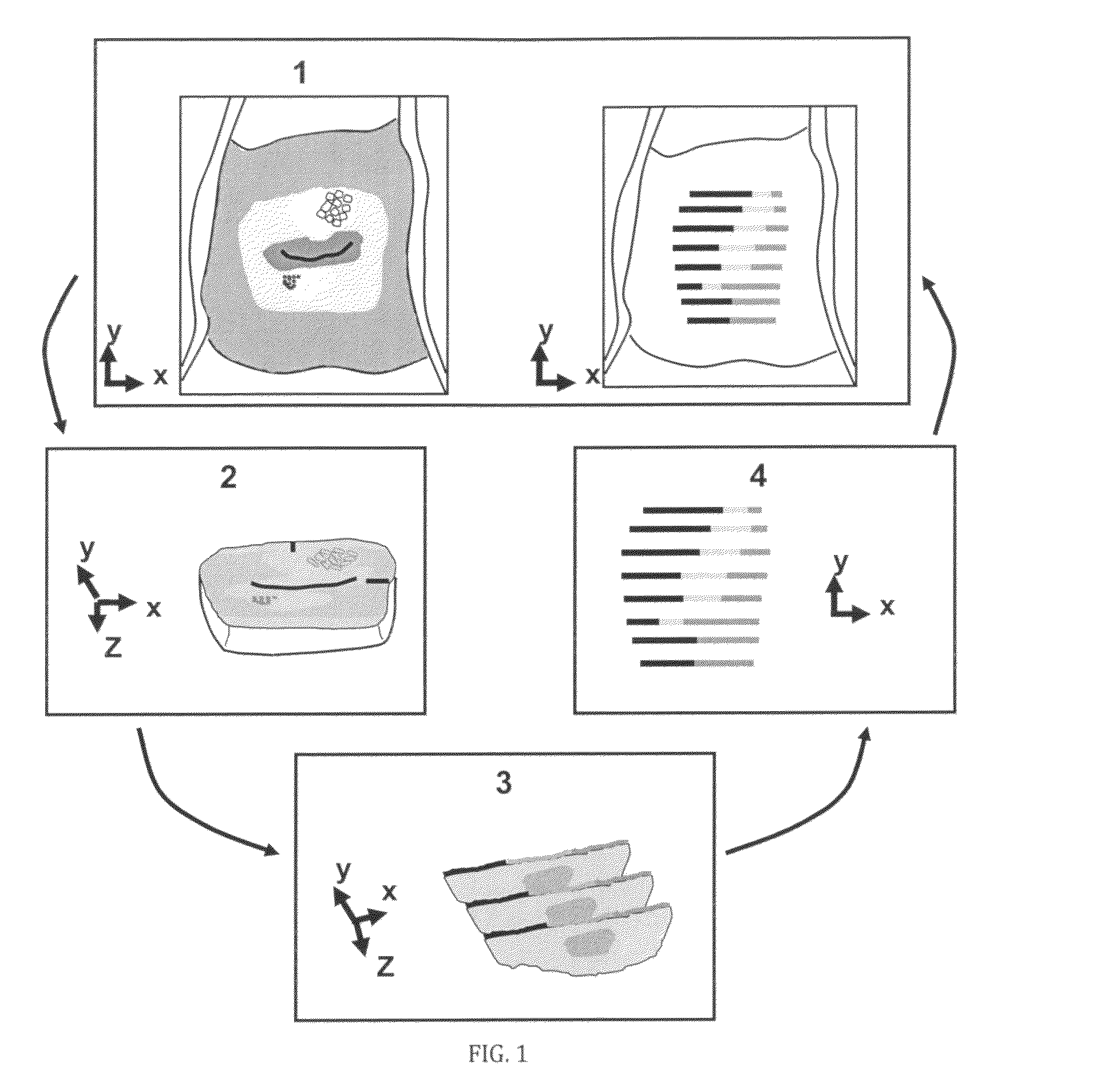
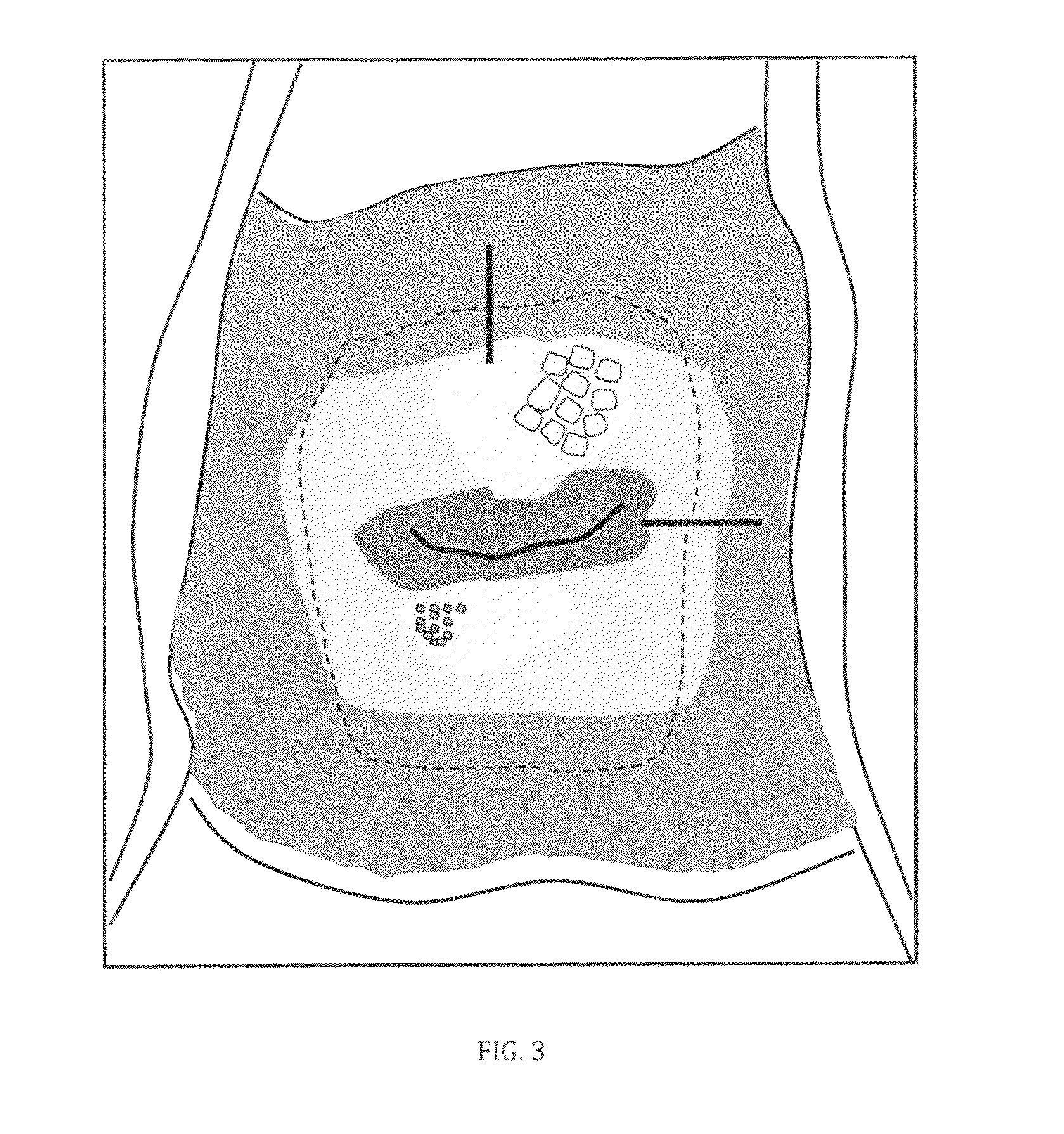
[0092]The slicing of specimen blocks having different colored needle tracks to form specimen slices having different colored dots allows for maintaining spatial orientation of a three-dimensional tissue specimen removed from an area of investigation. However, it may not be necessary to form specimen blocks before forming specimen slices, as specimen slices can be taken directly from the specimen. The fiduciary lines placed at the time of tissue excision, and the internal needle tracks, along with the precise slices of tissue at exacting intervals allows for the “z-axis” histology to accurately “underlie” the x and y axes. In order to perform the functions of preserving orientation and determining shrinkage, marks in the specimen must be part of the tissue, be inserted as soon as the tissue is excised, and be part of the specimen throughout the entire tissue processing. Marks that are only added after fixation and resulting dehydration cannot be used to determine and compensate for orientation and shrinkage. Further, if the marks are made with material having properties different from the properties of the sample, then such marks may not allow accurate restoration to compensate for orientation and shrinkage.
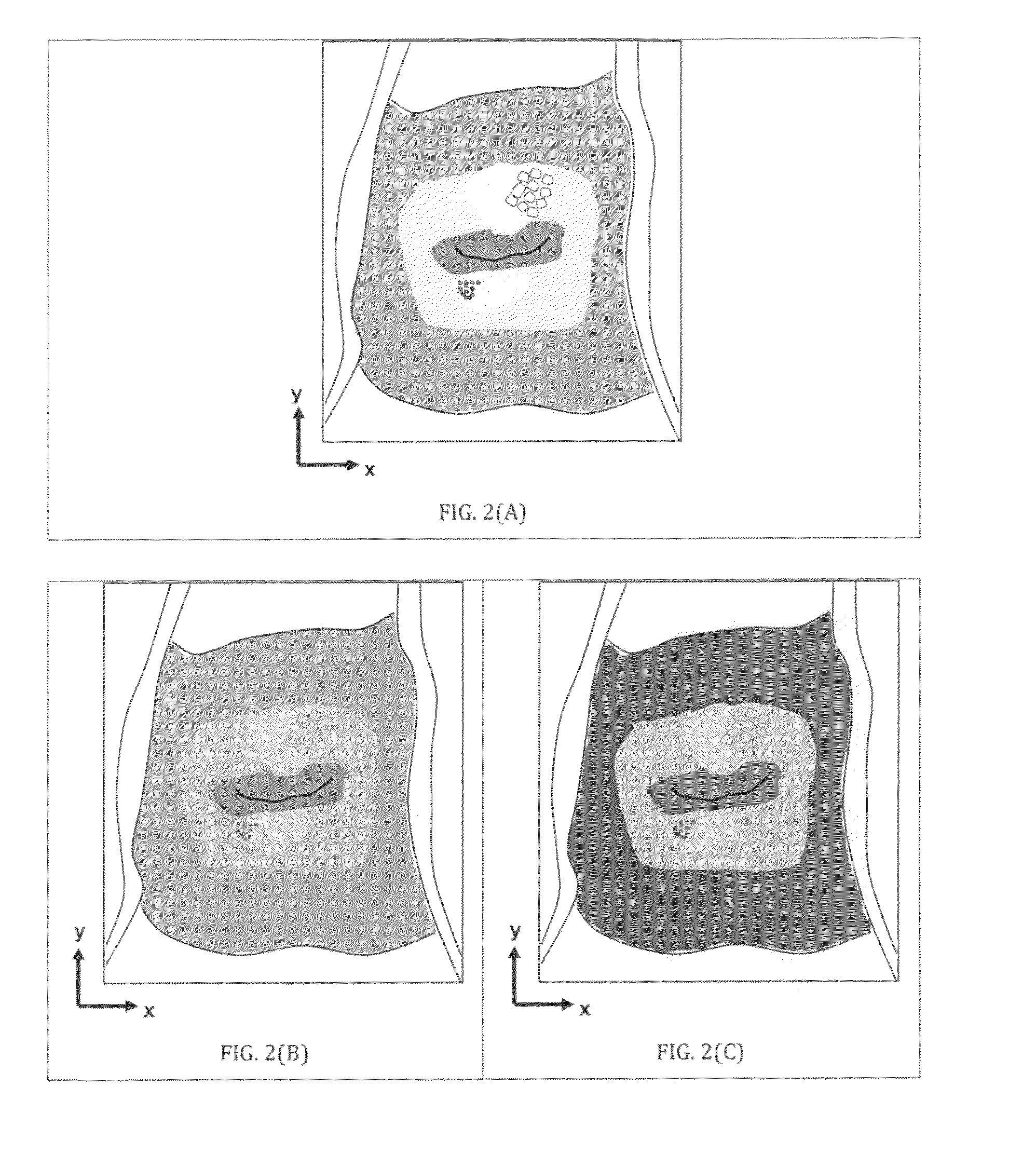
[0093]This ability to maintain spatial orientation at each level of tissue processing and accurately determine the location of the tissue slice as it relates to the overlying tissue photographed prior to tissue excision allows the subsequent histopathologic diagnoses to be annotated to the surface of each slice of the specimen, and mapped to the appropriate overlying x-y axis points. This becomes especially important when assigning topographic characteristics of the colposcopic appearance of the cervix in the two-dimensional (x-y) plane provided by the colposcopic photography immediately preceding the tissue excision. It also can become important in the evaluation of hyperspectral imaging as it relates to the spectrum of light emitted from the cervix following application of acetic acid. In essence, this method of tissue processing from the moment the tissue specimen leaves the body, to the point where annotations of the histopathologic “linear diagnosis” is rendered along each micron of the surface will allow the most accurate assignments of z-axis to the overlying x-y plane of the original sample “in situ”.

Problems solved by technology

While this hand-drawn representation of the cervix is far from adequate, the drawing does provide a limited reference point from which to compare the status of the cervix in future examinations.
An often undesired result is “false positives,” so that unnecessary biopsies are acquired and the specimen is determined normal, which increases the risk of infection, bleeding, physical and emotional discomfort and cost.
Therefore, it is not possible to pinpoint the exact location on the cervix of the reported pathology.
Otherwise, no matter how perfect the histopathology and the quality of the digital imagery, the two data sets cannot be matched and the true sensitivity and specificity of the computer aided detection system cannot be determined.

Method used


Image

  • Process for preserving three dimensional orientation to allow registering histopathological diagnoses of tissue to images of that tissue
  • Process for preserving three dimensional orientation to allow registering histopathological diagnoses of tissue to images of that tissue
  • Process for preserving three dimensional orientation to allow registering histopathological diagnoses of tissue to images of that tissue

Examples


Embodiment Construction

Overview

[0111]The presently preferred embodiments of the invention described herein disclose a systematic framework to maintain the spatial orientation at each level of tissue processing of a three-dimensional tissue specimen removed from an area of investigation, perform detailed histopathology analysis, and precisely map the location of the histopathology back to a digital image of the area of investigation acquired prior to removing the tissue specimen.

[0112]By utilizing tissue image acquisition, processing, and registration techniques in combination with mechanical and image processing tools and novel tissue processing procedures, the present invention accurately determines the location of the histopathology analysis (the ‘z-axis’) of the tissue specimen as it relates to the overlying tissue structure (the ‘x-y-axes’). The procedural steps of the presently preferred embodiments, as schematically illustrated in FIG. 1, and which will be described in detail in the following sectio...


PUM

PropertyMeasurementUnit
working distanceaaaaaaaaaa
distanceaaaaaaaaaa
distanceaaaaaaaaaa

Abstract

A process for maintaining 3 dimensional orientation between a tissue specimen and images of the area of investigation, to register histopathologic diagnoses of multiple locations within the specimen with corresponding locations on the surface of said area of investigation.

Description

[0001]This application claims the benefit of U.S. Provisional Patent Application No. 61 / 195,771 filed Oct. 9, 2008.TECHNICAL FIELD[0002]This invention relates to histopathology analysis and reconstruction and, more specifically, to maintaining the spatial orientation of three-dimensional tissue specimens removed from an area of investigation as an aid in the diagnosis of cancer and pre-cancerous lesions.BACKGROUND ART[0003]Although this invention is being disclosed in connection with cervical cancer, it is applicable to many other areas of medicine and other fields. Diagnosing cervical cancer in women is a multi-step procedure involving examination of the cervix, possible biopsy and follow-up (D. G. Ferris, J. T. Cox, D. M. O'Connor, V. C. Wright, and J. Foerster, Modern Colposcopy. Textbook and Atlas, pp. 1-699, American Society for Colposcopy and Cervical Pathology, 2004; 2. E. Burghardt, H. Pickel, and F. Girardi, Colposcopy—Cervical Pathology, Textbook and Atlas, Thieme, New Yor...

Claims


Application Information

Patent Timeline
Patent Type & Authority Patents(United States)
IPC IPC(8): G01N1/30
CPCG01N1/286
Inventor GUSTAFSSON, ULF PETEROYAMA, JODY C.BERGSTEN, SARA MARIAWHITESELL, ANDREW BEAUMONTLIEBERMAN, RICHARD W.ELDRED, MICHAEL PAUL
Owner CADES SCHUTTE A LIMITED LIABILITY LAW PARTNERSHIP
Why Eureka
Try Eureka