3D tissue model formation from non-parallel 2D images

a tissue model and 2d image technology, applied in the field of 3d tissue model formation, can solve the problems of poor sensitivity (53%) of the prostate biopsy procedure, the test cannot definitively diagnose pca, and the inability of physicians to directly target tumors, etc., and achieve the effect of easy clinical integration

Inactive Publication Date: 2013-08-27
THE UNIV OF WESTERN ONTARIO ROBARTS RES INST BUSINESS DEV OFFICE
View PDF5 Cites 26 Cited by
  • Summary
  • Abstract
  • Description
  • Claims
  • Application Information

AI Technical Summary

Benefits of technology

[0006]We have developed a 3D tissue biopsy system that can be easily integrated clinically, by tailoring our implementation in such a way that it requires minimal new hardware and few changes to the current clinical procedure. Constructing a 3D tissue model from a collection of 2D TRUS biopsy images provides the necessary 3D information for intra-biopsy needle guidance and accurate recording of biopsy locations. We have developed a 3D prostate shape reconstruction algorithm that adheres to the restrictions of the current image acquisition device and procedural techniques while requiring minimal new hardware.

Problems solved by technology

Techniques such as digital rectal exam and prostate-specific antigen (PSA) tests are effective for identifying patients who are at risk; however, these tests cannot definitively diagnose PCa.
Unfortunately, early stage PCa is often isoechoiec and not visible under the conventional ultrasound, or tumor foci are microscopic, so physicians are unable to target tumors directly.
Poor sensitivity (˜53%) plagues the prostate biopsy procedure, since tumors are not always targeted directly.
This inherent inaccuracy may lead to delays in cancer diagnosis and treatment, as well as increased patient anxiety and discomfort due to repeat biopsies.
This makes the procedure prone to spatial ambiguity, as the physician tries to envision the 3D location and orientation of their 2D TRUS view within the prostate.

Method used


Image

  • 3D tissue model formation from non-parallel 2D images
  • 3D tissue model formation from non-parallel 2D images
  • 3D tissue model formation from non-parallel 2D images

Examples


Embodiment Construction

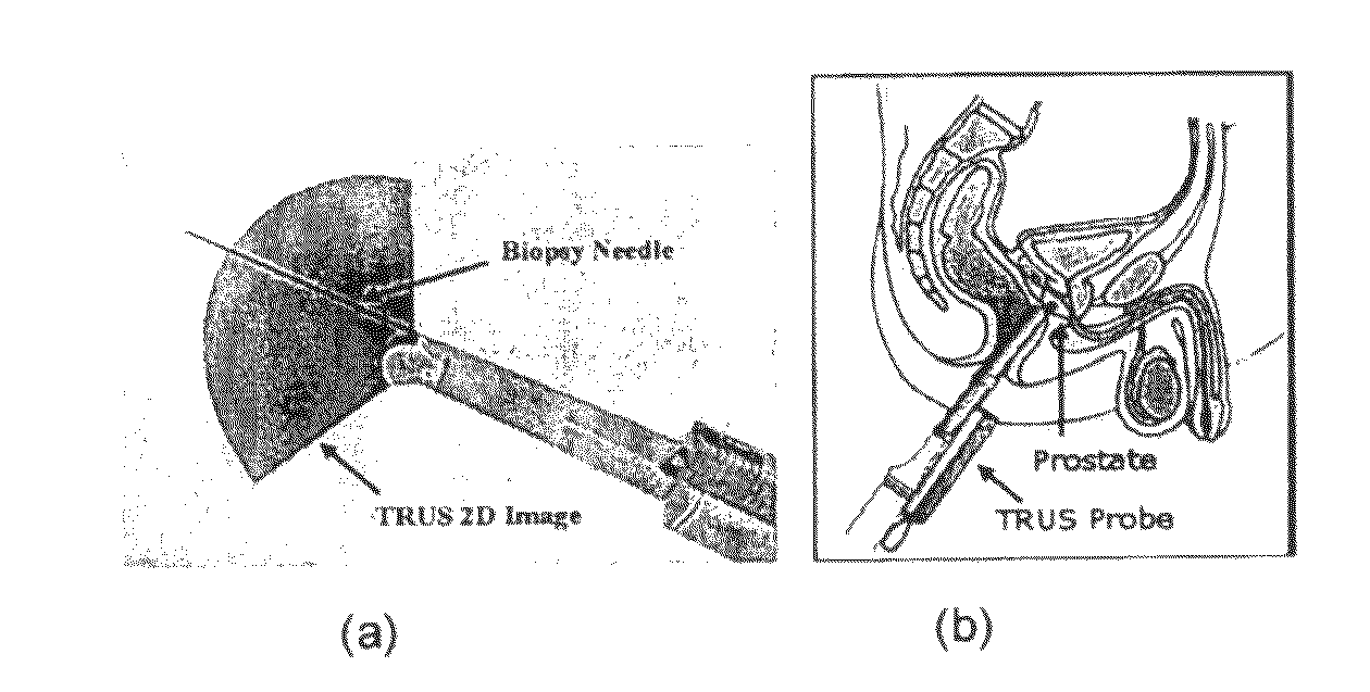
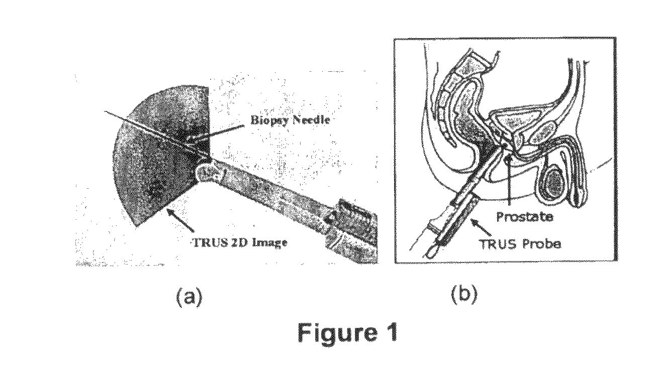
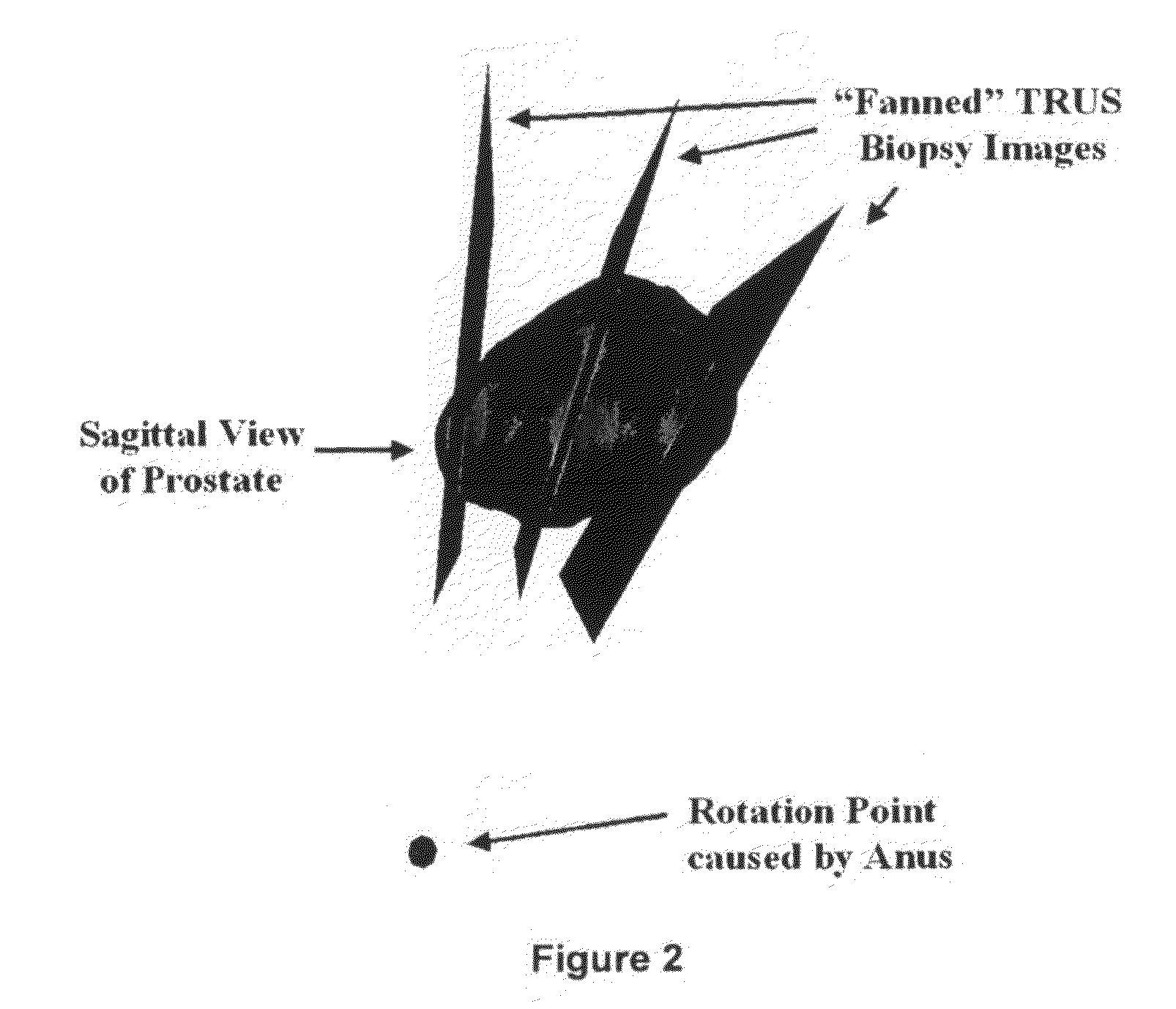
[0020]The TRUS probe currently used for prostate biopsies is designed with an “end-firing”, curvilinear ultrasound transducer, which allows the needle trajectory to remain within the 2D scan field of view during insertion (FIG. 1a). This configuration eliminates the possibility of acquiring parallel biopsy images either in the axial or sagittal planes as the probe is limited to rotational, fan-like movements pivoted around the anus (see FIG. 2). Using this probe, 3D image acquisition would require some form of mechanical assembly, as is done in 3D prostate ultrasound, or would require the physician to complete a continuous sweep of the prostate while the TRUS probe is tracked by a 6 degrees of freedom (DoF) spatial tracking device. The first option would not adhere to current clinical procedure and more importantly would remove the “free-hand” control and range of motion that many physicians prefer. The second option would have little impact on the biopsy procedure; however, to gene...


PUM


Abstract

Biopsy of the prostate using 2D transrectal ultrasound (TRUS) guidance is the current gold standard for diagnosis of prostate cancer; however, the current procedure is limited by using 2D biopsy tools to target 3D biopsy locations. We have discovered a technique for patient-specific 3D prostate model reconstruction from a sparse collection of non-parallel 2D TRUS biopsy images. Our method can be easily integrated with current TRUS biopsy equipment and could be incorporated into current clinical biopsy procedures for needle guidance without the need for expensive hardware additions. We have demonstrated the model reconstruction technique using simulated biopsy images from 3D TRUS prostate images of 10 biopsy patients. This technique of model reconstruction is not limited to the prostate, but can be applied to the reconstruction of any tissue acquired with non-parallel 2-dimensional ultrasound images.

Description

CLAIM OF PRIORITY[0001]This application is a national phase application pursuant to 35 U.S.C. §371 of International Application No. PCT / IB2008 / 003129, filed May 28, 2008, which claims priority to U.S. Provisional Patent Application No. 60 / 1940,427 filed on May 28, 2007.FIELD OF THE INVENTION[0002]We have developed algorithms, designs and methods for a 3D tissue modeling system that can be easily integrated clinically and improves on the range and accuracy of other 2D and 3D systems.BACKGROUND OF THE INVENTION[0003]Screening techniques for prostate adenocarcinoma (PCa) have increased diagnostic sensitivity to detect early stage diseases. Techniques such as digital rectal exam and prostate-specific antigen (PSA) tests are effective for identifying patients who are at risk; however, these tests cannot definitively diagnose PCa. Biopsy of the prostate is required for a definitive histopathological diagnosis and is usually critical in providing information that allows the prediction of t...

Claims


Application Information

Patent Timeline
Patent Type & Authority Patents(United States)
IPC IPC(8): G06K9/00A61B8/14
CPCG06T7/0067G06T17/10A61B8/0833A61B8/12G06T2207/10132G06T2207/20116G06T2207/30081A61B8/4245G06T7/564
Inventor COOL, DEREKFENSTER, AARONDOWNEY, DONALSHEREBRIN, SHI
Owner THE UNIV OF WESTERN ONTARIO ROBARTS RES INST BUSINESS DEV OFFICE
Why Eureka
Try Eureka