
Articular cartilage fixation device and method

a fixation device and cartilage technology, applied in the surgical field, can solve the problems of large defects that occur in articular cartilage that are easy to grow into small defects, and the progression of small defects to large defects is often unknowable, so as to facilitate the fixation of the device or implant, and maximize the pull-through force

Active Publication Date: 2013-12-03
SCHWARTZ BIOMEDICAL LLC
View PDF10 Cites 18 Cited by
  • Summary
  • Abstract
  • Description
  • Claims
  • Application Information

AI Technical Summary

Benefits of technology

[0014]It is an objective of the present invention to fixate a cartilage repair device (ox implant) or a cartilage flap to the subchondral bone to facilitate repair or regeneration of the chondral or osteochondral defect. The cartilage repair device (or implant) or cartilage flap is approximated and retained within the chondral or osteochondral defect for an adequate amount of time such that the cartilage repair device (or implant) can perform its function and facilitate the appropriate healing response.
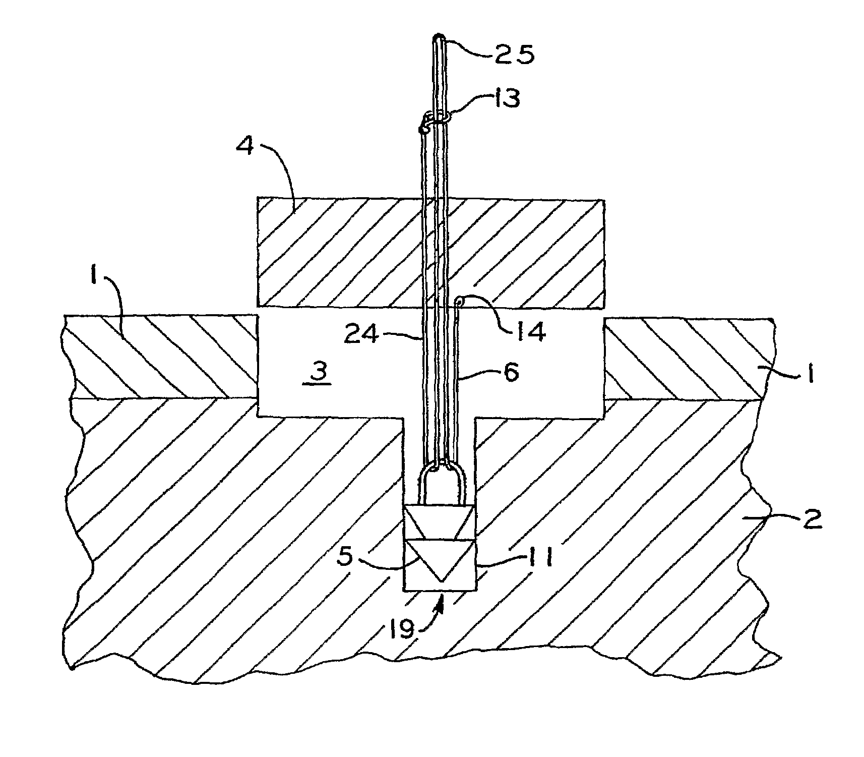
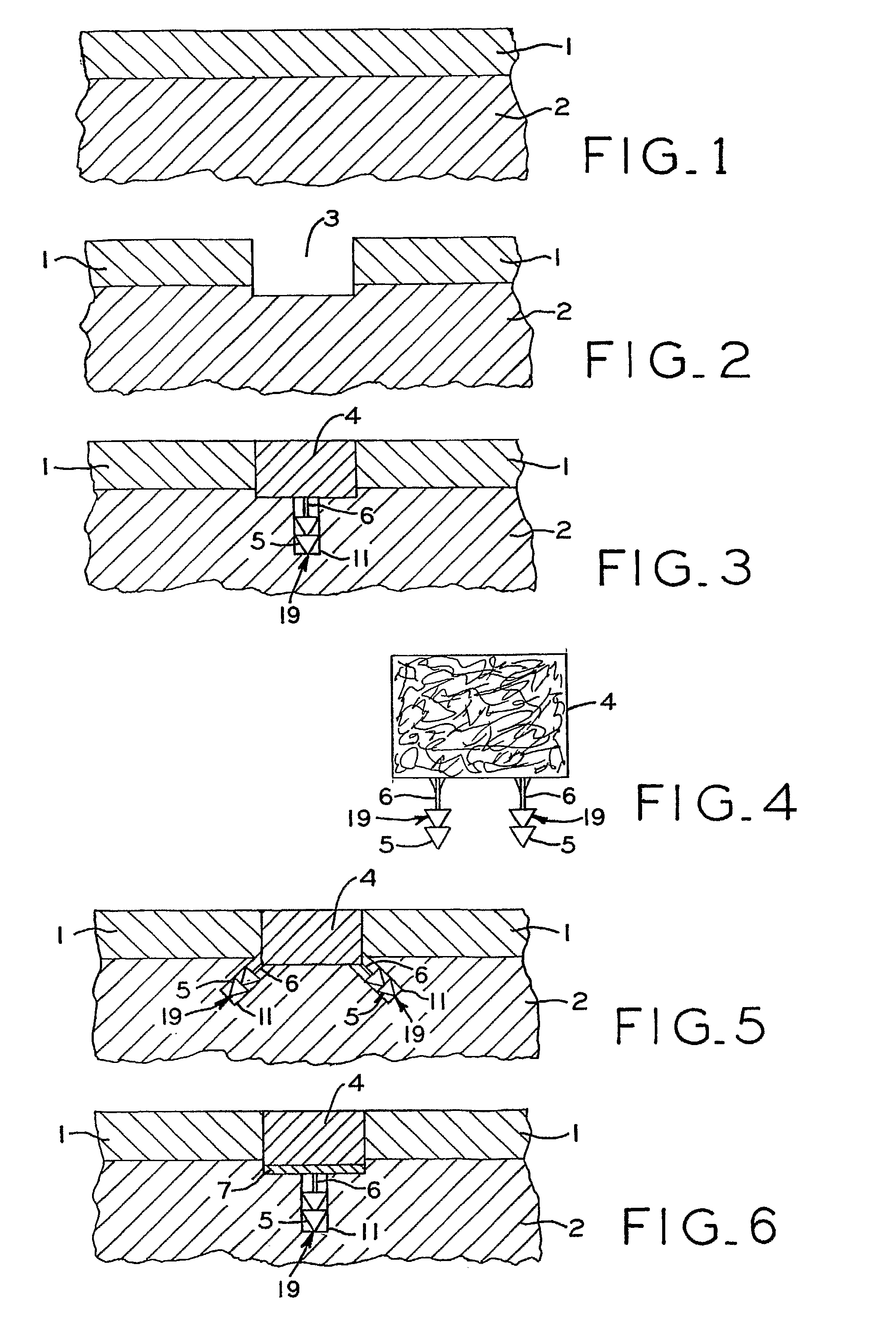
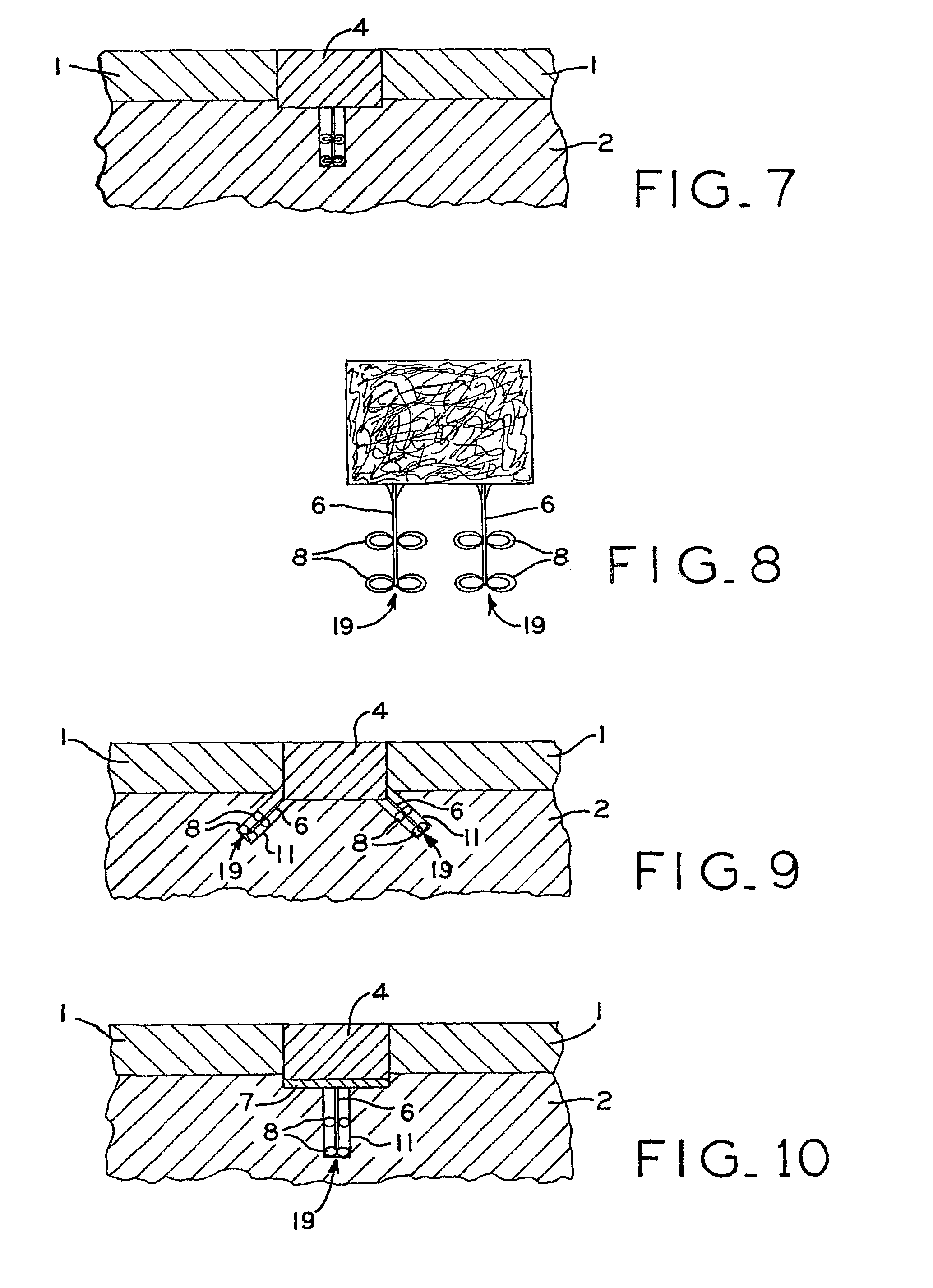
[0029]It is also an objective of the present invention to be comprised of a biocompatible flexible member that is looped through a biocompatible anchor. One of the proximal ends of the flexible member pass completely through the cartilage repair device or implant; whereas, the other proximal end of the flexible member is attached to the cartilage repair device or implant. The distal end of the looped flexible member passes through the anchor, and the anchor is positioned and fixed into the subchondral bone immediately below or adjacent to the chondral or osteochondral defect or is fixed into the adjacent cartilage. As the free proximal end of the flexible member is pulled, the proximal end of the flexible member that is attached to the cartilage repair device (or implant) advances distally along with the cartilage repair device (or implant) toward the biocompatible anchor as the biocompatible anchor allows the flexible member to slide through it (acting as a pulley) so that the cartilage repair device (or implant) is approximated into the chondral or osteochondral defect. When the cartilage repair device (or implant) is in the desired position, a wedging device is slid down or along the flexible member and pushed into the hole used for the biocompatible anchor such that the proximal end of the flexible member that is attached to the cartilage repair device (or implant) is locked in place. One or more of such flexible members and anchors could protrude from the cartilage repair device (or implant) to facilitate the fixation of the device or implant.
[0030]It is also an objective of the present invention for the cartilage repair device (or implant) to have a rigid or semi-rigid base that is attached to the cartilage repair device (or implant) and through which the flexible member is looped such that one proximal end of the flexible member is passed and the other proximal end of the flexible member is attached to the rigid or semi-rigid base. The distal end of the flexible member is passed through a biocompatible anchor such that the anchor acts as a pulley. The anchor could be cannulated or non-cannulated. The free proximal end of the flexible member passes completely through the cartilage repair device or implant. The biocompatible anchor is positioned into the subchondral bone immediately below or adjacent to the chondral or osteochondral defect or is fixed into the adjacent cartilage. As the free proximal end of the flexible member is pulled, the proximal end of the flexible member along with the rigid or semi-rigid base and cartilage repair device (or implant) advance distally as the biocompatible anchor allows the flexible member to slide through it (acting as a pulley) to approximate the cartilage repair device (or implant) into the chondral or osteochondral defect. When the cartilage repair device (or implant) is in the desired position, a wedging device is slid down or along the flexible member and pushed into the hole within the rigid or semi-rigid base or into the hole used for the biocompatible anchor such that the proximal end of the flexible member that is attached to the cartilage repair device (or implant) is locked in place. One or more of such flexible members and anchors could protrude from the cartilage repair device (or implant) to facilitate the fixation of the device or implant.
[0031]It is also an objective of the present invention to be comprised of a biocompatible anchor, a biocompatible flexible member that extends proximally from the biocompatible anchor to within or through the cartilage repair device or implant, and a biocompatible retaining component positioned on the proximal end of the flexible member. Prior to surgery, the retaining or stopping component is attached to the flexible member, and the distal end of the flexible member is attached to the biocompatible anchor. The anchor could be cannulated or non-cannulated. The retaining component could be composed of a nonwoven piece of biocompatible material that is attached to the flexible member or a woven piece of biocompatible material that is attached to the flexible member or a knitted piece of biocompatible material that is attached to the flexible member or any combination of the above. The purpose of the retaining or stopping component is to act as a shoulder device on the proximal end of the flexible member against which tension can be applied through the flexible member such that fixation can be accomplished with this device by maximizing the pull-through force of the retaining or stopping component.

Problems solved by technology

Therefore, small defects that occur in articular cartilage have a tendency to grow into larger defects.
Being an aneural tissue, this progression of defects from small to large often occurs without the knowledge of the patient because of the lack of pain.
When the defect reaches a size or level such that the patient begins to feel discomfort, defect progression often has occurred.
This treatment results in a fibrocartilage scar formation within the defect initially.
However, the biomechanical properties of the repair scar tissue are inferior to that of the adjacent normal articular cartilage; therefore, the repair tissue eventually wears away to expose the defect.
Since the repair tissue biomechanical properties are weaker than native articular cartilage, the articular cartilage surrounding the defect area tends to become overloaded because the repair tissue is not carrying as much load as normal articular cartilage would carry in the defect area.
The result often is that as time progresses a larger defect forms than was originally present during the initial surgery.
This procedure is very controversial because it is dependent on the skill of the surgeon and because the use of multiple plugs results in areas of the defect that are not covered.
So, the patient's body will typically fill the gaps between and around the plugs with inferior scar tissue.
Meniscus degeneration can also occur as a result of aging so that soft places develop in the tissue such that even common activities such as squatting can cause meniscal tears.
Meniscectomies typically provide immediate pain relief and restoration of knee function to the patient; however, with the absence of the meniscus, the long term effect on the knee can be cartilage wear on the condylar or tibial plateau surfaces and the eventual development of an arthritic condition such as osteoarthritis.

Method used


Image

  • Articular cartilage fixation device and method
  • Articular cartilage fixation device and method
  • Articular cartilage fixation device and method

Examples


Embodiment Construction

[0082]A variety of articular cartilage fixation devices 19 utilizing the principles of the present invention are illustrated in the accompanying drawings. The illustrated surgical devices 19 are intended for implantation in a patient for repairing a tissue of the body in the patient. The illustrated embodiments would most commonly be used in repairing articular cartilage, such as that in the knee, hip, shoulder, or ankle; however, the invention is not so limited. Articular cartilage and subchondral bone are illustrated at 1 and 2, respectively, in the accompanying drawings (FIGS. 1-3,5-7, 9-11, 13-15, 17-19, 21-23, 25-27, 29-31, 33-36, 40-42). An example of a cartilage and bone defect is shown in FIGS. 1-3,5-7, 9-11, 13-15, 17-19, 21-23, 25-27, 29-31, and 33-36, 40-42. The invention is also expected to be useful in the treatment of cartilage only defects (i.e. defects without the involvement of bone) and in the treatment of damaged or diseased cartilage in other body parts as well.

[...


PUM

PropertyMeasurementUnit
biocompatibleaaaaaaaaaa
biocompatible flexibleaaaaaaaaaa
flexibleaaaaaaaaaa

Abstract

An articular cartilage fixation device and method for repairing and regenerating diseased or injured soft tissue such as articular cartilage of the knee, hip, shoulder, and the like. A cartilage flap or cartilage repair device is retained within the chondral or osteochondral defect for a sufficient amount of time such that the cartilage repair device can perform its function and facilitate an appropriate healing response. A plurality of biocompatible anchors are attached to a plurality of flexible members and are shaped to seat into the tissue beneath or near the defect such that the cartilage flap is retained within the defect.

Description

CROSS-REFERENCE TO RELATED APPLICATIONS[0001]This application is a §371 filing of PCT application PCT / US05 / 06286 filed on Mar. 1, 2005, which claims priority from U.S. Provisional Application 60 / 549,748, filed on Mar. 3, 2004. The disclosures of these applications are included by reference herein in their entirety.FIELD OF INVENTION[0002]The present invention relates generally to surgical devices for approximating, repairing, or regenerating damaged or diseased soft tissue (i.e. articular cartilage, fibrocartilage, collagenous structures, ligaments, tendons, etc. . . . ) of joints (knee, hip, shoulder, temporomandibular joint, spine, fingers, ankle, toes, etc. . . . ), and to surgical methods using such devices.BACKGROUND OF INVENTION[0003]Cartilage is a functional tissue found in diarthrodial joints at the ends of opposing bones and participates in load transmittal and load distribution between the bone ends. In addition to load distribution, cartilage also provides a nearly fricti...

Claims


Application Information

Patent Timeline
Patent Type & Authority Patents(United States)
IPC IPC(8): A61F2/08A61B17/00A61B17/04A61B17/064A61F2/00A61F2/02A61F2/30
CPCA61F2/0811A61F2/30756A61B17/0401A61B17/0642A61B2017/00004A61B2017/0458A61B2017/0475A61B2017/0647A61F2002/0888A61F2002/30062A61F2002/30461A61F2002/30841A61F2002/30971A61F2210/0004A61F2220/0075
Inventor SCHWARTZ, HERBERT E.
Owner SCHWARTZ BIOMEDICAL LLC
Learn More