Osteoimplant and method of making same

a technology of implants and osteosis, applied in the field of osteosis devices, prosthesis, bandages, etc., can solve the problems of pain, muscle weakness and/or paralysis, and the deterioration of intervertebral discs

Inactive Publication Date: 2014-03-04
WARSAW ORTHOPEDIC INC
View PDF239 Cites 41 Cited by
  • Summary
  • Abstract
  • Description
  • Claims
  • Application Information

AI Technical Summary

Benefits of technology

[0049]The plug embodiment of the osteoimplant of the invention is configured to fit within the cavities of commercially available implants, e.g., spinal cages, and relatively well-defined cavities at a surgical site. The plugs are engineered to fit efficiently, are easy to insert and tend to remain where placed. An additional advantage of the plug is that in many of its configurations, it swells somewhat upon contact with body fluids thus ensuring good bone contact at the implant site even where the site is irregularly shaped. The plug, when made from demineralized bone, is generally osteoinductive and the fiber-like shape of the bone particles further provides a favorable scaffold for bone cell growth thus potentially combining osteoconductive and osteoinductive properties.
[0050]The plug can be formed in a range of shapes and sizes that fit commercially available osteoimplants, e.g., spinal cages, and relatively well-defined bone defects. Some designs of cages may require two or more shaped plugs. The present invention also provides efficient methods for manufacturing the preformed bone plug and methods of using the preformed bone plug for the surgical treatment and / or repair of a bone defect site.
[0057]Another particularly useful embodiment of the invention herein is an osteoimplant provided as a coherent aggregate, or matrix, of elongate bone particles possessing an open pore structure and a bulk density of less than about 0.3 g / cm3. The open pore structure of the aggregate renders the osteoimplant highly absorbent of surrounding liquids. The osteoimplant formed from the aggregate is flexible when dry (i.e., when containing less than about 5 weight percent water) and does not require time consuming rehydration prior to implantation. It can assume any desired shape and / or configuration and can be cut to the desired dimensions, e.g., with surgical scissors, before and / or after the aggregate has absorbed fluid. Even in the wetted / hydrated state, the osteoimplant will maintain its original shape and coherency and can be readily handled by the medical practitioner.
[0062]The osteoimplant of the invention can be readily applied to virtually any bone repair site in the body and can be utilized alone or in combination with one or more adjunct medical devices and / or procedures. The osteoimplant of the invention finds particular utility in the areas of dental reconstructive surgery and spinal fusion where substantial amounts of body fluid, e.g., saliva and / or blood, are frequently encountered or where autograft (e.g., local bone, marrow or iliac crest, etc.) is incorporated in the osteoimplant. The unique ability of the osteoimplant to absorb body fluids and still retain its original shape represents a significant advance in the medical field.

Problems solved by technology

Spinal discs may be displaced or damaged due to trauma, disease, or aging.
The extruded nucleus pulposus may press upon a spinal nerve, resulting in nerve damage, pain, numbness, muscle weakness and / or paralysis.
Intervertebral discs may also deteriorate due to the normal aging process or disease.
As a disc dehydrates and hardens, the disc space height will be reduced leading to instability of the spine, decreased mobility and pain.
Collapse of the disc space can cause instability of the spine, abnormal joint mechanics, premature development of arthritis or nerve damage in addition to severe pain.
Once fusion occurred, the hardware used to maintain the stability of the vertebrae became superfluous and became a permanent non-functional foreign body.
Moreover, the surgical procedures employed to implant a rod or plate to stabilize the spine while fusion was taking place were frequently lengthy and involved.
Success of the discectomy and fusion procedure requires the development of a contiguous growth of bone to create a solid mass because the implant may not withstand the compressive loads on the spine for the life of the patient.
Spinal fusion cages are typically designed to support vertebrae in the proper geometry during the fusion process and are not intended to provide a long term, permanent support.
While effective, it causes secondary damage (to the harvest site) and requires preparation before it can be used.
Furthermore, it is somewhat difficult to maintain in place due to its semi-fluid nature.
The more fluid forms such as powders, gels and pastes are relatively easy to implant at the repair site but difficult to maintain in place.
In addition, there is the possibility of wasted material anytime a standard material has to be adapted to fill a cage.

Method used


Image

  • Osteoimplant and method of making same
  • Osteoimplant and method of making same
  • Osteoimplant and method of making same

Examples


example 1

Process of Making a Species-Specific Osteoimplant with Defined Dimensions.

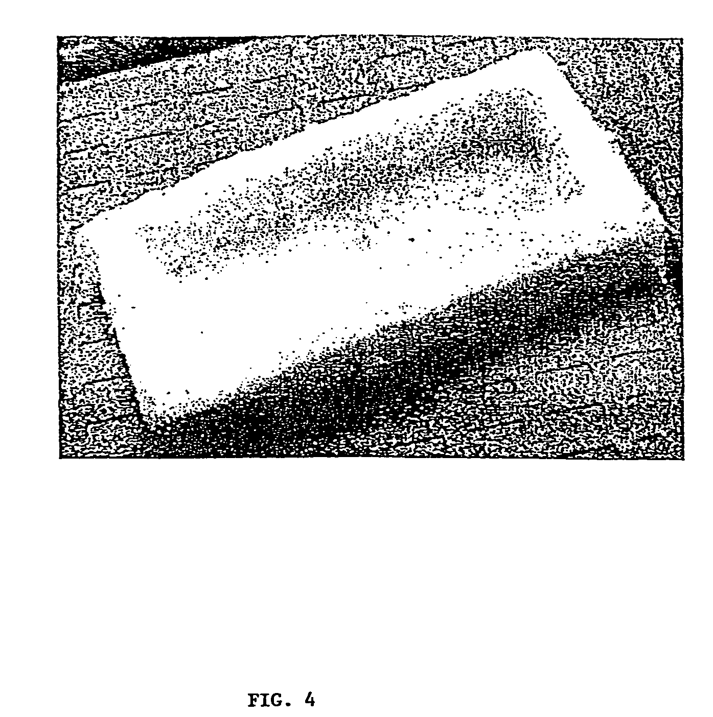
[0104]Species-specific (Rhesus Monkey) long bones were aseptically cleaned. The cortical bone was processed in the bone milling apparatus described in U.S. Pat. No. 5,607,269 to yield 65 grams of elongate bone particles. The elongate bone particles were placed in a reactor and allowed to soak for 5-10 minutes in 0.6N HCl plus 20-2000 ppm nonionic surfactant solution. Following drainage of the HCl / surfactant, 0.6N HCl at 15 ml per gram of total bone was introduced into the reactor along with the elongate bone particles. The reaction proceeded for 40-50 minutes. Following drainage through a sieve, the resulting demineralized elongate bone particles were rinsed three times with sterile, deionized water at 15 ml per gram of total bone, being replaced at 15 minute intervals. Following drainage of the water, the bone particles were covered in alcohol and allowed to soak for at least 30 minutes. The alcohol was then ...

example 2

Evidence of Osteoinduction by Grafton DBM in Non-Human Primate Spine Fusion.

[0107]While autogenous iliac crest bone graft remains the “gold standard”, much work continues to identify viable bone graft extenders, enhancers, and substitutes. While several demineralized bone matrix formulations have been shown to be variably osteoinductive in rodent ectopic bone assays, few have demonstrated efficacy in higher species and more challenging applications such as posterolateral spine fusion. To date, none have been tested in a non-human primate posterolateral spine fusion model which has been previously determined to be extremely challenging with less than 40% of animals achieving successful fusion with autogenous iliac crest bone graft. The purpose of this example was to test the osteoimplant described in Example 1 for evidence of osteoinduction and its use as an extender / enhancer for autogenous bone graft in a non-human primate.

[0108]Four skeletally mature rhesus macaques underwent singl...

example 3

Implantation of Osteoimplant in a Human Patient to Promote Spinal Fusion.

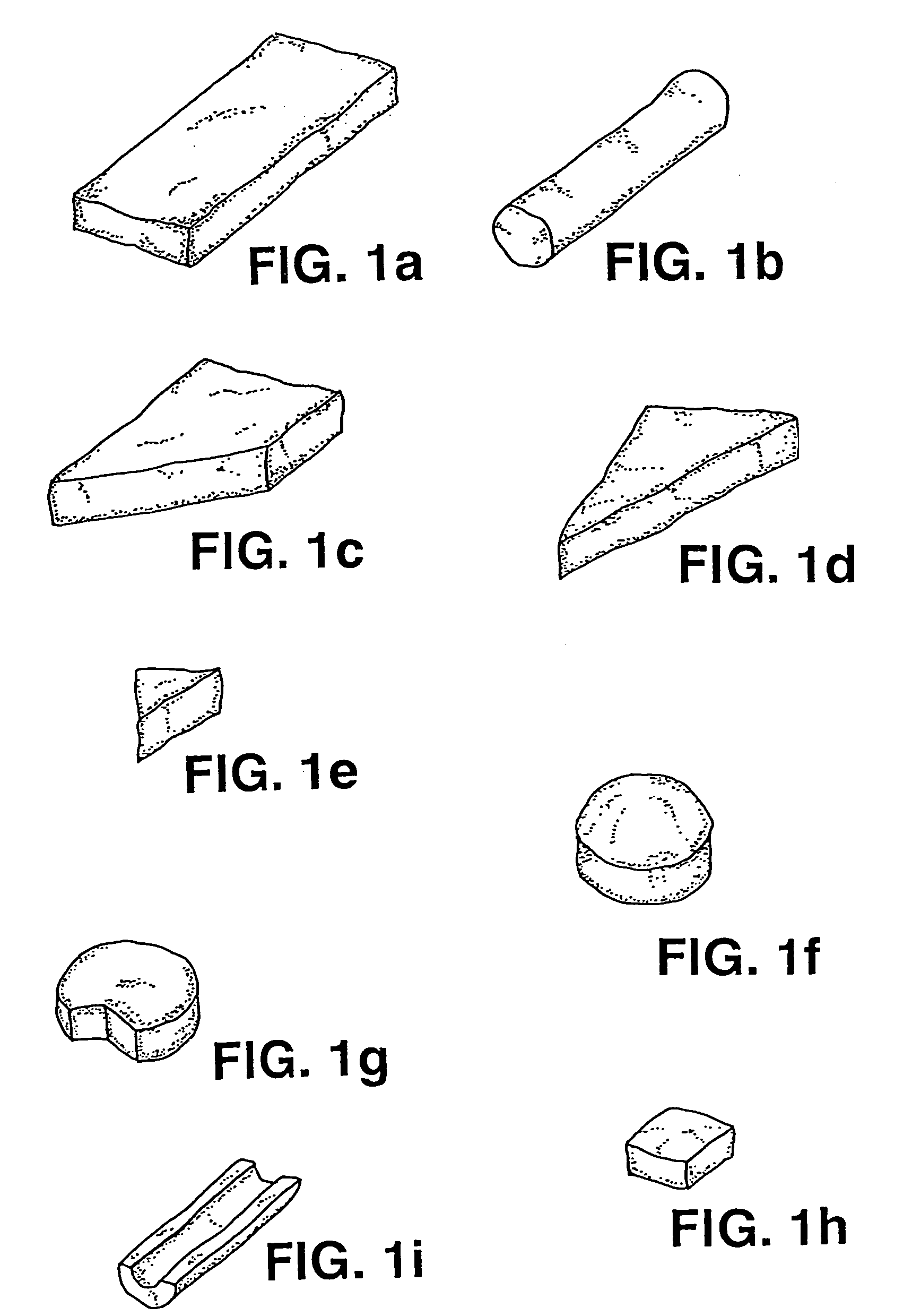
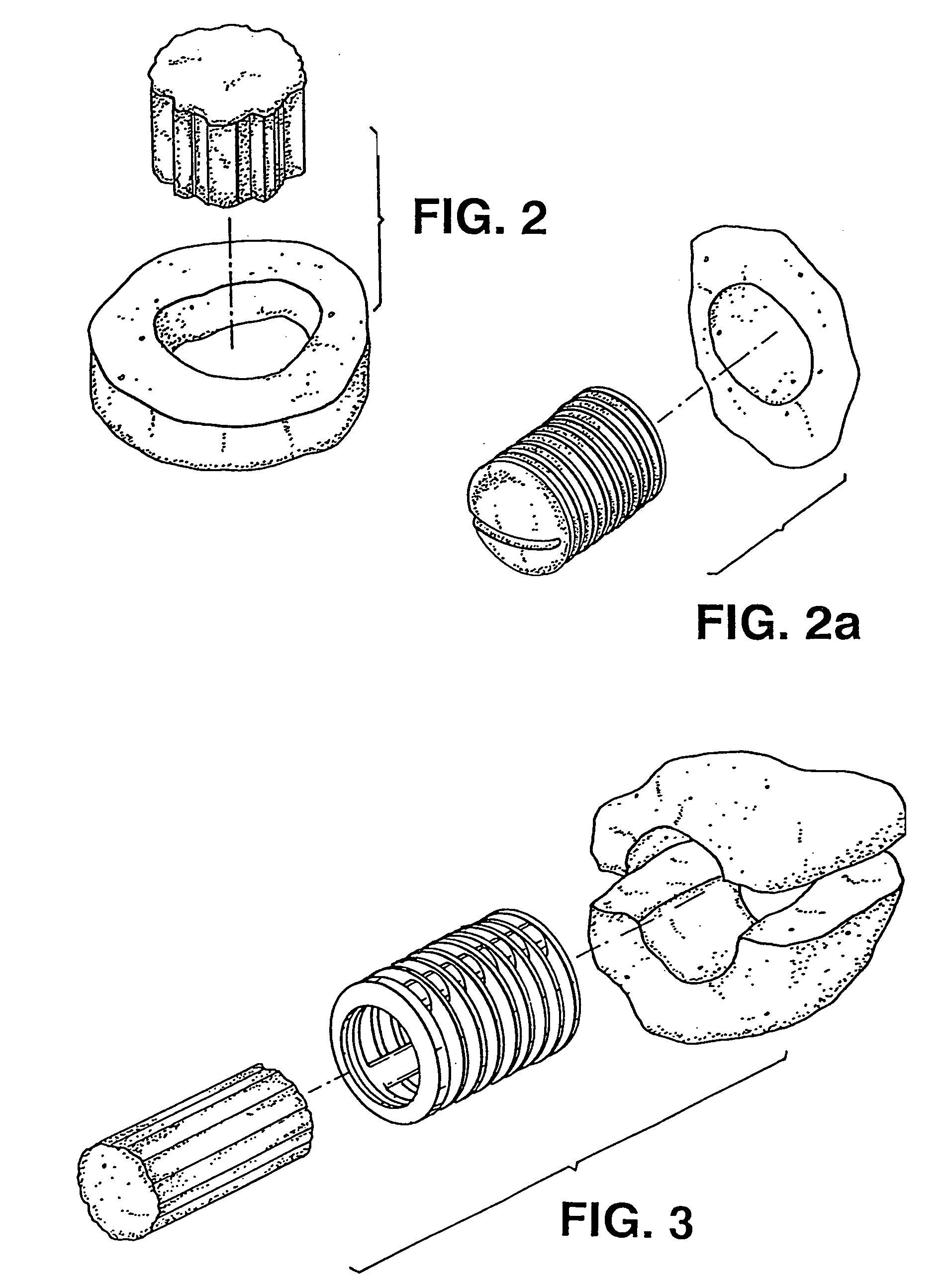
[0109]Human-specific osteoimplant was made in the same manner described in Example 1. However, the mold dimensions and final dimensions of the osteoimplant were altered to adjust to the approximate size required for human posterolateral spinal fusion procedure (known by those skilled in the art). The dimensions of the osteoimplant pieces were approximately 5.0 cm in length, 2.5 cm in width and approximately 1 cm in height with trough dimensions 4 cm in length, 1.5 cm in width and depth approximately 0.7 cm. The trough design specifically allowed for the surgeon to fill the center of the osteoimplant with autograft or allograft or both. Autograft is usually obtained from local bone at the site of the procedure, or marrow, or iliac crest or a combination. The fluids rapidly dispersed within the osteoimplant hydrating the osteoimplant. The osteoimplant is placed either trough down facing the decorticated transvers...


PUM

PropertyMeasurementUnit
weight percentaaaaaaaaaa

Abstract

An osteoimplant is provided which comprises a coherent aggregate of elongate bone particles, the osteoimplant possessing predetermined dimensions and shape. The osteoimplant is highly absorbent and sponge-like in nature. Also provided herein are a method of fabricating the osteoimplant and a method of repairing and / or treating bone defects utilizing the osteoimplant.

Description

CROSS REFERENCE TO RELATED APPLICATIONS[0001]This application is a divisional of U.S. patent application Ser. No. 10,137,862, filed on May 2, 2002, which is a continuation-in-part of International Application No. PCT / US01 / 22853, filed Jul. 19, 2001, and claims the 35 U.S.C. §119 (e) benefit of provisional applications 60 / 219,198, filed on Jul. 19, 2000 and 60 / 288,212 filed May 2, 2001. The entire contents of aforesaid applications 10 / 137,862, PCT / US01 / 22853, 60 / 219,198 and 60 / 288,212 are incorporated by reference herein.BACKGROUND OF THE INVENTION[0002]1. Field of the Invention[0003]This invention relates to an osteoimplant of predetermined dimensions and shape made up of a coherent aggregate of elongate bone particles and to a method for making the osteoimplant. Among its other applications, the osteoimplant can be fashioned as a plug for insertion in a space or cavity within an implant used in an orthopedic procedure, e.g., an intervertebral spacer employed in spinal fusion, or fo...

Claims


Application Information

Patent Timeline
Patent Type & Authority Patents(United States)
IPC IPC(8): A61F2/00A61F2/44A61F2/30A61F2/28C12N5/02A61L27/36A61K9/00A61L31/00A61F2/46A61B17/86
CPCA61F2002/30238A61F2230/0023A61L27/365A61F2002/30957A61F2230/0082A61F2/3094A61F2002/3028A61F2002/30131A61F2002/30153A61F2230/0006A61F2230/0067A61F2230/0069A61L27/3608A61F2210/0061A61L27/3683A61F2002/30225A61F2230/0021A61F2002/30154A61F2002/30158A61F2002/30224A61F2002/30075A61F2310/00365A61F2230/0013A61F2002/2839A61F2002/30205A61F2/442A61F2230/0026A61F2002/4649A61F2002/30733A61F2002/2835A61F2/4644A61F2230/0019A61F2230/0065A61B17/866A61F2002/30059A61F2/446A61L2430/02A61F2/28A61F2002/30261A61F2230/0063A61F2002/30156A61F2002/30113A61F2002/30879A61L27/3691A61F2002/2817A61F2002/302A61F2002/30892A61L31/005A61F2002/30891A61F2002/30237A61L27/38A61F2002/30971B29C41/22B29C41/46B29K2105/16B29K2511/06B29L2031/7532
Inventor MANRIQUE, ALBERTEDWARDS, JEAN T.SCARBOROUGH, NELSON L.BODEN, SCOTT D.TRAIANEDES, KATHYSHIMP, LAWRENCE A.RUSSELL, JAMES L.
Owner WARSAW ORTHOPEDIC INC
Why Eureka
Try Eureka