Method and apparatus for stain separation in digital pathology images

a digital pathology and stain separation technology, applied in the field of pathology image analysis, can solve the problems of inability to achieve the stain separation effect,

Active Publication Date: 2014-06-03
SONY CORP
View PDF5 Cites 23 Cited by
  • Summary
  • Abstract
  • Description
  • Claims
  • Application Information

AI Technical Summary

Problems solved by technology

The process is slow and expensive.
This approach is not ideal since the pure dye area may not exist or may be difficult to locate.
A second approach uses non-negative matrix factorization, but is a computationally complex and infeasible method.
A third approach is color analysis based, but does not account for stain color spectra overlap and leads to loss of stain co-localization information.

Method used


Image

  • Method and apparatus for stain separation in digital pathology images
  • Method and apparatus for stain separation in digital pathology images
  • Method and apparatus for stain separation in digital pathology images

Examples


Embodiment Construction

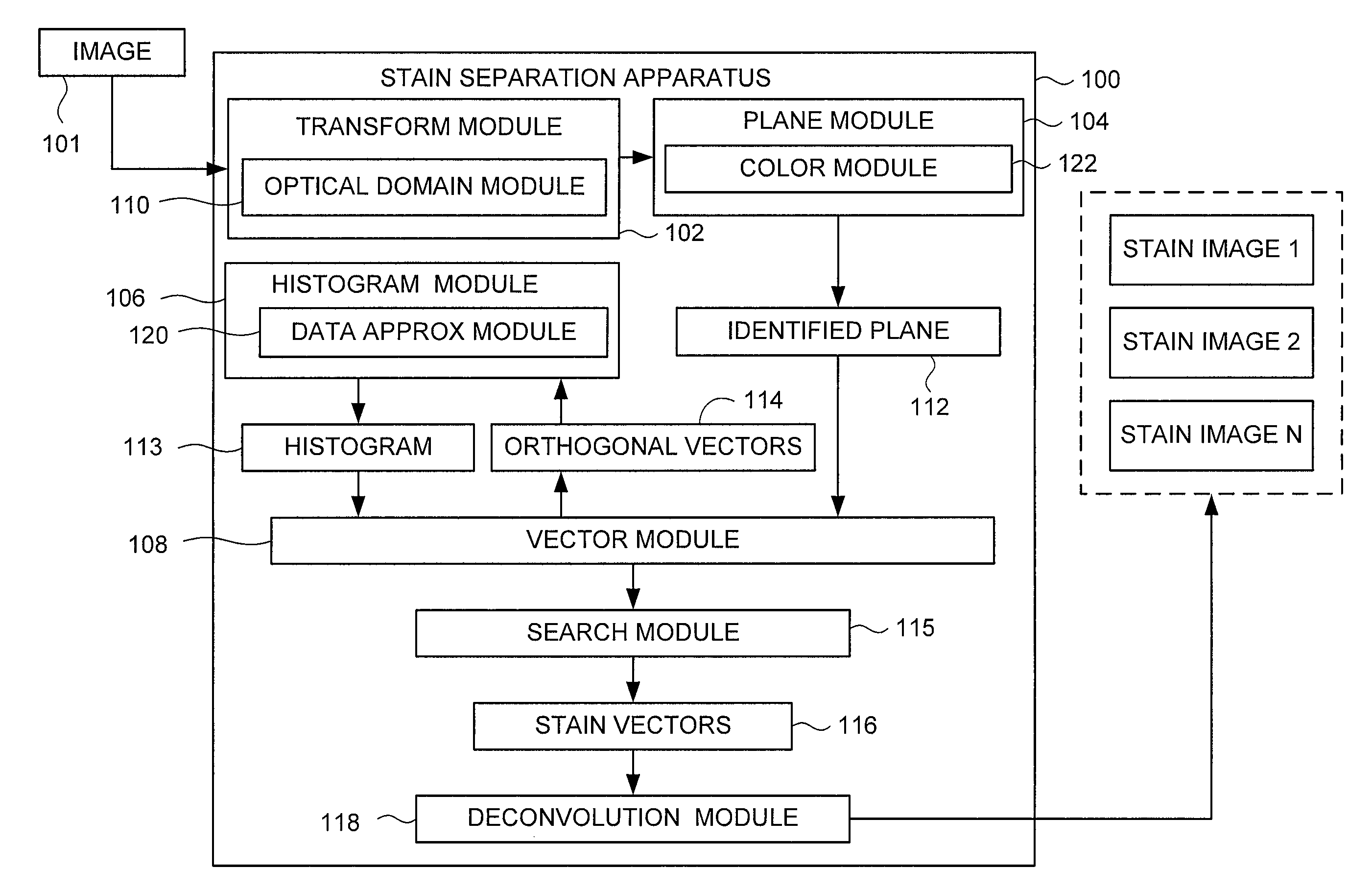
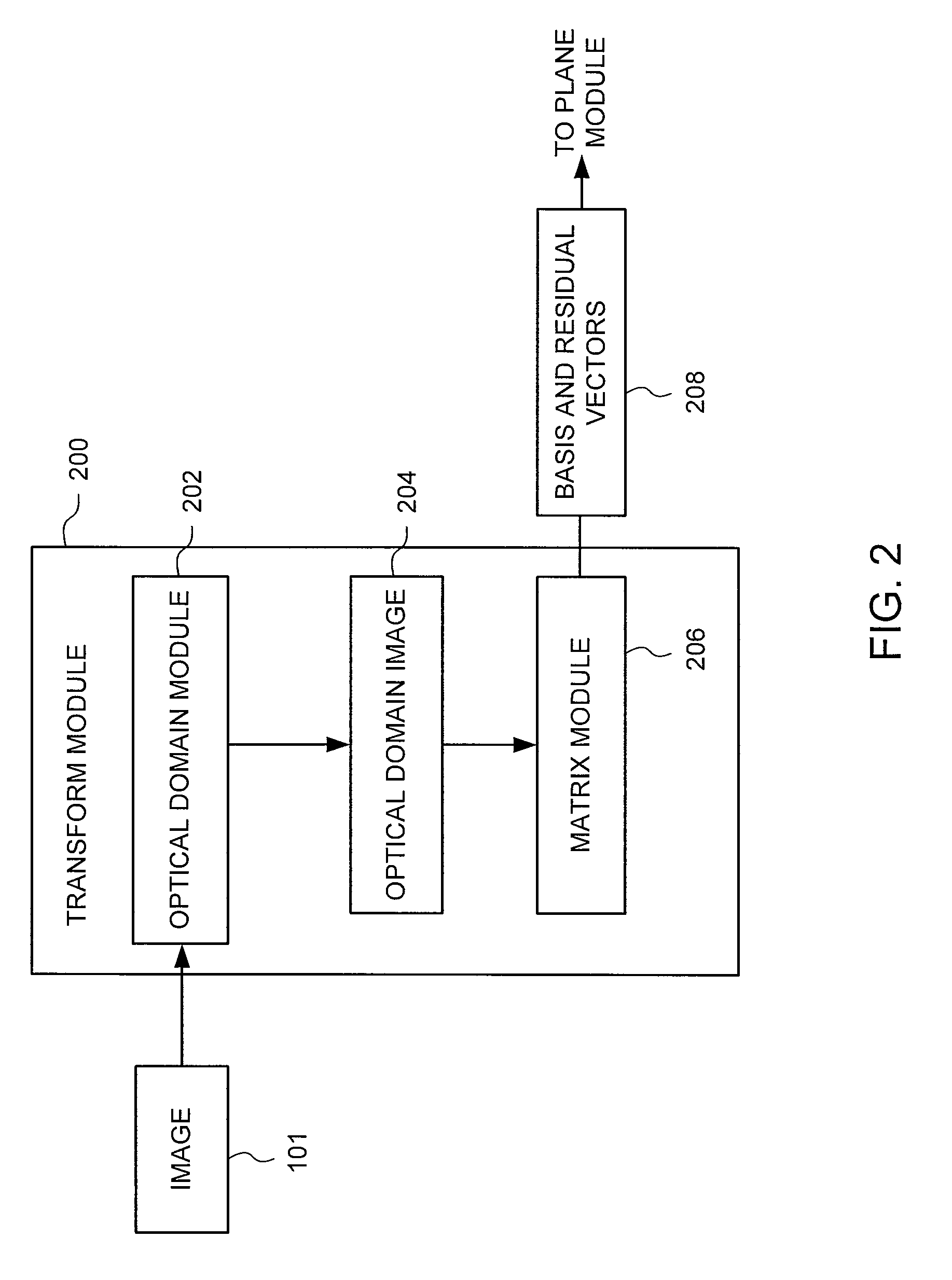
[0017]As explained further below, various embodiments of the invention disclose a method and apparatus for stain separation in digital pathology images. In one embodiment, a digital pathology image is converted from its color domain to the optical density domain color mode using a logarithmic conversion operation. The optical density is a logarithmic ratio of the radiation falling upon a material, to the radiation transmitted through a material. The digital pathology image often is stained with various stains to differentiate between particular specimens. Converting to the optical density image is a preprocessing step used to isolate the effects of the various stains. Basis vectors are determined for colors represented in the optical domain image, and a plane is constructed from the basis vectors. Two orthogonal vectors are extracted from the plane and a histogram is formed of the original data represented by the orthogonal vectors, thereby reducing the quantity of data to process. ...


PUM

PropertyMeasurementUnit
optical densityaaaaaaaaaa
color domainaaaaaaaaaa

Abstract

A stain separation system of digital pathology images that performs transforming a digital image from a first color domain to an optical domain to form an optical domain image (ODI), identifying a plane containing two or more basis vector which contain the pixels of the ODI, determining a plurality of orthogonal vector within the identified plane, forming a histogram of the digital image represented by the orthogonal vectors and determining one or more final stain vectors by searching for candidate vectors in the plane that minimize a cost function of the histogram.

Description

BACKGROUND[0001]1. Field of the Invention[0002]Embodiments of the present invention generally relate to pathology image analysis and, more particularly, to a method and apparatus for stain separation in digital pathology images.[0003]2. Description of the Related Art[0004]In the area of biology and medicine, understanding cells and their supporting structures in tissues, tracking their structure and distribution changes are very important. Histology, the study of the microscopic anatomy of tissues, is essential in disease diagnosis, medicinal development and many other fields. In histology, the process of examining a thin slice of tissue sample under a light microscope or electron microscope is usually performed. In order to visualize and differentiate the microscopic structure, one common approach is to stain the tissue sample with a combination of several dyes that have selective responses to different biological substances. In doing so, specified biological substances such as nuc...

Claims


Application Information

Patent Timeline
Patent Type & Authority Patents(United States)
IPC IPC(8): G01N33/48G06K9/00G01N33/50A61K49/00G06V10/26G06V10/56
CPCG06K9/4652G06K9/0014G06K9/34G06V20/695G06V10/26G06V10/56
Inventor LIU, MING-CHANGROBERTSON, MARK
Owner SONY CORP
Why Eureka
Try Eureka