
Non-invasive methods and systems for producing cardiac electrogram characteristic maps for use with catheter ablation of ventricular tachycardia

a technology of ventricular tachycardia and characteristic maps, which is applied in the field of non-invasive methods and systems of producing cardiac electrogram characteristic maps for use in catheter ablation of ventricular tachycardia, can solve the problems of scar transmurality extent, difficult to display tissue heterogeneity, and significantly prolonging the procedure by the end of the catheter

Active Publication Date: 2015-09-29
THE JOHN HOPKINS UNIV SCHOOL OF MEDICINE
View PDF6 Cites 11 Cited by
  • Summary
  • Abstract
  • Description
  • Claims
  • Application Information

AI Technical Summary

Benefits of technology

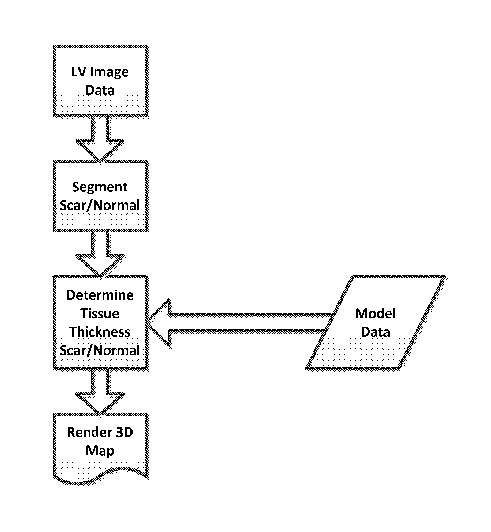
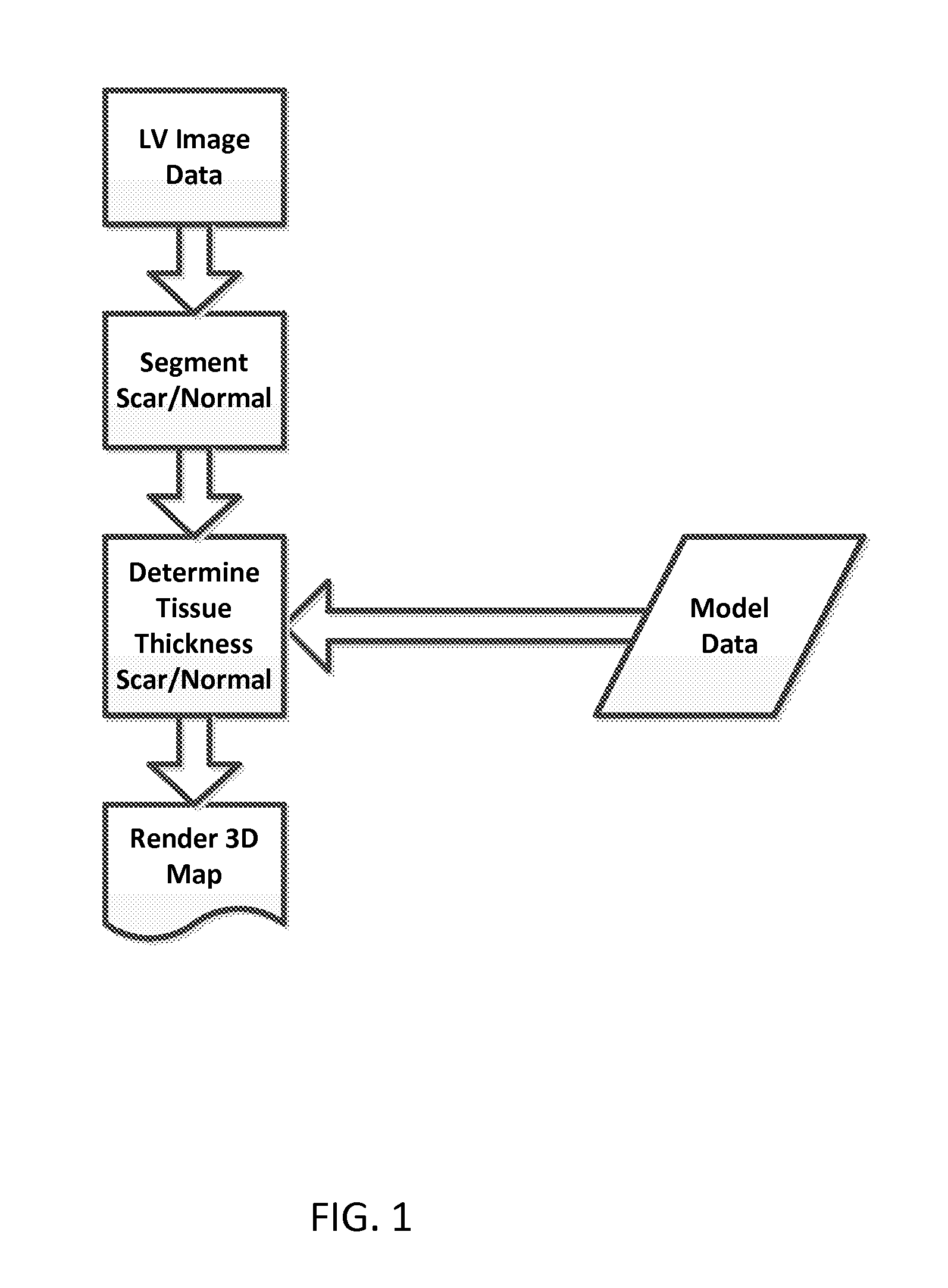
The patent is about a method and system for creating a three-dimensional map of the heart to help treat ventricular tachycardia. The system receives three-dimensional images of the left ventricle of the heart and breaks them into scar tissue, normal myocardium tissue, and left ventricle cavity regions. It also determines the thickness of these regions and uses predetermined data to associate each region with a specific electrogram characteristic. This information helps create a map that accurately depicts the electrical activity of the heart. This system can potentially improve the effectiveness and accuracy of catheter ablation, a procedure used to treat ventricular tachycardia.

Problems solved by technology

However, creation of invasive EAM significantly prolongs procedural time, and is limited by sampling density.
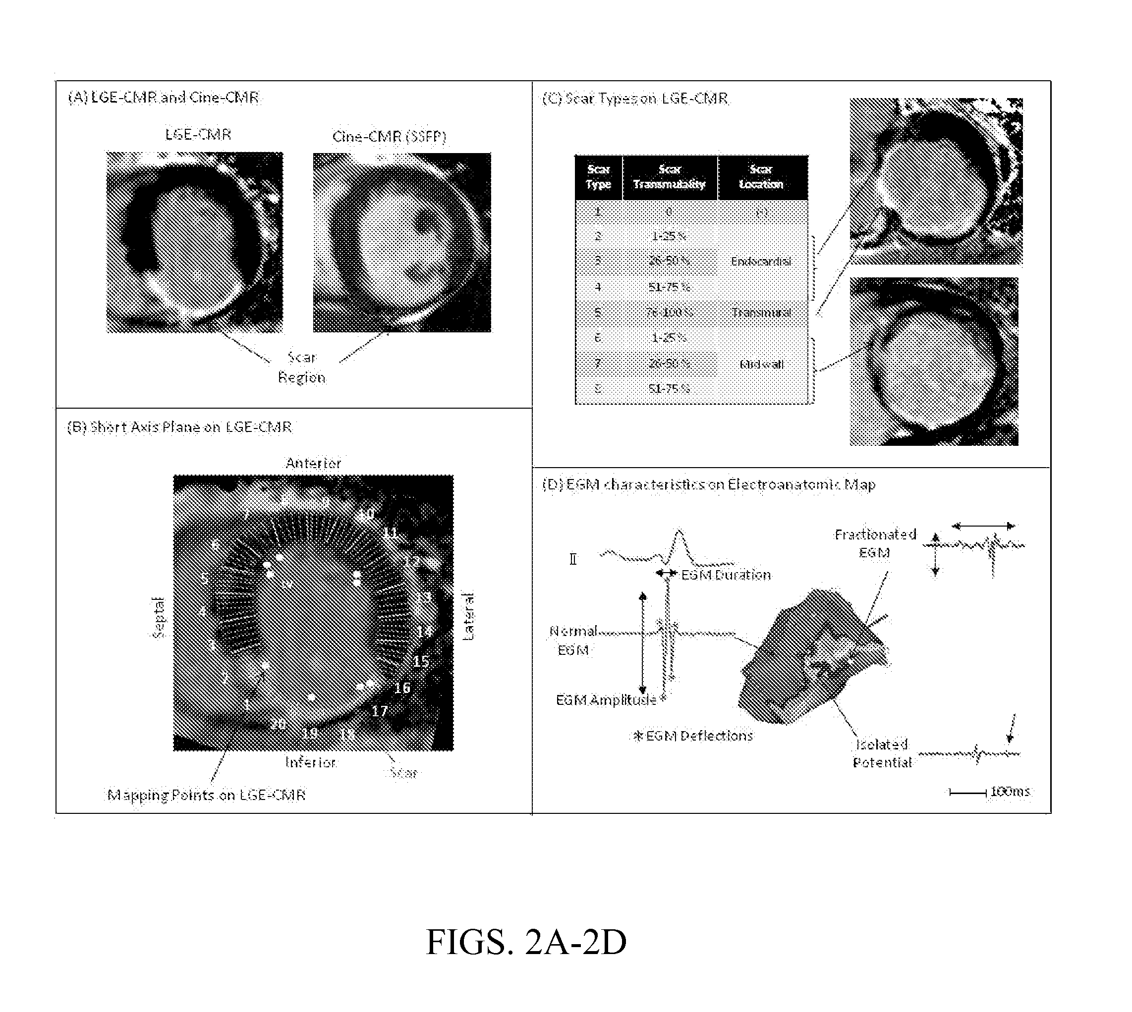
Scar maps can be directly imported into the procedure; however, the extent of scar transmurality and surviving muscle fibers (i.e. heterogeneity of tissues) are difficult to display.

Method used


Image

  • Non-invasive methods and systems for producing cardiac electrogram characteristic maps for use with catheter ablation of ventricular tachycardia
  • Non-invasive methods and systems for producing cardiac electrogram characteristic maps for use with catheter ablation of ventricular tachycardia
  • Non-invasive methods and systems for producing cardiac electrogram characteristic maps for use with catheter ablation of ventricular tachycardia

Examples


examples

[0035]The following examples are provided to help explain further concepts and details of some embodiments of the current invention. However, the general concepts of the current invention are not limited to the particular examples.

Methods

Study Patients

[0036]LGE-CMR images were acquired in 23 patients (23 males, 68±8 years old) with ischemic cardiomyopathy and scar-related VT prior to catheter ablation. The first 13 patients formed the retrospective training set for development of multivariate models, and the subsequent 10 patients formed the prospective test set for model validation. Catheter ablation was performed for recurrent ICD shocks or prior to ICD implantation for secondary prevention of VT.

CMR Studies

[0037]CMR was performed with a 1.5T magnetic resonance scanner (Avanto, Siemens, Erlangen, Germany). In patients with ICD systems, potential risks of exposure to magnetic fields were explained before CMR scans, and CMR images were obtained using our established safety protocol....


PUM


Abstract

A non-invasive method of producing a three-dimensional cardiac electrogram characteristic map for use in catheter ablation of ventricular tachycardia includes receiving left ventricle three-dimensional image data of a patient's heart; segmenting a left ventricle image of the patient's heart based on the left ventricle three-dimensional image data into scar tissue, normal myocardium tissue and left ventricle cavity regions; determining scar tissue thickness and normal myocardium tissue thickness for a plurality of portions of the left ventricle image of the patient's heart; receiving predetermined data that associate a value of at least one electrogram characteristic to each scar tissue thickness and each normal myocardium tissue thickness for the plurality of portions of said left ventricle image of said patient's heart; and generating the three-dimensional cardiac electrogram characteristic map of the at least one electrogram characteristic corresponding to the left ventricle image of the patient's heart based on the predetermined data.

Description

CROSS-REFERENCE OF RELATED APPLICATION[0001]This is a national stage application under 35 U.S.C. §371 of PCT / US2012 / 032768 filed Apr. 9, 2012, the entire contents of which are incorporated herein by reference and this application claims priority to U.S. Provisional Application No. 61 / 472,699 filed Apr. 7, 2011, the entire contents of which are hereby incorporated by reference.[0002]This invention was made with U.S. Government support of Grant No. K23HL089333, awarded by NIH. The U.S. Government has certain rights in this invention.BACKGROUND[0003]1. Field of Invention[0004]The field of the currently claimed embodiments of this invention relates to non-invasive methods and systems of producing cardiac electrogram characteristic maps for use in catheter ablation of ventricular tachycardia.[0005]2. Discussion of Related Art[0006]Radiofrequency catheter ablation is increasingly used as an adjunct to implantable cardioverter defibrillators (ICD) and medical therapy for management of scar...

Claims


Application Information

Patent Timeline
Patent Type & Authority Patents(United States)
IPC IPC(8): G06K9/00A61B5/00A61B5/0402A61B18/14
CPCA61B5/0044A61B5/0402A61B5/4836A61B5/7203A61B5/7278A61B18/1492A61B5/0036A61B5/318
Inventor NAZARIAN, SAMANMILLER, CHRISTOPHERSASAKI, TAKESHI
Owner THE JOHN HOPKINS UNIV SCHOOL OF MEDICINE
Learn More