Agglutination assay

a technology of agglutination and assay, which is applied in the field of agglutination assay, can solve the problems of difficult to achieve stable agglutination with smaller analytes

Inactive Publication Date: 2015-12-01
PLATFORM DIAGNOSTICS LTD
View PDF62 Cites 1 Cited by
  • Summary
  • Abstract
  • Description
  • Claims
  • Application Information

AI Technical Summary

Benefits of technology

[0063]In an advantage of the present invention, the reagents and sample may be applied to the porous carrier in any order and in any combination and there is no requirement for immobilization of a reagent at a fixed point, unlike the lateral flow assays of the art which are generally restricted to applying sample and reagents separately and in a fixed order. Any one or more of the reagents and / or sample may be combined prior to commencement of the assay, either in a separate vessel (such as a capillary or well), or in the porous carrier itself (for example in a region of porous carrier with large pores that permit free movement of agglutinated and non-agglutinated particles). In particular, it may be preferable to combine the hub, second binding partner, and / or detectable particle prior to the assay. Any mixtures may be incubated for a short period of time, for example 1-6 minutes, preferably 2-5 minutes, prior to application to the porous carrier.
, the reagents and sample may be applied to the porous carrier in any order and in any combination and there is no requirement for immobilization of a reagent at a fixed point, unlike the lateral flow assays of the art which are generally restricted to applying sample and reagents separately and in a fixed order. Any one or more of the reagents and / or sample may be combined prior to commencement of the assay, either in a separate vessel (such as a capillary or well), or in the porous carrier itself (for example in a region of porous carrier with large pores that permit free movement of agglutinated and non-agglutinated particles). In particular, it may be preferable to combine the hub, second binding partner, and / or detectable particle prior to the assay. Any mixtures may be incubated for a short period of time, for

Problems solved by technology

Although reasonable agglutination can be achieved using large, multi-epitopic analytes where multiple binding events are possible, it can be difficult to achieve stable agglutination with smaller analytes which may contain only a few epitopes.

Method used


Image

  • Agglutination assay
  • Agglutination assay
  • Agglutination assay

Examples


example 1

Preparation of Soluble Hub Reagent 1

[0133]1. Desalt the anti-hCG (alpha-subunit) into 0.1 M phosphate pH 7.5 buffer, using a 1.6×15 cm G25M Sephadex column, and determine concentration and yield.

[0134]2. Activate the anti-hCG antibody, using 8 molar equivalents of NHS-PEG-MAL. Incubate the reaction mixture at 20° C. for two hours. Quench the reaction with 100 molar equivalents of glycine and desalt the maleimide-activated anti-hCG into 5 mM EDTA, PBS pH 7.3 buffer using two shots down a 1.6×15 cm G50F Sephadex column. Determine concentration and yield of activated antibody.

[0135]3. Activate a 500 kDalton aminodextran using 1000 molar equivalents of 2-Iminothiolane (2-IT). Incubate the reaction mixture at 20° C. for 110 minutes. Desalt the thiol activated aminodextran into 5 mM EDTA, PBS pH 7.3 buffer, using G25M Sephadex media. Determine incorporation ratio of thiol:aminodextran using the Ellman's assay.

[0136]4. Add 25 Molar equivalents of the maleimide-activated anti-hCG antibody t...

example 2

Preparation of Soluble Hub Reagent 2

[0137]1. Desalt the anti-hCG (alpha-subunit) into 0.1 M phosphate pH 7.5 buffer, using a 1.6×15 cm G25M Sephadex column, and determine concentration and yield.

[0138]2. Activate the anti-hCG antibody, using 8 molar equivalents of NHS-PEG-MAL. Incubate the reaction mixture at 20° C. for two hours. Quench the reaction with 100 molar equivalents of glycine and desalt the maleimide-activated anti-hCG into 5 mM EDTA, PBS pH 7.3 buffer using two shots down a 1.6×15 cm G50F Sephadex column. Determine concentration and yield of activated antibody.

[0139]3. Activate a 500 kDalton aminodextran using 1000 molar equivalents of 2-Iminothiolane (2-IT). Incubate the reaction mixture at 20° C. for 110 minutes. Desalt the thiol activated aminodextran into 5 mM EDTA, PBS pH 7.3 buffer, using G25M Sephadex media. Determine incorporation ratio of thiol:aminodextran using the Ellman's assay.

[0140]4. Add 25 Molar equivalents of the maleimide-activated anti-hCG antibody t...

example 3

Preparation of Membrane Strips for Tests Using Wet Reagents

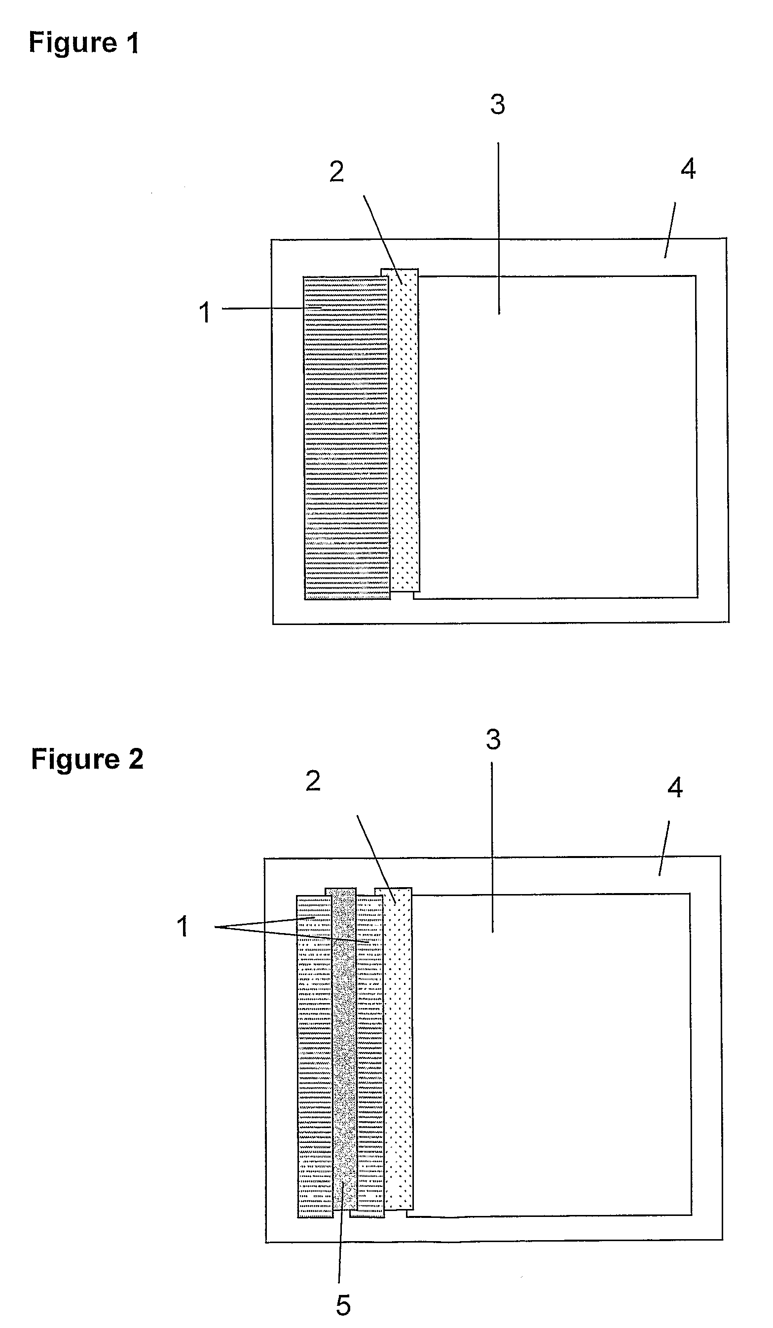
[0142]Membrane materials were cut to size as follows:[0143](i) Wick, e.g. Surewick G028-14 (Millipore), 30 mm×60 mm.[0144](ii) Agglutinate trapping membrane, e.g. Fusion 5 (Whatman), 5 mm×60 mm.[0145](iii) Absorbent sink, e.g. Absorbent Pad 222 (Ahlstrom), 55 mm×60 mm.[0146](iv) Self-adhesive plastic (×2) e.g. 0.04″ Clear polyester with D / C hydrophilic PSA (G&L) 70 mm×100 mm

[0147]A composite ‘card’ of the above materials was assembled as shown in FIG. 1. Adjacent membrane materials were overlapped by approximately 1 mm, to ensure good fluid transfer between successive sections of the strip. The second sheet of self-adhesive plastic was applied firmly to the upper surface. The resulting ‘card’ was sliced into 5 mm strips and the plastic trimmed to allow reagents and sample to enter the wick.


PUM

PropertyMeasurementUnit
diameteraaaaaaaaaa
diameteraaaaaaaaaa
lengthaaaaaaaaaa

Abstract

The invention relates to agglutination assays and related kits, reagents and devices. In particular methods of assaying small analytes having few epitopes are disclosed, by means of using hub moieties to which multiple analytes may be bound by a first epitope, together with a further moiety capable of binding a second analyte epitope and which is also capable of binding to a detectable particle. Stable agglutinated complexes may be so formed, which may used as the basis for various assay formats.

Description

REFERENCE TO RELATED APPLICATIONS[0001]This application is the US national phase entry of International Patent Application No. PCT / GB2006 / 004204, filed Nov. 13, 2006, which claims priority to UK Patent Applications No. 0523124.6, filed Nov. 12, 2005 and 0610973.0 filed Jun. 3, 2006.FIELD OF THE INVENTION[0002]The present invention relates to an agglutination assay for detection of analytes in a sample. In particular, the invention relates to porous carrier-based assays and assay devices, kits comprising means for conducting such assays, and assay methods for the detection of analyte in a sample.BACKGROUND TO THE INVENTION[0003]Immunoassays are a well established technique for detecting and quantifying analytes in samples. They are particularly useful for detecting and / or measuring substances in biological samples as an aid to disease diagnosis and prognosis, and for predicting a patient's response to therapy. Techniques such as the radioimmunoassay and enzyme immunoassay, which revo...

Claims


Application Information

Patent Timeline
Patent Type & Authority Patents(United States)
IPC IPC(8): G01N33/558G01N33/563G01N33/543
CPCG01N33/558G01N33/54313
Inventor RUDDELL, CAROLYN JENNIFERALLEN, GERALD JOHNEVANS, DOUGLAS ROBERTBURKE, ELIZABETH
Owner PLATFORM DIAGNOSTICS LTD
Why Eureka
Try Eureka