Patient-specific craniofacial implants

a craniofacial implant and patient-specific technology, applied in the field of patient-specific craniofacial implants, can solve the problems of voids of different sizes and shapes in bone and soft tissue, voids left in the frontal, parietal and/or temporal areas of the craniofacial skeleton that need repair, and lack of adequate aesthetic reconstruction of the area through bone void filling. to achieve the effect of restoring proper appearan

Active Publication Date: 2015-12-22
HOWMEDICA OSTEONICS CORP
View PDF28 Cites 15 Cited by
  • Summary
  • Abstract
  • Description
  • Claims
  • Application Information

AI Technical Summary

Benefits of technology

[0009]The patient-specific, dual-purpose temporal implants of the present invention are designed to fill a void in the skull while also simultaneously augmenting the pterional / temporal area. Preferably, alloplastic material is used in the construction of such patient-specific temporal implants. The mechanical properties of the alloplastic material used to construct these implants will allow them to cover the void and to recreate the soft tissue bulk that is preferably present in the temporal area prior to any injury and / or atrophy occurring in a patient. The patient-specific implants of the present invention are designed to fill a cranial void while also restoring aesthetic symmetry by augmenting the temporal area of the skull to counter temporal hollowing. Therefore, the patient-specific cranial implants of the present invention may be used to replace a bony void left by a craniotomy either as a preemptive or prophylactic solution to addressing aesthetic asymmetry that may occur due to atrophy of temporal muscle or overlying fat pad of the bony void.
[0019]A second aspect of the present invention is a craniofacial implant comprising a base portion having an outer surface curved in a medial to lateral direction, a superior to inferior direction, and a posterior to anterior direction, and a curved augment portion protruding outwardly from the outer surface of the base portion in the medial to lateral direction to restore proper appearance anywhere along the temporal skeleton when both a hard tissue deformity and soft tissue deformity co-exist. The craniofacial implant of the present invention could also be designed for soft-tissue only defects, providing the surgeon a new method to camouflage assymetries—both from congential and acquired etiologies.

Problems solved by technology

Certain congenital conditions and acquired deformities related to surgery, irradiation, and / or trauma may result in varying sized and shaped voids in bone and soft tissue.
For example, severe impacts to the head could leave the frontal, parietal and / or temporal areas of the craniofacial skeleton in need of repair.
Bone flaps, standard or customized alloplastic implants, titanium mesh, and liquid methyl methacrylate that are used to fill bone voids do not account for soft tissue atrophy in the temporal area of the skull.
Thus, these bone void filling means do not provide an adequate aesthetic reconstruction of this area.
Both aesthetic and reconstructive procedures that violate the temporal fat pad may result in temporal hollowing.
In some common instances, the temporalis muscle cannot be re-inserted since the bone flap is unable to be placed back into position, thereby leaving an absent fixation point adjacent the temporal crest.
Many patients who have undergone neurosurgical and / or temporal procedures may lose the integrity of the temporalis muscle during temporal or pterional craniotomy surgery.
Both aesthetic and reconstructive procedures that violate the temporal fat pad, such as coronal incisional approaches, for instance, may also result in temporal hollowing, which can happen if a surgeon devascularizes the area with dissection or if the area has received irradiation or tissue resection for oncological treatment.
Thus, these solutions are problematic because a revision surgery is generally required to correct the deformity, and the patient will likely exhibit aesthetic asymmetry prior to the revision surgery.

Method used


Image

  • Patient-specific craniofacial implants
  • Patient-specific craniofacial implants
  • Patient-specific craniofacial implants

Examples


Embodiment Construction

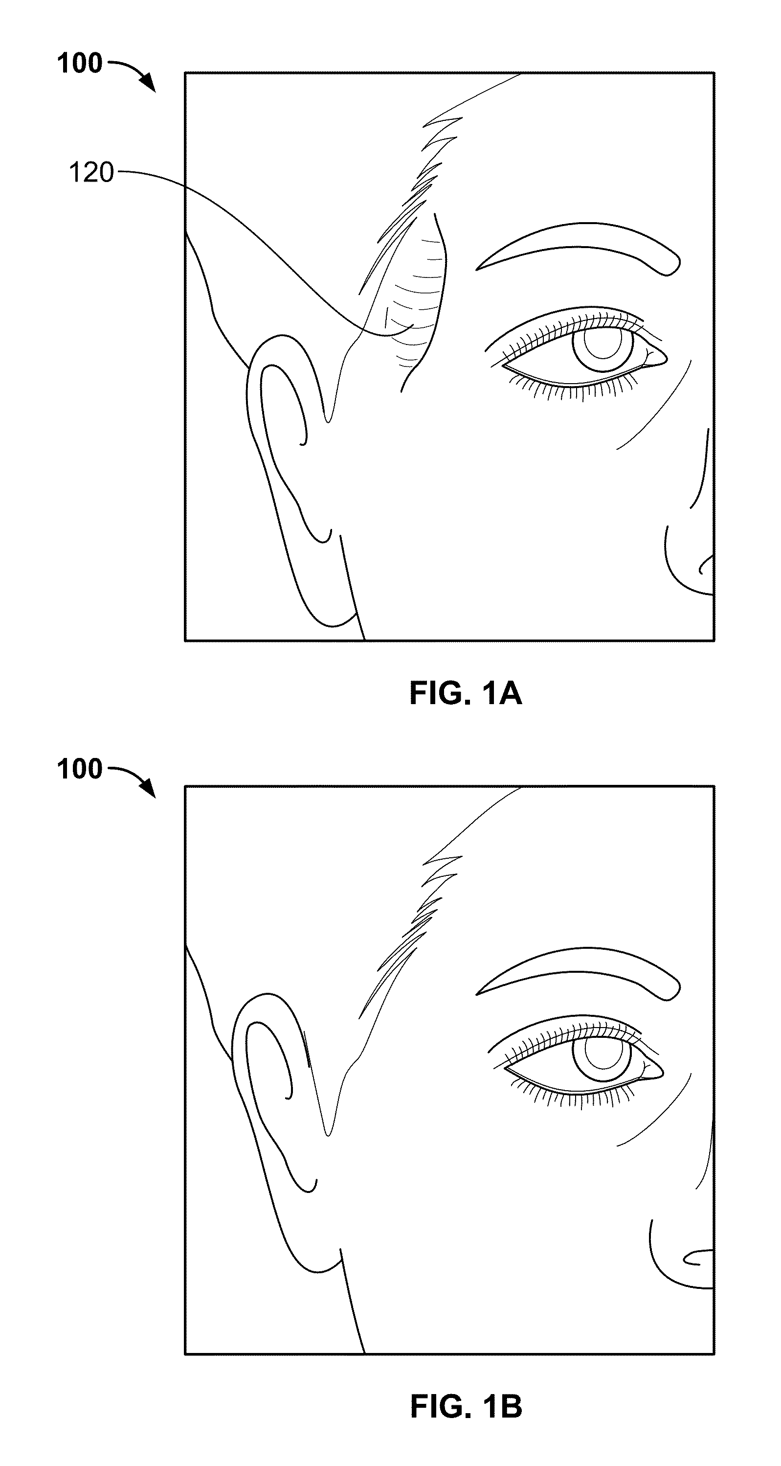
[0053]FIG. 1A is a perspective view of a patient's face 100 exhibiting temporal hollowing 120 in the pterional region of the skull of the patient. The size, shape and location of such temporal hollowing of a patient may differ based on patient's anatomy as well as the type of injury and / or the amount of tissue atrophy incurred by the patient in this region.

[0054]FIG. 1B is a perspective view of the patient's face with a repaired pterional region such that the temporal hollowing has been corrected and is no longer present. After an initial surgery to correct a bony void is performed, a subsequent procedure using a pterional graft, PMMA, filler, absorbable material or tissue engineered substrate, for example, may be performed in order to repair the soft tissue defect. The subsequent procedure may be conducted via injection of PMMA percutaneously into this region or by placing a pterional flap through a small incision made in the skin. However, a patient will likely exhibit aesthetic a...


PUM


Abstract

Disclosed herein are patient-specific craniofacial implants structured for filling bone voids in the cranium as well as for simultaneously providing soft tissue reconstruction and / or augmentation for improved aesthetic symmetry and appearance. Pterional voids or defects generally result from a chromic skull deformity along with a compromised temporalis muscle or soft tissue distortion from previous surgery. When muscle atrophy occurs in the pterion, temporal hollowing generally results where there would be soft tissue but for the atrophy. The patient-specific temporal implants herein are configured to have an augmented region adjacent the temporal region of the cranium in order to account for and correct any such temporal hollowing.

Description

FIELD OF THE INVENTION[0001]The present invention relates to patient-specific craniofacial implants and methods of designing such implants for filling both bone and soft tissue in the temporal region of a patient's head.BACKGROUND OF THE INVENTION[0002]Certain congenital conditions and acquired deformities related to surgery, irradiation, and / or trauma may result in varying sized and shaped voids in bone and soft tissue. For example, severe impacts to the head could leave the frontal, parietal and / or temporal areas of the craniofacial skeleton in need of repair. Cranial bone voids are commonly filled throughout the anterior, middle, and posterior cranial vaults, for example, using autologous bone flaps, standard or customized alloplastic implants, titanium mesh, biologic absorbable materials, tissue engineered substrates and / or liquid methyl methacrylate in order to provide much needed cerebral protection. However, in times where secondary cranioplasty requires a bone substitute, te...

Claims


Application Information

Patent Timeline
Patent Type & Authority Patents(United States)
IPC IPC(8): A61F2/28A61F2/00A61F2/30
CPCA61F2/2875A61F2/0059A61F2240/002A61F2002/30948A61F2002/30952A61F2/30942A61F2/28
Inventor GORDON, CHADBRYNDZA, JOHN ROCCOBISIC, TAHIR
Owner HOWMEDICA OSTEONICS CORP
Why Eureka
Try Eureka