Device and method for viewing a body lumen

a technology of body lumen and device, which is applied in the field of in vivo viewing, can solve the problems of limited angle of view of imager or the accessibility of such a sensor located at the endoscope tip, behind, to remote or concealed portions and limit the caregiver from viewing what he is performing at all times and from all angles. to achieve the effect of better viewing and/or imaging of the body lumen

Active Publication Date: 2016-05-17
GIVEN IMAGING LTD
View PDF60 Cites 3 Cited by
  • Summary
  • Abstract
  • Description
  • Claims
  • Application Information

AI Technical Summary

Benefits of technology

[0012]Embodiments of the present invention provide a device and methods for better viewing and / or imaging a body lumen.
[0014]In some embodiments the imaging device may have a shape configured for fully or partially wrapping the outer surface of a second device, e.g. an endoscope, colonoscope, Double-Balloon Endoscopy™ system, catheter, needle, laparoscope etc. In some embodiments, the shape of the imaging device's housing may be designed to have a curve which may fit an outer section of different devices having a spherical, square or other shaped profile. In some embodiments, the in vivo imaging device may be connected to an endoscope type device. According to some embodiments, the imaging device is connected to the endoscope through at least two semi rigid cables of a predetermined length. Typically, the number of connectors between the capsule and the endoscope is at least two, for easier and more stabilized positioning of the capsule in relation to the endoscope.

Problems solved by technology

The angle of view afforded by an imager or the accessibility of such a sensor that is located at the endoscope tip, or behind the optical window of the capsule, to remote or concealed portions of the body lumen, such as the colon in the gastrointestinal (GI) tract, which also has many folds inside, is limited.
In an area which includes many folds, e.g. the colon, the endoscope's imager may not be able to view a pathology placed within a fold, which limits the caregiver from viewing what he is performing at all times and from all angles.

Method used


Image

  • Device and method for viewing a body lumen
  • Device and method for viewing a body lumen
  • Device and method for viewing a body lumen

Examples


Embodiment Construction

[0032]In the following detailed description, numerous specific details are set forth in order to provide a thorough understanding of the invention. However, it will be understood by those skilled in the art that the present invention may be practiced without these specific details. In other instances, well-known methods, procedures, and components have not been described in detail so as to not obscure the present invention.

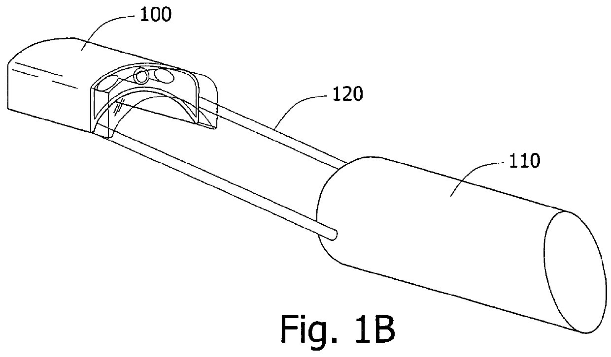
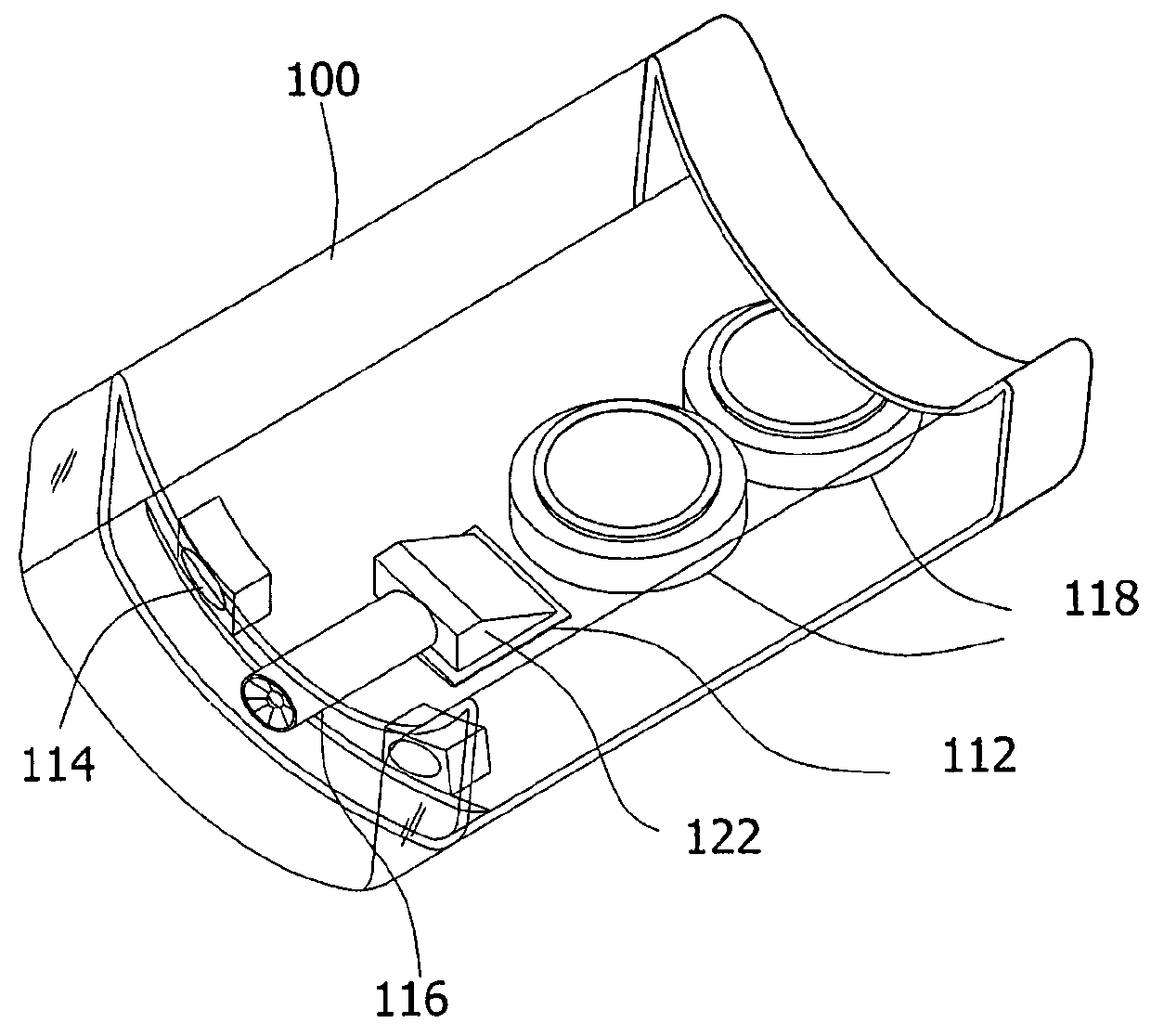
[0033]Some embodiments of the present invention are directed to a typically swallowable in-vivo device. Some embodiments are directed to a capsule endoscope connected to an endoscope that may actively progress through a body lumen, e.g., the gastro-intestinal (GI) tract. In some embodiments, the in vivo imaging device may include in addition to an imaging unit or an imager, other sensors, for example, a pH sensor, a temperature sensor, a pressure sensor, sensors of other in-vivo parameters, or the like. Devices systems and methods according to some embodiments of ...


PUM


Abstract

A device and method for viewing a body lumen are provided. An assembly of a capsule endoscope and an endoscope is inserted into a body lumen. The capsule endoscope is positioned within the body lumen at a predetermined distance from the endoscope, and images are obtained from opposite sides of a desired location in the body lumen. In vivo procedures may be performed, while images of the procedure are obtained from opposite sides of the procedure location.

Description

CROSS-REFERENCE TO RELATED APPLICATIONS[0001]This application claims the benefit of U.S. Provisional Patent Application No. 60 / 929,923, filed on Jul. 18, 2007, which is incorporated in its entirety herein by reference.FIELD OF THE INVENTION[0002]The present invention relates to the field of in vivo viewing. More specifically, the present invention relates to an in vivo device and methods for viewing a body lumen through either an endoscope or a capsule endoscope, or through both.BACKGROUND OF THE INVENTION[0003]Medical procedures in body lumens and cavities, such as gastroenterology procedures and laparoscopic surgery procedures, may require specifically designed medical devices. Typically, the devices include a performing end (distal end) functionally coupled to a controlling end (proximal end). The performing end, which is inserted into the body, is operated and manipulated by the controlling end, which is accessible to an external operator.[0004]In some cases the device further i...

Claims


Application Information

Patent Timeline
Patent Type & Authority Patents(United States)
IPC IPC(8): A61B1/00A61B1/06A61B1/04A61B1/05
CPCA61B1/04A61B1/041A61B1/00183A61B1/053A61B1/00101
Inventor GILAD, ZVIKAPASCAL, AMIT
Owner GIVEN IMAGING LTD
Why Eureka
Try Eureka