Image processing apparatus for processing a tomogram of an eye to be examined, image processing method, and computer-readable storage medium
a tomogram and image processing technology, applied in the field of image processing apparatus, image processing method, computer program, can solve the problem of insufficient contrast between the oct image and the other image, and achieve the effect of high precision
- Summary
- Abstract
- Description
- Claims
- Application Information
AI Technical Summary
Benefits of technology
Problems solved by technology
Method used
Image
Examples
first embodiment
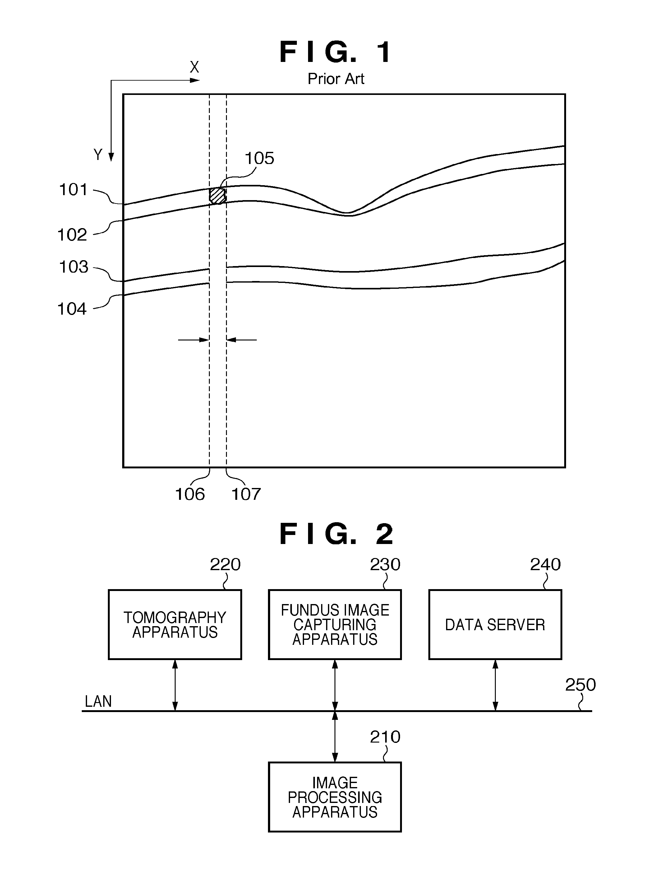
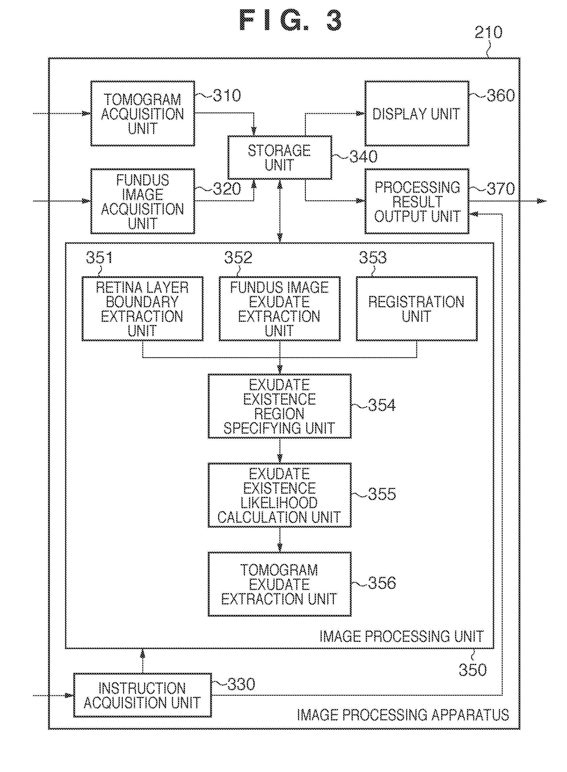
[0024]The apparatus arrangement of an image processing system according to the first embodiment will be described below with reference to FIG. 2. An image processing apparatus 210 applies image processing to images captured by a tomography apparatus 220 and fundus image capturing apparatus 230, and images stored in a data server 240. The detailed arrangement of the image processing apparatus 210 is as shown in FIG. 3. The tomography apparatus 220 obtains a tomogram of an eye portion, and includes, for example, a time domain OCT or Fourier domain OCT. The tomography apparatus 220 three-dimensionally obtains a tomogram of an eye portion of an object (patient: not shown) according to operations of an operator (engineer or doctor: not shown). The acquired tomogram is transmitted to the image processing apparatus 210 and data server 240.
[0025]The fundus image capturing apparatus 230 captures a two-dimensional (2D) image of an eye fundus portion. The fundus image capturing apparatus 230 t...
second embodiment
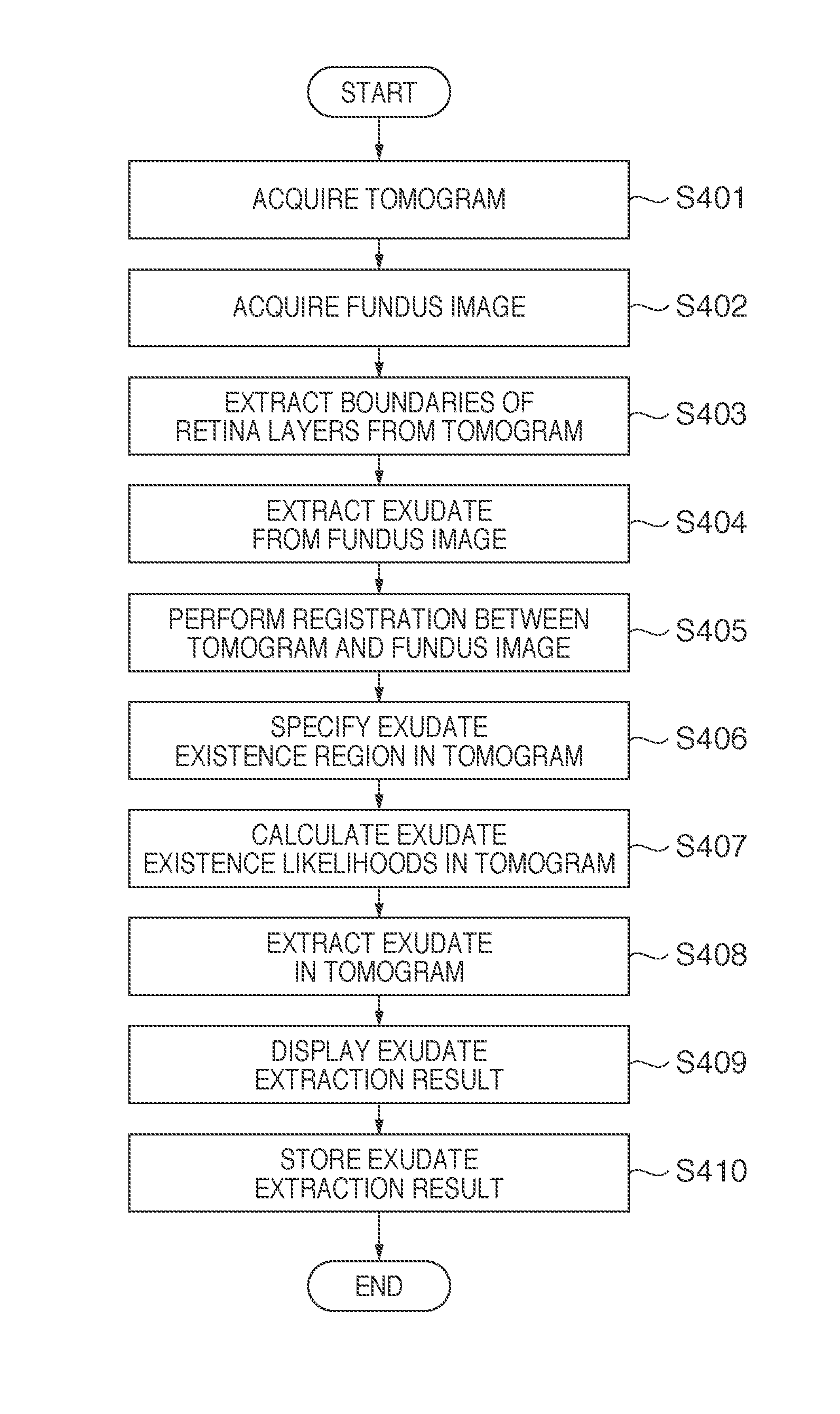
[0052]This embodiment will explain an example of a modification in which exudate existence likelihoods in step S407 of the first embodiment are set based on previous knowledge about correlation information between the distances between the boundaries of retina layers and existence likelihoods. The apparatus arrangement of an image processing system corresponding to this embodiment is the same as that shown in FIGS. 2 and 3. In the first embodiment, a highest existence likelihood is assigned to a position separated from a retinal pigment epithelium boundary by a given distance RPE-EXUDATE, and smaller existence likelihoods are assigned with increasing distance from this position. However, an inner limiting membrane—retinal pigment epithelium boundary distance varies depending on objects. Even in a single object, the distance varies depending on locations in an eye fundus. For this reason, the aforementioned existence likelihood setting method is unlikely to appropriately assign likel...
third embodiment
[0059]This embodiment will explain an example in which extraction error determination about a region extracted by exudate extraction in a tomogram in step S408 in FIG. 4 is made. In step S406, a region to which exudate extraction processing is to be applied is limited using an exudate region acquired from a fundus image in addition to retina layer extraction results. For this reason, when a region other than an exudate in the fundus image is erroneously extracted as an exudate, the exudate extraction processing is unwantedly applied to a region where no exudate exists in the tomogram. This embodiment will be described in detail below.
[0060]FIG. 9 is a view for explaining the aforementioned extraction error. Referring to FIG. 9, reference numeral 901 denotes an inner limiting membrane; 910, a boundary between an optic nerve fiber layer and its underlying layer; 902, a junction between inner and outer photoreceptor segments; and 903, retinal pigment epithelium boundary. Also, referenc...
PUM
 Abstract
Description
Claims
Application Information
 - R&D
- Intellectual Property
- Life Sciences
- Materials
- Tech Scout
- Unparalleled Data Quality
- Higher Quality Content
- 60% Fewer Hallucinations
Browse by: Latest US Patents, China's latest patents, Technical Efficacy Thesaurus, Application Domain, Technology Topic, Popular Technical Reports.
© 2025 PatSnap. All rights reserved.Legal|Privacy policy|Modern Slavery Act Transparency Statement|Sitemap|About US| Contact US: help@patsnap.com


