
Measuring blood flow and venous capacitance

a technology of venous capacitance and blood flow, which is applied in the field of measuring blood flow and venous capacitance, can solve the problems of non-linear pressure versus volume relationship, low pressure level, and inability to increase the pressure of the contained blood

Active Publication Date: 2016-10-25
ITAMAR MEDICAL LTD
View PDF14 Cites 4 Cited by
  • Summary
  • Abstract
  • Description
  • Claims
  • Application Information

AI Technical Summary

Benefits of technology

Enables non-invasive, quantitative measurements of volumetric blood flow and venous capacitance at any body level, overcoming the limitations of traditional VOP methods by preventing venous distension and maintaining linearity of the pressure-volume relationship.

Problems solved by technology

However as the veins become increasingly filled, and the venous wall begins to be stretched, the ability of the veins to expand without increasing the pressure of the contained blood tends to be reduced, upon pressure increases, thereby resulting in a non-linear pressure versus volume relationship.
Nevertheless, a substantial portion of the volume of blood which the veins are able to accommodate from their fully emptied state, occurs without the venous wall being stretched, and this occurs at a very low level of pressure.
A major limitation of VOP, as it is currently practiced, is that it is restricted to measurements in body parts which are maintained above heart level, for the following reason: The combination of extremely high venous compliance described above, and the generation of hydrostatic gradients in the vascular system due to vertical displacement relative to heart level, means that the veins in body parts which are below heart level easily become filled with blood and therefore lose the capacity to freely accommodate additional volumes of blood.
When venous blood vessels become filled due to hydrostatic pressure gradients, the partially filled veins cannot be accurately used for VOP measurements due to the relative reduction in the ability to further expand and the lack of linearity of the pressure volume relationship because of vessel wall stretching.
While the filling of the veins due to such hydrostatic pressures can be prevented by the application of a sufficient level of external counter pressure to the measurement site (so as to counterbalance the intra-venous pressure), traditional VOP volumetric collecting cups are mechanically incapable of being pressurized to any appreciable degree without being forced off the measurement site.
They are therefore unable to provide the necessary degree of pressure to counterbalance the intra-venous pressure, and thus cannot be used to reliably measure BF below heart level.
Substitute collecting devices, such as mercury in silastic rings or circumferential strain gauges, provide no actual pressure field to the bulk of the tissues distal to the venous occlusion cuff, so that resting venous distention cannot be counterbalanced by any of these methods.
Furthermore, since they do not actually measure volume but rather an index of circumference, they are not capable of being accurately calibrated to provide an index of volume.
Because of these limitations, traditional VOP devices are not able to modify the local venous transmural blood pressure so as to allow BF measurements or venous capacitance at specific pressure levels to be made when the measured body part is below heart level, and therefore, as noted above, VOP measurements have generally been limited to body regions which are located above the subject's heart or are deliberately elevated above heart level.

Method used


Image

  • Measuring blood flow and venous capacitance
  • Measuring blood flow and venous capacitance
  • Measuring blood flow and venous capacitance

Examples


Embodiment Construction

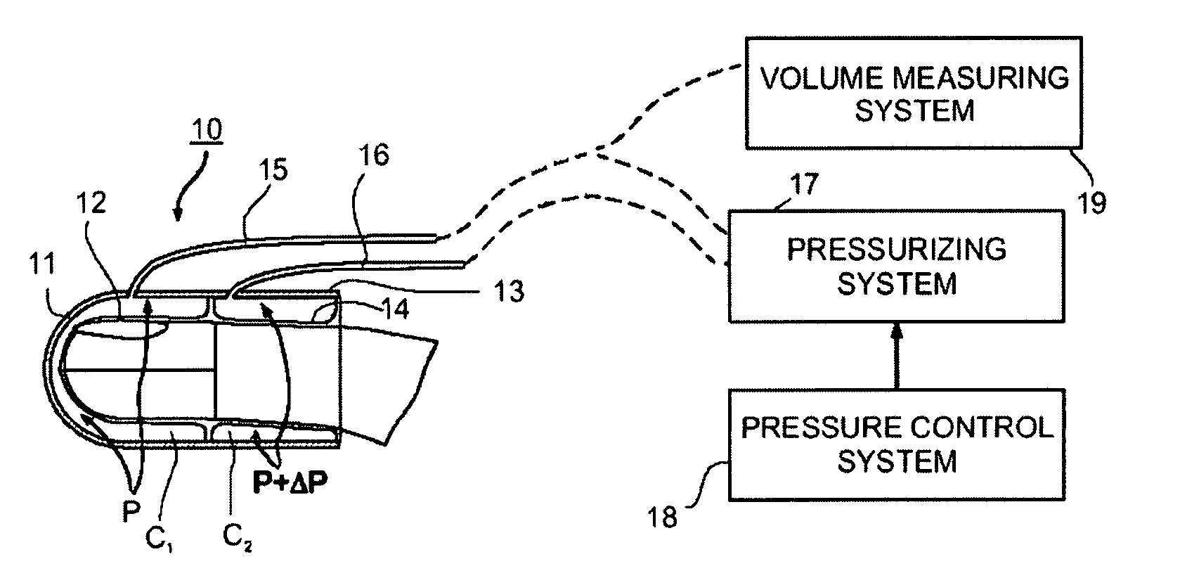
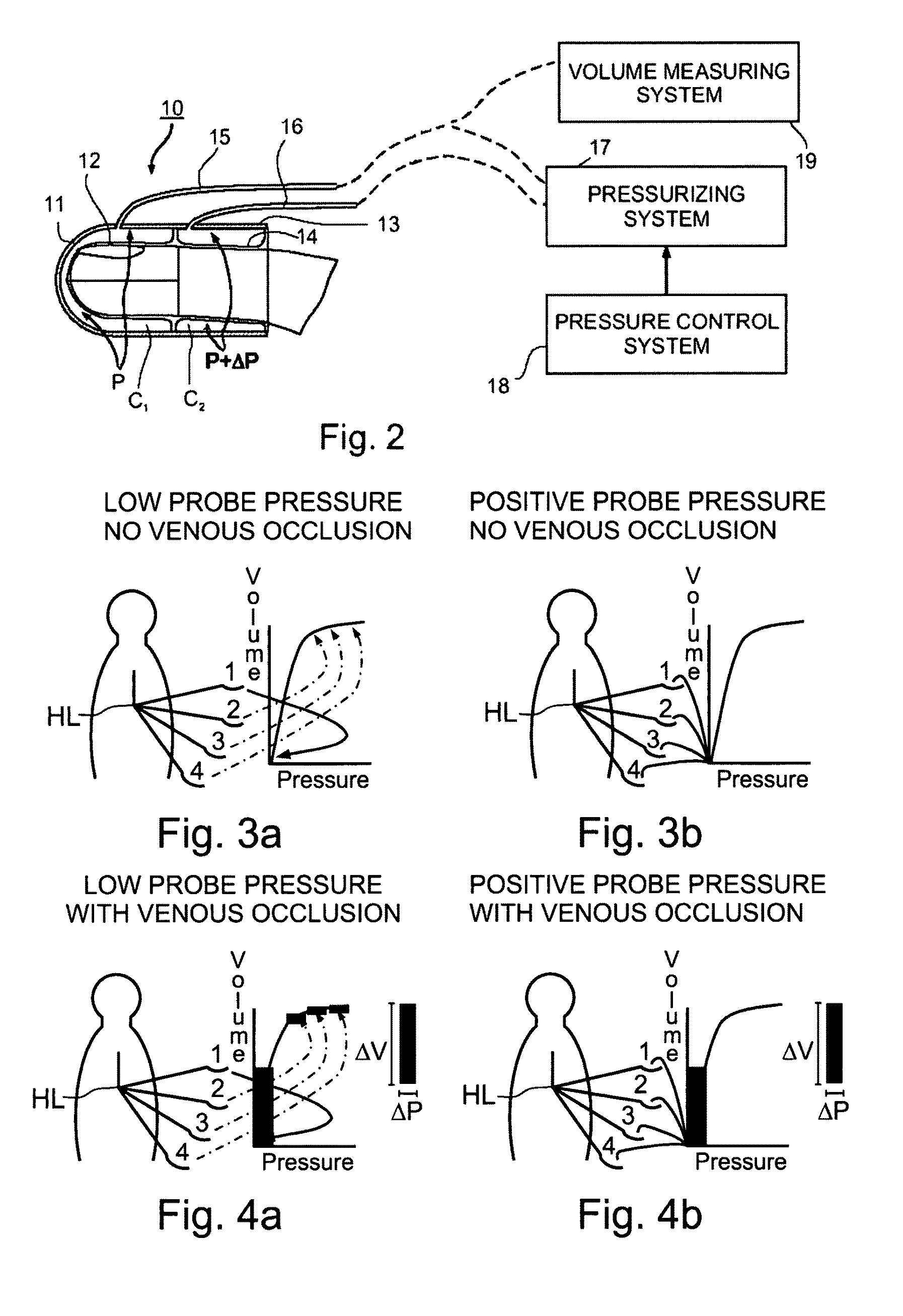
[0042]FIG. 2 illustrates one form of apparatus which may be used for non-invasively measuring a physiological parameter of a subject in accordance with the present invention. The apparatus illustrated in FIG. 2 includes a peripheral arterial tonometry (PAT) probe constructed as described in the above-cited patents to be applied to a selected body region, a pressurizing system controlled to pressurize different sections of the probe in accordance with the present invention, and a measuring system for measuring volume changes in the body region as a result of the controlled pressurization changes applied to the body region. In the example illustrated in FIG. 2, the body region is a finger (or toe) of the subject.

[0043]Thus, the apparatus illustrated in FIG. 2 includes a PAT probe, generally designated 10, serving as the venous occlusion plethysmographic device. Probe 10 includes a housing having a first section 11 configured to enclose the distal tip of the finger and carrying, on its...


PUM


Abstract

Non-invasively measuring a physiological parameter by applying to a subject's body part a venous occlusion plethysmographic device including a first section having a first fluid chamber and at least a second region having a second fluid chamber to engage a first region of the body part and a second region of the body part, proximal of the first region; pressurizing the first chamber to a pressure sufficient to compensate for hydrostatic pressure added to the inherent venous pressure at the maximal vertical lowering level of the body part relative to the subject's heart level; intermittently raising and lowering the pressure in the second fluid chamber to a pressure above or equal to the pressure in the first chamber, but below the arterial blood pressure; measuring changes in volume of the first region of the body part; and utilizing the measured volume changes to provide a measure of the physiological parameter.

Description

RELATED APPLICATIONS[0001]This Application is a National Phase of PCT Patent Application No. PCT / IL2005 / 000993 having International Filing Date of Sep. 15, 2005, which claims the benefit of U.S. Provisional Patent Application No. 60 / 609,813 filed on Sep. 15, 2004. The contents of the above Applications are all incorporated herein by reference.FIELD AND BACKGROUND OF THE PRESENT INVENTION[0002]The present invention relates to a method and apparatus for non-invasively measuring certain physiological parameters of a subject. The invention is particularly useful in venous occlusion plethysmography (VOP) for measuring volumetric blood flow, or other physiological parameters derived from volumetric blood flow, as well as providing certain-quantitative indices of venous compliance, and is therefore described below with respect to such applications.[0003]Venous occlusion plethysmography is a known method for non-invasively measuring a physiological parameter of a subject, particularly volum...

Claims


Application Information

Patent Timeline
Patent Type & Authority Patents(United States)
IPC IPC(8): A61B5/02A61B5/022A61B5/029A61B5/00
CPCA61B5/02241A61B5/029A61B5/6826A61B5/6838A61B5/7285A61B2560/0261
Inventor SCHNALL, ROBERT P.
Owner ITAMAR MEDICAL LTD
Learn More