Methods, systems and devices for treatment of cerebrospinal venous insufficiency and multiple sclerosis

a cerebrospinal vein and multiple sclerosis technology, applied in the field of biology and medicine, can solve the problems of ms being difficult to diagnose in its early stages, neuroaxonal apoptosis and degeneration, and unable to cure ms, so as to improve the and improve the effect of flow through the stenotic body lumen

Active Publication Date: 2019-06-06
EXPLORAMED DEV LLC
View PDF0 Cites 0 Cited by
  • Summary
  • Abstract
  • Description
  • Claims
  • Application Information

AI Technical Summary

Benefits of technology

[0021]In accordance with one aspect of the present invention, there are provided endovascular devices that are implanted within a stenotic region of a body lumen, such stent devices being modifiable (e.g., “tunable”) following initial implantation to optimize their efficacy and minimize untoward effects. For example, the present invention includes endovascular stents that are a) initially constrainable in a collapsed configuration having a first diameter suitable for advancement into the stenotic region of the body lumen, b) self-expandable to a second diameter when unconstrained and c) further pressure-expandable in whole or in part to diameter(s) larger than the second diameter. Such stents are of a suitable design and construction. For example, in one embodiment, such stent comprises one or more self-expanding components (e.g., zig-zag, mesh ring(s), helical members, braided sections, etc.) with one or more plastically deformable constraining member(s) (e.g., stretchable filaments) connected thereto. The plastically deformable constraining member(s) initially constrain the stent so that it cannot self-expand beyond the second diameter. Thereafter, if it is desired to further expand all or, importantly, specific region(s) of the stent beyond the second diameter, a balloon or other expandable member may be positioned within the stent and used to plastically deform one or more of the constraining member(s) to further expand the stent (or selected region(s) thereof) to at least a third diameter which is larger than the second diameter. The stent is up-sized in specific regions so that it fully opposes the wall of the vein along the length of the stent and provides anchoring sections in which the up-sized regions are upstream or downstream of narrow venous sections such that under sizing does not occur as is common with conventional stents.
[0034]In accordance with yet another aspect of the present invention, there are provided methods for improving flow through a stenotic body lumen in a human or animal subject (e.g., the lumen of a vein in a patient suffering from CCVI, MS or another neurodegenerative disease), comprising the step of delivering energy to a wall of the stenotic body lumen or to tissue located near the stenotic body lumen or to obstructive matter located within the stenotic body lumen to cause shrinkage, contraction or other movement of tissue or obstructive matter in a manner that improves fluid flow or equalization of pressure through the stenotic body lumen. The energy is delivered from a catheter or other delivery device that is outside or inserted into the body lumen or from an endovascular or exovascular device implanted within or near the body lumen. The energy comprises any suitable type of energy including but not limited to heat, light, laser light, infrared light, a plasma field, sound, ultrasound, electromagnetic radiation and ionizing radiation.
[0035]In accordance with yet another aspect of the present invention, there are provided methods for improving flow through a stenotic body lumen in a human or animal subject (e.g., the lumen of a vein in a patient suffering from CCVI, MS or another neurodegenerative disease), comprising the step of delivering a quantity of material (e.g., a gel or foam) to a location within the subject's body so that the material exerts force(s) which enlarge the stenotic body lumen or deter compression or contraction of the stenotic body lumen (e.g., shield the vein so that forces of muscular contraction do not cause the vein to collapse). In some embodiments, the material may undergo a change in volume or shape after it has been delivered. For example, the material may contract (e.g., shrink) in situ so as to pull on adjacent tissue, causing enlargement of the stenotic body lumen.
[0036]In accordance with yet another aspect of the present invention, there are provided methods for improving flow through a stenotic body lumen in a human or animal subject (e.g., the lumen of a vein in a patient suffering from CCVI, MS or another neurodegenerative disease), comprising the step of implanting within the body lumen a device that comprises two or more strut members extending between two or more radially expandable ring or partial-ring members.
[0038]In accordance with yet another aspect of the present invention, there are provided methods for improving flow through a stenotic body lumen in a human or animal subject (e.g., the lumen of a vein in a patient suffering from CCVI, MS or another neurodegenerative disease), comprising the step of implanting magnets at locations in or near the stenotic body lumen. For example, the magnets are positioned on opposite sides of the body lumen and repel one another so as to dilate the body lumen.
[0041]In accordance with yet another aspect of the present invention, there are provided methods and apparatus whereby endovascular devices, such as stents, are prevented or deterred from moving or migrating from a desired implantation site within a blood vessel. In some embodiments, the endovascular device may comprise a radially expandable (e.g., self-expanding or pressure-deformable) scaffold member with incorporated or associated means (e.g., apparatus, materials, attributes and / or operational features) for deterring unwanted movement or migration after implantation. The means (e.g., apparatus, materials, attributes, operational features and / or method steps) which may be incorporated in, associated with or used in an endovascular device to deter unwanted movement or migration of the endovascular device include but are not limited to; a) protrusions, members, hooks, barbs, ridges, surface features, bumps, or other projections or members on the endovascular device that enter, embed in, penetrate and / or frictionally engage the wall of the blood vessel in which the endovascular device is positioned, b) magnets on the endovascular device and / or in the blood vessel wall and / or outside of the blood vessel which create a magnetic attraction to deter unwanted migration or movement of the endovascular device, c) adhesives, tissue glues or other materials which cause the endovascular device to adhere to or remain in contact with the adjacent blood vessel wall, d) the application of endothelial seeking, growth factors, cells, cell growth promoters or fenestrations, passageways or surface features into or onto which tissue (e.g., endothelium) may grow such that the endovascular device or any part thereof becomes encapsulated, overgrown, covered, fully or partially surrounded or otherwise engaged by cells or tissue (e.g., endothelial tissue) in a manner which deters its unwanted movement or migration and / or e) design / radial expansion attributes of the device and / or method steps during implantation of the device which cause the device to remain in sufficient abutment or engagement with the blood vessel wall to deter its unwanted movement or migration, including in cases where implanted in a vein or capacitance vessel, the ability of the device to expand and remain in coaptation or abutment with the vein wall even when the vein undergoes physiological dilation or radial expansion.

Problems solved by technology

Multiple sclerosis (MS) causes demyelination and scarring of the axons, which in turn results in impaired conduction of action potentials, neuroaxonal apoptosis and degeneration.
MS is difficult to diagnose in its early stages.
There is no known cure for MS and the specific pathophysiology by which MS progresses is not fully understood.
MS is not considered a hereditary disease but certain genetic variations are associated with increased risk of developing MS.
The patient's immune system can then activate those T lymphocytes, causing autoimmune damage to the neural cells.
However, this initial study, the devices and techniques employed were generally designed for treatment of other types of obstructions in other lumens of the body and, thus, are less than optimal for use in this application.
Also, other investigators have failed to confirm any increased incidence of CCVI in MS patients.
One recent publication has reported that the re-stenosis rate following angioplasty without stenting was unacceptably high (approximately 50% for internal jugular vein occlusions) and that such re-stenosis was often accompanied by worsening neurological symptoms.
Generally, use of these stents is less than optimal because a number of adverse complications can occur.
None of the existing methods, devices or systems are believed to be optimal for treating all cases of chronic cerebrospinal venous insufficiency in MS patients.
There is an absence in the market of dedicated devices of the proper size, length and functional characteristics for treating CCVI.
Using existing stents at the level of the internal jugular vein and / or azygous vein creates risk of displacement, migration along the vein, stenosis, thrombosis, thromboembolism, migration thru and / or perforation of the venous wall and pulmonary migration, thus affecting venous outflow.

Method used


Image

  • Methods, systems and devices for treatment of cerebrospinal venous insufficiency and multiple sclerosis
  • Methods, systems and devices for treatment of cerebrospinal venous insufficiency and multiple sclerosis
  • Methods, systems and devices for treatment of cerebrospinal venous insufficiency and multiple sclerosis

Examples


Embodiment Construction

[0104]The following detailed description and the accompanying drawings to which it refers are intended to describe some, but not necessarily all, examples or embodiments of the invention. The described embodiments are to be considered in all respects only as illustrative and not restrictive. The contents of this detailed description and the accompanying drawings do not limit the scope of the invention in any way.

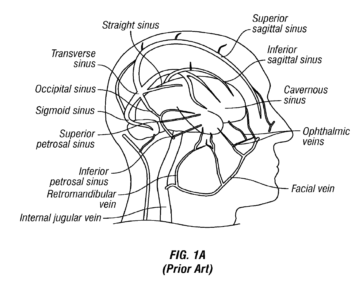
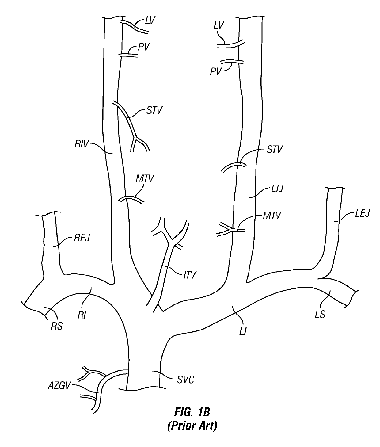
[0105]FIG. 1A is a schematic diagram of the major veins of the human brain with the names of specific veins labeled thereon. FIG. 1B is a schematic diagram showing major veins of the neck and upper thorax through which venous blood drains from the veins of the brain. Reference letters are used to indicate specific veins, as follows:

LabelVesselSVCSuperior Vena CavaRIRight InominateLILeft InominateRSRight SubclavianLSLeft SubclavianRIJRight Internal JugularLIKLeft Internal JugularREJRight External JugularLEJLeft External JugularITVInferior Thyroid VeinsMTVMiddle Thyroid VeinsS...


PUM


Abstract

Methods and devices for relieving stenoses in, or otherwise improving blood flow through, body lumens. Although applicable in a variety of different body lumens, the methods and devices of this invention are specifically useable for relieving stenoses in, or otherwise improving blood flow through, veins which drain blood from the brain for treatment of multiple sclerosis or other neurodegenerative disorders that are caused, triggered or exacerbated by venous occlusion or venous insufficiency.

Description

RELATED APPLICATION[0001]This application claims priority to U.S. Provisional Patent Application Ser. No. 61 / 303,274 filed Feb. 10, 2010, the entire disclosure of which is expressly incorporated herein by reference.FIELD OF THE INVENTION[0002]The present invention relates generally to biology and medicine and more particularly to methods, systems and devices for treating venous obstruction or venous insufficiency and for treatment of neurological or other disorders, such as multiple sclerosis (MS), which may be associated with venous obstruction or insufficiency.BACKGROUND OF THE INVENTION[0003]Pursuant to 37 CFR 1.71(e), this patent document contains material which is subject to copyright protection. The copyright owner has no objection to facsimile reproduction of the complete patent document or the patent disclosure, as it appears in the Patent and Trademark Office patent file or records, but otherwise reserves all copyright rights whatsoever.[0004]Nerve cells of the central nerv...

Claims


Application Information

Patent Timeline
Patent Type & Authority Applications(United States)
IPC IPC(8): A61F2/06A61F2/915A61F2/82A61F2/89
CPCA61F2/06A61F2/915A61F2230/0054A61F2/885A61F2220/0016A61F2/89A61F2220/0008A61F2002/825
Inventor MAKOWER, JOSHUABRIGHT, II, EARL A.BENDER, THEODORE M.CANTANESE, III, JOSEPHCHANG, JOHN Y.KING, THOMASSLONE, CLINTONLAMSON, THEODORE C.IMRAAN, AZIZ
Owner EXPLORAMED DEV LLC
Why Eureka
Try Eureka