Differentiation marker and differentiation control of eye cell
a technology of differentiation control and differentiation marker, applied in the field of cells, can solve the problems of insufficient donation of cornea in japan, no ability to regenerate itself, etc., and achieve the effect of high proliferation ability, effective identification and collection
- Summary
- Abstract
- Description
- Claims
- Application Information
AI Technical Summary
Problems solved by technology
Method used
Image

Examples
example 1
Expression of Stem Cell Marker in Whole Layer Human Corneal Tissue
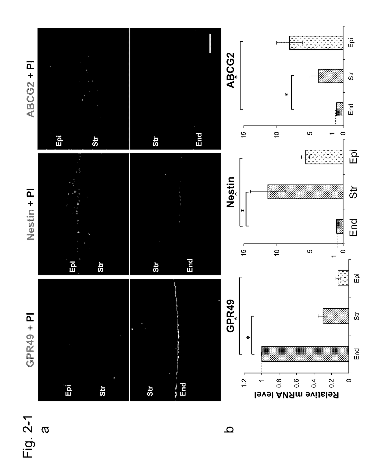
[0646]An in vivo expression pattern of GPR49 / LGR5 in human CEC was investigated by an indirect immunostaining method. For comparison, Nestin and ATP binding set subfamily G member 2 (ABCG2) which were markers of an immature cell and a precursor cell were also investigated.
[0647]In order to retrieve a protein to be expressed specifically for corneal endothelial cells, immunostaining was comprehensively performed using the already reported stem cell marker (FIG. 2a). As a result, strong expression of GPR49 / LGR5 was recognized specifically for corneal endothelial cells. As a comparison subject, Nestin (Lendahl U et al., Cell, 1990) which was an undifferentiated cell marker and ABCG2 (Chen et al., Stem Cells, 2004) which was expressed specifically for limbus corneae basal cells were used.
[0648]When CEC of these tissues were investigated in this manner, strong expression of GPR49 / LGR5 was observed, particularly, in a perip...
example 2
Localization of Expression of GPR49 / LGR5 in Human Corneal Whole Tissue Section
[0652]Then, the present inventors studied a localization pattern of GPR49 / LGR5 using the whole mount immunofluorescence method.
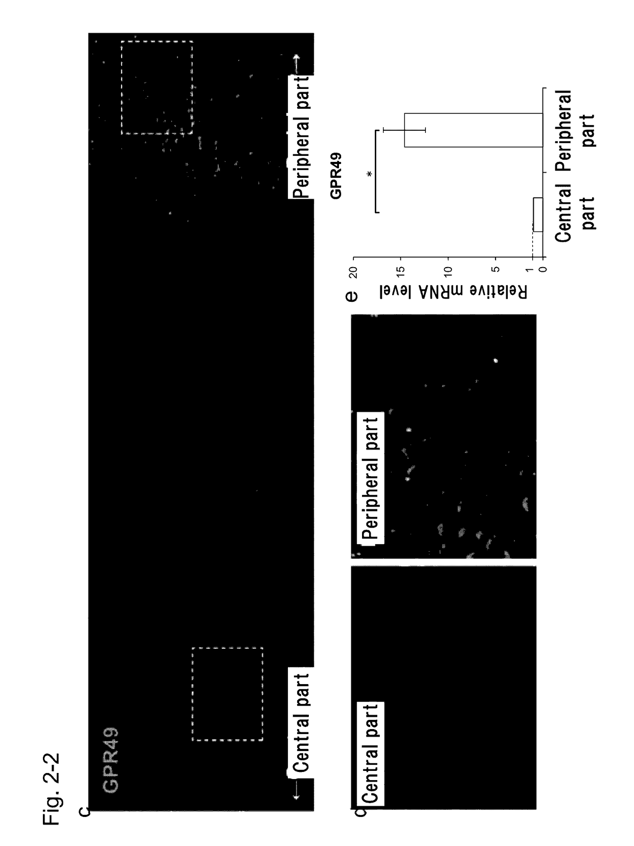
[0653]In the present Example, in order to investigate localization of expression of GPR49 / LGR5 in a corneal endothelial tissue, a Descemet's membrane containing corneal endothelial cells was peeled from a corneal tissue, and the amount of expression was compared and studied by immunostaining and real time PCR (FIG. 2c).
[0654]As a result of the immunostaining, as shown in FIGS. 2d to e, strong expression of GPR49 / LGR5 was recognized in a cell membrane and a cytoplasm of an endothelial cell in a corneal periphery (FIG. 2d). In addition, the amount of expression of GPR49 / LGR5 mRNA was also elevated in a peripheral corneal endothelium as compared with the central portion (FIG. 2e).
[0655]It was found that expression of GPR49 / LGR5 is increased in a peripheral region, in CEC, and that the...
example 3
Expression of GPR49 / LGR5 in Cultured Human Corneal Endothelial Cell
[0656]In the present Example, expression of GPR49 / LGR5 in a cultured human corneal endothelial cell was investigated.
[0657]A human corneal endothelial cell is poor in the proliferation ability in a living body, and cell culture outside a living body is also extremely difficult. Previously, a variety of culture methods have been studied, but a subculture method in the state where a hexagonal cobblestone cell form is maintained has not been established. Then, the amount of expression of GPR49 / LGR5 upon culture of a human corneal endothelial cell was studied by the existing method. As a comparison subject, Nestin was used.
[0658]As a result of immunostain, in a corneal tissue (in vivo), very strong expression was observed in both of. GPR49 / LGR5 and Nestin (FIG. 3a). In a primary cultured cell, expression of GPR49 / LGR5 was not recognized, but expression of Nestin was confirmed. In a subculture cell (in vitro P0), expressi...
PUM
 Abstract
Description
Claims
Application Information
 - R&D Engineer
- R&D Manager
- IP Professional
- Industry Leading Data Capabilities
- Powerful AI technology
- Patent DNA Extraction
Browse by: Latest US Patents, China's latest patents, Technical Efficacy Thesaurus, Application Domain, Technology Topic.
© 2024 PatSnap. All rights reserved.Legal|Privacy policy|Modern Slavery Act Transparency Statement|Sitemap